Original Article
BMSCs-induced CD45RB
+dendritic cells suppress T cell activity
Fei Hua*, Ziying Yang*, Xiaomei Teng, Yueqiu Chen, Haoyue Huang, Daguang Cui, Yunfeng Zhao, Zhenya Shen
Department of Cardiovascular Surgery, Institute of Cardiovascular Science, The First Affiliated Hospital of Soochow University, Suzhou 215006, Jiangsu, China. *Equal contributors and co-first authors.
Received September 4, 2016; Accepted November 29, 2016; Epub February 15, 2017; Published February 28, 2017
Abstract: Mesenchymal stem cells (MSCs) exhibit strong immunoregulatory capabilities, however the mechanism by which MSCs regulate T cell function via modulating dendritic cell (DC) activity is poorly understood. Rat MSCs and DCs were isolated from bone marrow, and CD4+ T cells were isolated from the spleen. DCs were co-cultured with Lipopolysaccharide and MSCs, and after 5 days, DC cell surface markers were assessed by flow cytometry.
CD45RB+ DCs were then isolated and expression of costimulatory molecules, Foxp3, and antigen uptake capability
was assessed. We also incubated CD4+ T cells in the presence or absence of conA, CD45RB+ DCs, immature DCs (imDCs) and mature DCs (mDCs) and then measured T cell proliferation, T-bet, GATA-3, RORγt, and Foxp3 mRNA
content by qPCR, and supernatant cytokine content by cytometric bead array. MSC-DC co-culture induced CD45RB
and ILT4 expression, and reduced CD86 and MHC-II expression. These purified CD45RB+ DCs exhibit surface
mark-er expression pattmark-erns, antigen uptake ability, Foxp3 expression typical of imDCs. Prolifmark-eration of T cells incubated with imDCs and CD45RB+ DCs was significantly slower than those incubated with mDCs. T cells co-cultured with
imDCs and CD45RB+ DCs expressed significantly higher levels of GATA-3 and Foxp3 mRNA and lower levels of T-bet and RORγ mRNA, and released higher levels of IL-4 and lower levels of IL-2, IFN-γ, IL-17A, IL-6 and TNF-α than those
co-cultured with mDCs. Rat bone marrow derived MSC induce CD45RB+ DC differentiation, therefore promoting
dif-ferentiation of T cells to Th2 and CD4+Foxp3+ T cells.
Keywords: Mesenchymal stem cells, dendritic cells, CD45RB, CD4+ T cells, immunosuppression
Introduction
The capacity of mesenchymal stem cells (MS- Cs) to differentiate into mesenchymal lineages and exert immune-regulatory effect suggests that MSC transplantation may confer clinical
benefits to a range of conditions [1-6]. Speci-fically, MSCs can regulate innate and acquired
immune responses by directly modifying T and B cell responses, or indirectly affecting differ-entiation of T and/or B cells, or myeloid-derived cells (e.g. dendritic cells, DCs) and macropha-
ges [7, 8]. These extraordinary capabilities sug -gest that transplantation of MSCs may repre-sent a clinically useful treatment for graft
ver-sus host disease (GVHD) [7]. Although the
mec-hanisms by which MSC modulate the behaviors of T cells, B cells and macrophages have been well studied, little is known about the mecha-nism by which MSCs regulate DC activity.
As the most important antigen presenting cells in the body, DCs process and present antigens mainly to naive and memory T cells. However, in order to initiate an immune response, DCs must mature; immature DCs are not only incapable of adequately activating T cells but can also
induce tolerance [9]. MSCs can regulate the
recruitment, maturation, and function of DCs,
and have been reported to significantly reduce
monocyte differentiation into DCs,
downregu-lateCD40, CD80, CD86, and MHC II [10-12]. MSCs also inhibit DC secretion of IL-12, the defi -ciency of which may induce T cell anergy and
tolerance [13]. Recently, Sadeghiet al. discov-ered that bone marrow derived MSCs (BM- MSCs) can inhibit the maturation of splenic DCs, and thus drive responding T cells toward
Th2 cytokine responses [14]. However, the pre -cise mechanism by which MSCs regulate T cells
DCs represent a heterogeneous population, expressing a range of different types and levels
of surface markers [15]. For example, DCs
ex-pressing high levels of CD11c and no CD45RB are termed CD11chighCD45RB- DCs, and are found to mainly secrete IL-12. In contrast, CD- 11clowCD45RB+ DCs are found to mainly secre-
te IL-10 [16-18]. CD11clowCD45RB+ have been reported to suppress immune responses by inducing tolerance and differentiation of regu-latory T cells (Tregs) in vivo [19]. Based on these
observations, we postulated that co-incubation of DCs with MSCs may induce maturation of CD11clowCD45RB+ DCs, which can subsequent-ly regulate the behavior of T cells and exert an immunosuppressive effect. In this study, we demonstrated that co-incubation of DCs with MSCs induced maturation of DCs expressing higher levels of CD45RB and ILT4, and lower levels of CD86 and MHC-II. These CD45RB+ DCs exhibit surface marker expression pat-terns and antigen uptake capability more simi-lar to immature DCs. Isolated CD45RB+ DCs inhibited the proliferation of CD4+ T cells, in- duced the differentiation of CD4+ T cells to Th2 and Tregs.
Methods and materials
Rats
Wistar rats (weight: 200-250 g) were provided by the Laboratory Animal Center of Soochow University (Suzhou, China). Animals were
main-tained under specific pathogen free and stan -dard conditions. All experimental procedures involving animals were approved by the animal ethical committee of Soochow University.
Isolation and culture of cells
MSCs were isolated from rat bone marrow as
previously described [20, 21]. Briefly, bone
marrow cells were isolated from femurs and tibias of Wistar rats. Isolated cells were
cul-tured in flasks in DMEM (Gibco, USA) supple -mented with 10% fetal bovine serum (FBS, Gibco, USA) in a CO2 incubator at 37°C. After 3 days, non-adherent cells were removed. Adhe-
rent cells were grown to 85% confluency, then
trypsinized and passaged. At passage 3, os- teogenic and adipogenic differentiation was assessed by measurement of MSC surface expression of CD90, hematopoietic markers CD45 and monocyte cell marker CD11b/c by
flow cytometry, as previously described [22].
Immature DCs were also derived from the femur and tibia bone marrow of Wistar rats.
Briefly, bone marrow was collected from femurs and passed through a 100-μm pore size mesh to remove fibrous tissue. Red blood cells were
lysed, and the remaining cells were cultured at 1×10⁶ cells/ml in RPMI 1640 medium (Hyclone, USA) supplemented with cytokines (10 ng/ml each of mouse GM-CSF and IL-4; R&D, USA). After 5 days, cells were harvested and the im- mature DCs (imDCs) phenotypewas analyzed
by flow cytometry (Becton Dickson, USA) [23].
Naive CD4+ T cells were isolated from the rat
spleen as previously described [24]. Briefly,
spleen cells were labeled with CD11b,
TER-119, Gr-1, I-A/I-E, CD8α, B220, and Gr-1 direct -ed mAbs (BD Pharmingen), and opsoniz-ed cells were removed by Dynabeads (Dynal Biotech). Purity of the remaining CD4+ cells was consis-tently 90-95%.
Co-incubation of MSCs or T cells with DCs
To investigate the effect of MSCs on DC matu-ration, 106 freshly harvested imDCs were plat-ed in 6-well plates with the same number of MSCs at a 1:1 ratio. MSCs were not irradiated before co-culture. The mixed cells were incu-bated in 2 ml of RPMI 1640 medium supple-mented with 10% FBS, stimulated with
lipopoly-saccharide (LPS) (200 ng/mL final concentra -tion, Sigma, USA) and in presence or absen- ce of MSC for 5 days. Expression of PDL1, ILT4, CD1d, CD14, CD45RA, CD45RB, CD40, CD80, CD86 and MHC-II was then assessed by
flow cytometry, and imDCs, MSCs+LPS-induced
DCs (CD45RB+ DCs) and LPS-induced DCs (ma- ture DC) were isolated. Immature DC and ma- ture DCs were separated according to CD86/ CD80 expression level, and CD45RB+ DCs were separated according to CD45RB expression,
via fluorescence-activated cell sorting.
To explore the effects of imDCs, mDCs and CD45RB+ DCs on CD4+ T cells, DCs were co-incubated with CD4+ T cells at a 1:10 ratio in RPMI 1640 medium supplemented with 10%
FBS and conA (10 ng/ml final concentration,
Sigma, USA) in 6-well plates for 3 days. CD4+ T
DC antigen uptake assay
A total of 1.0×10⁶ imDCs, CD45RB+ DCs or mDCs were incubated with latex beads/FITC-OVA (DC/Bead at ratio of 1:20000) at 37°C. After 1, 3, 6 or 24 h, cells were harvested and antigen uptake was assessed. After quenching the unincorporated FITC signal with 0.4%
try-pan blue, uptake was quantified as mean cell-associated fluorescence intensity measured by flow cytometry.
Real-time qRT-PCR
The FOXP3 mRNA content of imDCs, CD45RB+
DCs or mDCs and the T-bet, GATA-3, RORγt,
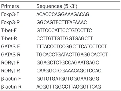
Foxp3 mRNA content of T cells co-incubated with or without these DCs were determined by qPCR using Revert Aid First Strand cDNA Synthesis Kit (Thermo, USA) and SYBR Green Realtime PCR Master Mix (Life, USA) following the manufacturer’s instructions using the prim-ers listed in Table 1.
Western blot
FOXP3 expression in imDCs, CD45RB+ DCs or mDCs was determined by western blot
(ebiosci-ence), as previously described [25, 26].
Proliferation test
The effect of DCs on T cell proliferation was
determined by CCK-8 assay. Briefly, imDCs,
CD45RB+ DCs and mDCs were inactivated by co-incubation with mitomycin (25 ug/mL, Sig- ma, USA) for 45 min at 37°C in 5% CO2. Then mitomycin was removed and cells were washed twice with PBS. Then DCs were plated in 96-well plates at density of 1×10⁵/well and co-incubat-ed with or without T cells (1×10⁵/well) and conA (10 ng/ml) at 37°C in 5% CO2 for 5 days. CCK-8
(10 μl/well) was added and the plates were
incubated for additional 4 h. The OD values at 450 nm were determined by a microplate read-er (CliniBio128C, Austria).
Detection of cytokines
The cytokine content of co-culture supernatant was assessed by CBA using the human Th1/ Th2/Th17 kit (BD Biosciences, San Jose, CA, USA) according to the manufacturer
instruc-tions. We measured IL-2, IL-10, IL-4, IL-6, IFN-γ, TNF-α, and IL-17A.
Statistical analysis
Data was presented as Mean ± SD, and ana-lyzed using ANOVA post hoc LSD test with SPSS 18.0 and Graphpad Prism 5. P<0.05 was
con-sidered statistically significant.
Results
Characterization of rat BM-MSCs
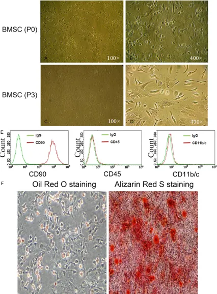
Isolated bone BM-MSCs were initially observed to be round (Figure 1A and 1B). After 3 pas-sages, cells attached well and became strip-like in appearance (Figure 1C and 1D). Expr- ession of CD90 and lack of CD45 and CD11b/ cexpressed in these cells indicated that they were MSCs (Figure 1E). Successful adipocyte or bone differentiation of these cells, induced by incubation with the corresponding mediu- ms, was validated by Oil Red O and Alizarin Red S staining (Figure 1F), confirming that
these cells were capable of adipocyte and bone differentiation.
Co-incubation with BM-MSCs induces DCs ex-pression of CD45RB and ILT4, and reduces DC
expression of CD86 and MHC-II
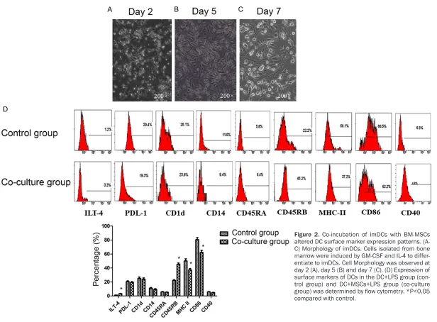
Cells (precursor cells of imDCs) isolated from bone marrow were incubated with GM-CSF and IL-4 for 7 days, and successful differentiation
to imDCs was confirmed by flow cytometry
(Figure 2A-C). These imDCs were then incubat-ed with LPS in the presence or absence of BM-MSCs. Incubation with LPS alone induced imDC maturation to mDCs. However, when im- DCs were co-incubated with BM-MSCs and LPS, cell surface markers of these DCs differed from mDCs. As shown in Figure 2D,
co-incuba-tion of imDCs with MSCs significantly increased
the fraction of DCs expressing CD45RB and ILT4, and reduced the fraction of DCs express-ing CD86 and MHC-II (Figure 2D). Expression of PLD-1, CD45RA, CD1D and CD14 did not differ
[image:3.612.91.289.82.231.2]significantly between imDCs and mDCs.
Table 1. Primers used in qPCR
Figure 1. Isolation and characterization of rat BM-MSCs. A and B. Morphology of MSCs at passage 0. C and D.
Figure 2. Co-incubation of imDCs with BM-MSCs altered DC surface marker expression patterns. (A-C) Morphology of imDCs. Cells isolated from bone marrow were induced by GM-CSF and IL-4 to differ-entiate to imDCs. Cell Morphology was observed at day 2 (A), day 5 (B) and day 7 (C). (D) Expression of surface markers of DCs in the DC+LPS group (con-trol group) and DC+MSCs+LPS group (co-culture
group) was determined by flow cytometry. *P<0.05
CD45RB+ DCs shows similar characteristics to
imDCs
The CD45RB+ DCs induced by co-incubation with BM-MSCs and LPS were isolated using
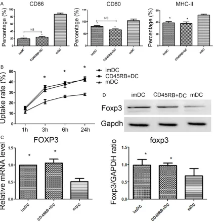
flow cytometry. Purified CD45RB+ DCs exhibit similar characteristics to imDCs, including sur-face marker expression pattern (Figure 3A), antigen uptake ability (Figure 3B), Foxp3 mRNA content (Figure 3C) and Foxp3 protein content
(Figure 3D). These features differ significantly
from those of mDCs, suggesting that BM-MSCs may inhibit the LPS-induced differentiation of imDCs to regulatory DCs.
CD45RB+ DCs and imDCs inhibit the
prolifera-tion of CD4+ T cells
[image:6.612.96.519.72.514.2]Cell morphology indicted that proliferation of T cells cultured with imDCs+conA and CD45RB+ Figure 3. Surface markers and foxp3 expression in CD45RB+ DCs. (A) CD80, CD86 and MHC-II expression in DCs was determined by flow cytometry. *P<0.01 compared with mDC group, NS, no significant difference (B) Antigen
uptake of DCs. imDCs, CD45RB+ DCs or mDCs were incubated with latex beads/FITC-OVA. Uptake was quantified by measuring cell-associated fluorescence intensity by flow cytometry. *P<0.05 compared with mDC group. (C) Foxp3
DCs+conA was significantly lower than in cells
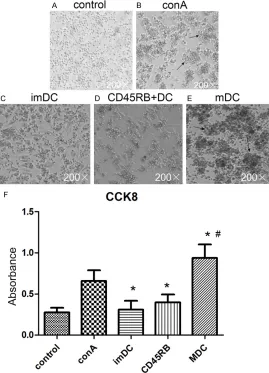
incubated with conA (Figure 4A-E). CCK-8 as-
say confirmed that proliferation rate was sig-nificantly slower in T cells incubated with
im-DCs and CD45RB+ DCs than those incubated
with conA alone, but significantly faster in T
cells incubated with mDCs+conA than those incubated with conA alone, which indicated that mDCs promoted the proliferation of T cells (Figure 4F).
imDCs and CD45RB+ DCs promote
differentia-tion of T cell to Th2 and CD4+Foxp3+ T cells
After co-incubation with different DCs for 3 days, T cells were isolated, and T-bet, GATA-3,
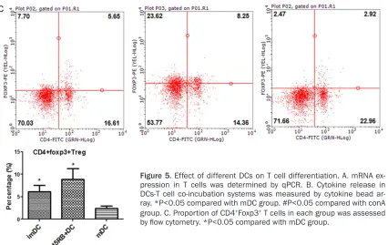
was higher than that in mDC+conA group (Figure 5C).
Discussion
[image:7.612.91.360.74.447.2]We postulated that co-incubation of DCs with MSCs may induce maturation of CD11clowCD- 45RB+ DCs, exert an immunosuppressive effect on T cells. To investigate this hypothesis we iso-lated rat BM-MSCs and prepared imDCs, and investigated the effect of MSCs on DC differen-tiation in vitro. Co-incubation with MSCs and LPS induced DC expression of CD45RB and ILT4, and down-regulated expression of CD86 and MHC-II. Isolation and characterization of Figure 4. Effect of different DCs on proliferation of CD4+ T cells. (A-E) T
cell morphology. T cells were incubated alone (A) orintheor presence (B) of conA. Arrows indicate proiliferating T cell colonies. T cells were co-incu-bated with imDC+conA (C), CD45RB+DC+conA (D) and mDC+conA (E). T
cell morphology was captured by light microscope. (F) T cell proliferation was determined by CCK-8 assay. *P<0.05 compared with conA group. #P<0.05 compared with imDC+conA and CD45RB+DC+conA groups.
RORγ, and Foxp3 mRNA content
was measured by qPCR, indicat-ing differentiation of T cells to Th1, Th2, Th17 or Treg, respec-tively. Compared with cells incu-bated with conA or mDCs+conA, T cells co-cultured with imDCs+ conA and CD45RB+ DCs+conA ex-
pressed significantly higher lev -els of GATA-3 and Foxp3 mRNA and lower levels of T-bet mRNA (Figure 5A). T cells co-cultured with mDCs+conA expressed
sig-nificantly higher levels of T-bet and RORγ mRNA than those
co-cultured with imDCs+conA or CD- 45RB+DCs+conA (Figure 5A). T-
bet, GATA-3, RORγ, and Foxp3
mRNA content did not differ
sig-nificantly between T cells co-cul -tured with imDCs+conA and CD- 45RB+DCs+conA.
Moreover, T cells co-cultured with imDCs+conA and CD45RB+ DCs+ conA release higher levels of IL-4
and lower levels of IL-2, IFN-γ, IL-17A, IL-6 and TNF-α (Figure 5B). Interestingly, IL-10 released from CD45RB+ DCs+conA group was much high than from other group, IL-10 was the only cyto-kine expressed differently from imDCs+conA and CD45RB+ DCs+ conA group.
CD45RB+ DCs indicated that MSCs-DC co- incubation inhibited DC maturation. CD45RB+ DCs exhibited similar surface biomarker pat-tern, antigen uptake ability and Foxp3 expres-sion to imDCs.
Proliferation of T cells incubated with imDCs and CD45RB+DCs was significantly slower than that of T cells incubated with mDCs. T cells co-cultured with imDCs and CD45RB+ DCs ex-
pressed significantly higher levels of GATA-3 and Foxp3 and lower levels of T-bet and RORγ
than T cells co-cultured with mDCs. Co-incu- bation of T cells with CD45RB+ DCs enhanced release of IL-4 and reduced release of IL-2,
IFN-γ, IL-17A, IL-6 and TNF-α, indicating promotion
of T0 to Th2 differentiation, and production of Tregs.
MSCs were previously reported to exhibit immu-nosuppressive effects by directly modulating T cell survival, proliferation and differentiation
[7], and MSCs have also been reported to mod -ulate the behavior of T cells indirectly, specially via DCs. MSCs can inhibit differentiation of
monocytes into imDCs [13] by blocking mono
-cyte cell cycle at the G0 phase [27]. MSCs also
inhibited maturation of imDCs and furthermore
downregulated expression of CD86/CD80 and
MHC-II [10], and reduce secretion of TNF-α and
IL-12p70 from immature DCs, while increasing
production of IL-10 [28, 29]. Our results con
-firmed that BM-MSCs downregulate DC expres -sion of CD86 and MHC-II.
We found that imDCs-MSC co-incubation pro-moted up-regulated DC expression of CD45RB+.
Purified CD45RB+ DCs exhibited similar surface marker expression, antigen uptake and Foxp3 expression to imDCs. These observations are consistent with previous reports that BM-MSCs inhibited maturation of imDCs into mDCs. We also discovered that imDCs and CD45RB+ DCs expressed higher levels of Foxp3 than mDCs. Foxp3 is a typical Treg biomarker and immunosuppressive factor, but Foxp3 expres-sion in DCs was rarely evaluated. Our results
suggest that in DCs Foxp3 may act as a specific
biomarker distinguishing imDCs and CD45RB+ DCs from mDCs.
[image:9.612.99.525.70.339.2]cannot activate T cells, and, in fact, induce immune tolerance. We found that both imDCs and CD45RB+ DCs inhibited proliferation of T cells, prompting differentiation towards Th2, and increasing the proportion of CD4+Foxp3+ T cells. However DC45RB+ DCs were not identi-cal to imDCs. T cells incubated with CD45RB+ DCs secreted more IL-10 than those incubated with imDCs or mDCs. This observation sug-gests that the mechanism by which CD45RB+
DCs influence T cell responses differs, at least
in part, from the effect of imDCs. The immuno-suppressive effect of IL-10 has been previously
reported [16, 19, 30], thus we conclude that
MSCs may exert immunosuppressive effects by inducing CD45RB+ DCs differentiation.
The mechanism by which of CD45RB+ DCs differentiate is not well understood. Delgado
et al. previously reported the vasoactive intesti-nal peptide (VIP) and the pituitary adenylate cyclase-activating polypeptide (PACAP) induced CD45RB+ DCs by stimulating theVPAC1
recep-tor and protein kinase A [31]. In future studies,
the pathway by which MSCs induce CD45RB+ DC maturation will be further investigated. Taken together, we conclude that co-incubation of MSCs and DCs inhibited maturation of im- DCs into mDCs and induced differentiation of CD45RB+ DCs. CD45RB+ DCs further inhibited proliferation of CD4+ T cells, and induced dif-ferentiation of CD4+ T cells to Th2 and Tregs.
Acknowledgements
This study was supported by National Natural Science Foundation of China (No. 81401316). Disclosure of conflict of interest
None.
Address correspondence to: Zhenya Shen, Depart- ment of Cardiovascular Surgery, Institute of Car-
diovascular Science, The First Affiliated Hospital of
Soochow University, 188 Shizi Street, Suzhou 215- 006, Jiangsu, China. Tel: +86-13606210812; Fax: +86-21-57643271; E-mail: [email protected]
References
[1] Zhao K and Liu Q. The clinical application of mesenchymal stromal cells in hematopoietic stem cell transplantation. J Hematol Oncol 2016; 9: 46.
[2] Najar M, Raicevic G, Fayyad-Kazan H, Bron D, Toungouz M and Lagneaux L. Mesenchymal stromal cells and immunomodulation: a gath-ering of regulatory immune cells. Cytotherapy 2016; 18: 160-171.
[3] Mamidi MK, Das AK, Zakaria Z and Bhonde R. Mesenchymal stromal cells for cartilage repair in osteoarthritis. Osteoarthritis Cartilage 2016; 24: 1307-1316.
[4] Malhotra S, Hu MS, Marshall CD, Leavitt T, Cheung AT, Gonzalez JG, Kaur H, Lorenz HP and Longaker MT. Mesenchymal stromal cells as cell-based therapeutics for wound healing. Stem Cells Int 2016; 2016: 4157934.
[5] Ezquer F, Bruna F, Calligaris S, Conget P and Ezquer M. Multipotent mesenchymal stromal cells: a promising strategy to manage alcoholic liver disease. World J Gastroenterol 2016; 22: 24-36.
[6] Thanunchai M, Hongeng S and Thitithanya-nont A. Mesenchymal stromal cells and viral infection. Stem Cells Int 2015; 2015: 860950.
[7] Castro-Manrreza ME and Montesinos JJ. Im-munoregulation by mesenchymal stem cells: biological aspects and clinical applications. J Immunol Res 2015; 2015: 394917.
[8] Uccelli A and de Rosbo NK. The immunomodu-latory function of mesenchymal stem cells: mode of action and pathways. Ann N Y Acad Sci 2015; 1351: 114-126.
[9] Liu YJ, Kanzler H, Soumelis V and Gilliet M. Dendritic cell lineage, plasticity and cross-reg-ulation. Nat Immunol 2001; 2: 585-589.
[10] Jiang XX, Zhang Y, Liu B, Zhang SX, Wu Y, Yu XD and Mao N. Human mesenchymal stem cells inhibit differentiation and function of mono-cyte-derived dendritic cells. Blood 2005; 105: 4120-4126.
[11] Maccario R, Podesta M, Moretta A, Cometa A, Comoli P, Montagna D, Daudt L, Ibatici A, Piag-gio G, Pozzi S, Frassoni F and Locatelli F. Inter-action of human mesenchymal stem cells with
cells involved in alloantigen-specific immune
response favors the differentiation of CD4+ T-cell subsets expressing a regulatory/suppres-sive phenotype. Haematologica 2005; 90: 516-525.
[12] Spaggiari GM, Abdelrazik H, Becchetti F and Moretta L. MSCs inhibit monocyte-derived DC maturation and function by selectively interfer-ing with the generation of immature DCs: cen-tral role of MSC-derived prostaglandin E2. Blood 2009; 113: 6576-6583.
[13] Nauta AJ, Kruisselbrink AB, Lurvink E, Wil-lemze R and Fibbe WE. Mesenchymal stem cells inhibit generation and function of both CD34+-derived and monocyte-derived dendrit-ic cells. J Immunol 2006; 177: 2080-2087.
effects of mice mesenchymal stem cells on maturation and activation of dendritic cells. Iran J Immunol 2014; 11: 177-188.
[15] Banchereau J and Steinman RM. Dendritic cells and the control of immunity. Nature 1998; 392: 245-252.
[16] Wakkach A, Fournier N, Brun V, Breittmayer JP, Cottrez F and Groux H. Characterization of den-dritic cells that induce tolerance and T regula-tory 1 cell differentiation in vivo. Immunity 2003; 18: 605-617.
[17] Fujita S, Seino K, Sato K, Sato Y, Eizumi K, Ya-mashita N, Taniguchi M and Sato K. Regulatory dendritic cells act as regulators of acute lethal
systemic inflammatory response. Blood 2006;
107: 3656-3664.
[18] Liu QY, Yao YM, Zhang SW, Yan YH and Wu X. Naturally existing CD11c(low)CD45RB(high) dendritic cells protect mice from acute severe
inflammatory response induced by thermal in -jury. Immunobiology 2011; 216: 47-53.
[19] Svensson M, Maroof A, Ato M and Kaye PM. Stromal cells direct local differentiation of regulatory dendritic cells. Immunity 2004; 21: 805-816.
[20] He J, Teng X, Yu Y, Huang H, Ye W, Ding Y and Shen Z. Injection of Sca-1+/CD45+/CD31+ mouse bone mesenchymal stromal-like cells improves cardiac function in a mouse myocar-dial infarct model. Differentiation 2013; 86: 57-64.
[21] Huang H, He J, Teng X, Yu Y, Ye W, Hu Y and Shen Z. Combined intrathymic and intrave-nous injection of mesenchymal stem cells can prolong the survival of rat cardiac allograft as-sociated with decrease in miR-155 expression. J Surg Res 2013; 185: 896-903.
[22] Dominici M, Le Blanc K, Mueller I, Slaper- Cortenbach I, Marini F, Krause D, Deans R, Ke-ating A, Prockop D and Horwitz E. Minimal
cri-teria for defining multipotent mesenchymal
stromal cells. The international society for cel-lular therapy position statement. Cytotherapy 2006; 8: 315-317.
[23] Cahill EF, Tobin LM, Carty F, Mahon BP and English K. Jagged-1 is required for the expan-sion of CD4+ CD25+ FoxP3+ regulatory T cells and tolerogenic dendritic cells by murine mes-enchymal stromal cells. Stem Cell Res Ther 2015; 6: 19.
[24] Turnquist HR, Raimondi G, Zahorchak AF, Fischer RT, Wang Z and Thomson AW. Rapamy-cin-conditioned dendritic cells are poor stimu-lators of allogeneic CD4+ T cells, but enrich for
antigen-specific Foxp3+ T regulatory cells and
promote organ transplant tolerance. J Immu-nol 2007; 178: 7018-7031.
[25] Fontenot JD, Gavin MA and Rudensky AY. Foxp3 programs the development and function of CD4+CD25+ regulatory T cells. Nat Immu-nol 2003; 4: 330-336.
[26] Ono M, Yaguchi H, Ohkura N, Kitabayashi I, Nagamura Y, Nomura T, Miyachi Y, Tsukada T and Sakaguchi S. Foxp3 controls regulatory T-cell function by interacting with AML1/Runx1. Nature 2007; 446: 685-689.
[27] Ramasamy R, Fazekasova H, Lam EW, Soeiro I, Lombardi G and Dazzi F. Mesenchymal stem cells inhibit dendritic cell differentiation and function by preventing entry into the cell cycle. Transplantation 2007; 83: 71-76.
[28] Aggarwal S and Pittenger MF. Human mesen-chymal stem cells modulate allogeneic im-mune cell responses. Blood 2005; 105: 1815-1822.
[29] Kronsteiner B, Peterbauer-Scherb A, Grillari-Voglauer R, Redl H, Gabriel C, van Griensven M and Wolbank S. Human mesenchymal stem cells and renal tubular epithelial cells
differen-tially influence monocyte-derived dendritic cell
differentiation and maturation. Cell Immunol 2011; 267: 30-38.
[30] Liu QY, Yao YM, Zhang SW and Sheng ZY. As-tragalus polysaccharides regulate T cell-medi-ated immunity via CD11c(high)CD45RB(low) DCs in vitro. J Ethnopharmacol 2011; 136: 457-464.