Original Article
Profiling of plasma circulating miRNA in coronary heart
disease patients detected by next-generation
small RNA sequencing
Zhiming Zhou1, Jun Chen2, Taishun Wu1, Qingcheng Liu1, Huiwen Song3, Shunchang Sun4
1Baoan Center for Disease Control and Prevention of Shenzhen, Shenzhen 518101, China; 2Department of Cardiovascular Diseases, Shenzhen Baoan Hospital, Southern Medical University, Shenzhen 518101, China; 3Department of Cardiovascular Diseases, Minhang Hospital, Fudan University, Shanghai 201199, China; 4Department of Laboratory Medicine, Ruijin Hospital North, School of Medicine, Shanghai Jiao Tong University, Shanghai 201801, China
Received May 13, 2015; Accepted September 11, 2016; Epub May 15, 2017; Published May 30, 2017
Abstract: MiRNA levels in plasma have emerged as potential novel noninvasive biomarkers for some diseases. We aimed to identify a miRNA expression profile in plasma of patients with coronary heart disease. The levels of plasma miRNA from a cohort of 49 patients with coronary heart disease and 13 healthy controls were detected by small RNA deep sequencing, the differentially expressed miRNAs were validated using the reverse transcription real-time quantitative polymerase chain reaction from an additional cohort of 135 hospitalized patients and 140 controls. Five miRNAs were differentially expressed, 30c-2-3p and 5091 were upregulated and 125b-5p, miR-501-3p, and miR-31-5p downregulated in plasma of patients with coronary heart disease than in controls. This study identified 5 plasma miRNAs that might play an important role in the pathogenesis of coronary heart disease. The level of miR-30c-2-3p was 132 folds upregulation (P=0.000), it suggests that miR-30c-2-3p may be a potential noninvasive biomarker for the diagnosis of coronary heart disease, however, with further research.
Keywords: miRNA, plasma, profiling, coronary heart disease
Introduction
Coronary heart disease (CHD), involving athero-sclerosis and myocardial infarction, is a leading cause of morbidity and mortality in the world. Although, pathogenesis of CHD is still not fully understood, it is considered that vascular endo- thelial cell injury, platelet reactivity, vascular smooth muscle cell proliferation, lipid infiltra-tion, and increased synthesis of connective tis-sue are the main pathological processes of CHD [1]. Forty-six CHD susceptibility loci identi-fied through GWAS do not appear to be mediat-ing risk through effects on traditional risk fac-tors, such as blood pressure and lipid levels, in subjects of European descent [2]. MicroRNA (miRNA) is reported as a group of short, single-stranded, noncoding RNA that can function as endogenous RNA interference to regulate ex- pression of the targeted genes. MiRNAs play an essential role in fundamental biological
character-ize miRNA profiles in plasma from CHD patients. Plasma samples from CHD patients and healthy individuals in a Chinese Han population were assessed for miRNA expression. We character-ize a plasma miRNA profile in CHD that may serve as a non-invasive approach in early diag-nosis for CHD in the future.
Materials and methods
Patients and controls
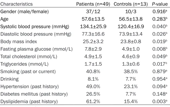
[image:2.612.92.389.97.285.2]A cohort of 49 hospitalized CHD patients (ba- seline clinical characteristics summarized in Table 1) and 13 healthy volunteers were recruit-ed in next-generation small RNA sequencing study, an additional cohort of 135 hospitalized CHD patients (baseline data summarized in Table 2) and 140 controls were recruited in quantitative reverse transcription-polymerase chain reaction analysis during June 2013 to July 2014 from Shenzhen Baoan Hospital, Southern Medical University. All CHD cases were diagnosed according to the following re- quirements: coronary angiography showed that stenosis of more than 50% was present in at least 1 of 3 main blood arteries (left circumflex artery, right coronary artery, and left anterior descending coronary artery) and stenosis can be revealed in 2 left main coronary arteries; patients suffered from symptoms of unstable angina pectoris: angina was present when at rest and lasted over 20 minutes, or newly occ- ured (<2 months) severe angina, or aggravation
nal disease, liver disease, connective tissue disease, and tumors were excluded. Healthy volunteers had no history or any symptom of CHD. As control, we used healthy control sub-jects without other documented disorders. Physical examination, laboratory tests, ECG and other preliminary tests were performed for these control subjects. The study was approved by the ethics committee of Shenzhen Baoan Hospital, Southern Medical University. Written consent was obtained from all patients and control individuals.
RNA isolation
Five-ml peripheral blood was collected from each participant using a drying tube containing EDTA as anticoagulant. The whole blood was separated into plasma and cellular fractions by centrifugation at 3,000 rpm and 4°C for 10 min, followed by 12000 rpm for 15 min to com-pletely remove cell debris. Plasma was again centrifuged at 25,000 rpm and 4°C for 10 min to remove additional cellular nucleic acids attached to cell debris, then kept plasma fro-zen at -80°C. Total RNA isolation from plasma samples was performed with the miRNeasy serum/plasma kit (Qiagen) according to the manufacturer’s protocol. After the initial dena-turizing step, we spiked in synthetic C.elegans miR-39 for all plasma in order to control varia-tions in RNA extraction and purification proce-dures because of the absence of homologous sequences in humans. To assess the quality of Table 1. Baseline clinical characteristics of CHD patients recruited in
the small RNA sequencing cohort
Characteristics Patients (n=49) Controls (n=13) P-value Gender (male/female) 37/12 10/3 0.916a
Age 57.6±13.5 56.5±13.8 0.283b
Systolic blood pressure (mmHg) 134.1±25.9 120.4±16.9 0.040b
Diastolic blood pressure (mmHg) 77.3±16.6 73.9±13.4 0.026b
Body mass index 25.2±3.2 23.8±0.8 0.019b
Fasting plasma glucose (mmol/L) 7.8±2.9 4.9±1.0 0.008b
Total cholesterol (mmol/L) 4.9±1.5 4.6±0.9 0.049b
Triglycerides (mmol/L) 1.7±1.5 1.3±0.6 0.017b
Smoking (past or current) 40.8% 38.5% 0.879a
Drinking 8.1% 7.7% 0.954a
Hypertension (past history) 49.0% 23.1% 0.094a
Diabetes mellitus (past history) 26.5% 7.7% 0.148a
Dyslipidemia (past history) 61.2% 15.4% 0.003a a: Chi square test; b: t test.
RNA, the purified RNA was measured with using Agilent 2100 Bioanalyzer and ABI Step-One-Plus real-time PCR.
Small RNA sequencing
Sixty-two smRNA libraries were constructed from RNAs of 49 CHD patients and 13 healthy volunteers by using a smRNA sample prep kit (Illumina, USA) following the standard proto-cols. Briefly, the appropriate fractions ranged from 18nt to 30nt were separated, purified via 15% (w/v) denaturing polyacrylamide gel elec-trophoresis and then linked to RNA adap- tors (5’-GTTCAGAGTTCTACAGTCCGACGATC and 3’-TCGTATGCCGTCTTCTGCTTG) using T4 RNA ligase and were subsequently reverse tran-scribed into cDNA by Superscript II Reverse Transcriptase. The cDNA fragments were ampli-fied, and the RT-PCR products were sequenced by the Illumina Hiseq 2000 (Illumina, San Die- go, CA, USA) according to Illumina’s protocol at Beijing Genomics Institute-Shenzhen.
Data analysis
The smRNA sequencing data were analyzed referring to the methods described in the previ-ous publications [8]. Briefly, after deleting 5’ adaptor and trimming 3’ acceptor sequences, filtering low quality reads (Q<10, the quality value was calculated by Q=ASCII character code-64) and cleaning up contaminated reads, the occurrence of each unique read was
count-aligned against the sequences of other non-coding RNAs (rRNAs, tRNAs, snRNAs, snoRNAs, scRNAs) presented on Rfam (http://rfam. sanger.ac.uk/) database, repeats database as well as piRNA database. The known miRNAs were annotated by aligning to miRBase 18.0 (http://www.mirbase.org/index.shtml). The re- maining reads that cannot be annotated were used to predict novel miRNAs by Mireap (http:// sourceforge.net/projects/mireap/). In consid-eration of a few smRNA tags might map to more than one category, this study followed the priority rules described in the previous publication to guarantee that every unique smRNA was mapped to unique annotation as follows: miRNA, Rfam, repeats, piRNA and mRNA [9]. All miRNA profiles were normalized using the geometric mean for each sample over all miRNAs. Profiles of the most differen-tially expressed miRNAs were subjected to hierarchical clustering to create a heat map. The predicted target genes of differentially expressed and miRNAs were subjected to analysis of gene ontology terms for gene ontology (GO) analysis. The predicted miRNA targets were mapped to the signaling path- way annotation databases downloaded from KEGG (http://www.genome.jp/kegg/) for path-way analysis.
Quantitative RT-PCR assay
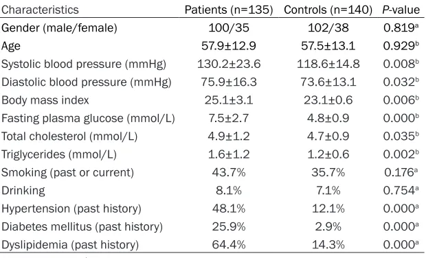
[image:3.612.92.402.97.285.2]To confirm the expression of miRNAs identified by the deep sequencing approach, reverse Table 2. Baseline clinical characteristics of CHD patients enrolled in the
validated cohort
Characteristics Patients (n=135) Controls (n=140) P-value Gender (male/female) 100/35 102/38 0.819a
Age 57.9±12.9 57.5±13.1 0.929b
Systolic blood pressure (mmHg) 130.2±23.6 118.6±14.8 0.008b
Diastolic blood pressure (mmHg) 75.9±16.3 73.6±13.1 0.032b
Body mass index 25.1±3.1 23.1±0.6 0.006b
Fasting plasma glucose (mmol/L) 7.5±2.7 4.8±0.9 0.000b
Total cholesterol (mmol/L) 4.9±1.2 4.7±0.9 0.035b
Triglycerides (mmol/L) 1.6±1.2 1.2±0.6 0.002b
Smoking (past or current) 43.7% 35.7% 0.176a
Drinking 8.1% 7.1% 0.754a
Hypertension (past history) 48.1% 12.1% 0.000a
Diabetes mellitus (past history) 25.9% 2.9% 0.000a
Dyslipidemia (past history) 64.4% 14.3% 0.000a a: Chi square test; b: t test.
transcription real-time quantitative polymerase chain reaction (RT-qPCR) analysis was per-formed using the total RNA samples from another 135 hospitalized CHD patients and 140 controls. The expressions of miRNAs were
[image:4.612.98.374.79.330.2]group, delta Ct values from two groups were evaluated using the t-Student test. Quantitative data from qRT-PCR were compared using unpaired t-tests. P<0.05 was considered statis-tically significant.
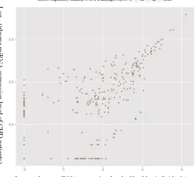
[image:4.612.93.373.418.500.2]Figure 1. The miRNA expression profiles in CHD patients and healthy indi-viduals are presented by next-generation RNA sequencing technology.
Table 3. Summary of differentially expressed plasma miRNAs by next-generation RNA sequencing technologya
miR ID log2Ratio (CHD/healthy) Probability Statusb
miR-30c-2-3p 16.37457256 0.80200747 Upregulated miR-5091 15.67550423 0.797619048 Upregulated miR-125b-5p -10.62370057 0.795238095 Downregulated miR-501-3p -10.93135424 0.791923436 Downregulated miR-31-5p -9.953194374 0.783753501 Downregulated
a: RNA sequencing cohort including 49 CHD patients and 13 healthy controls; bstatus: probability ≥0.78 & abs (log
2 (Y/X)) ≥1.0 as regulated.
quantified by All-in-One mi- RNA qRT-PCR Detection Kit (GeneCopoeia, Rockville, USA) following reverse transcription according to the manufactur-er’s instructions. Reactions were loaded onto a 96-well plate and run in triplicate on a LightCycler 480 Real-Time PCR System (Roche). The reactions were firstly incubat-ed at 95°C for 10 minutes, followed by 40 cycles of de- naturation at 95°C for 10 seconds, annealing at 60°C for 20 seconds, then 10 sec-onds of extension at 72°C. A melting curve analysis was performed immediately after qRT-PCR cycling to monitor the qRT-PCR reaction. The primers used for qRT-PCR in this study are as following: miR-30c-2-3p: CTGGGAGAA- GGCTGTTTACTCT, miR-5091: CTGGGAGAAGGCTGTTTACTCT, miR-125b-5p: TCCCTGAGAC- CCTAACTTGTGA, miR-501-3p: AATGCACCCGGGCAAGGATT- CT, miR-31-5p: AGGCAAGATG- CTGGCATAGCT, RNU6-2: CA- AAGTCAGTGCAGGTAGGCTTA and SNORD44: CCTGGATGAT- GATAAGCAAATGCTG. The sn- RNA levels of RNU6-2 and SNORD44 were used as in- ternal controls. MiRNA ex- pression was normalized to snRNAs of RNU6-2 and SN- ORD44 and to get the rela- tive abundance. Averages of five independent experiments each performed in triplicate with standard errors were presented. The delta Ct meth-od was used to determine relative number of copies of miRNA. To search for differen-tially expressed miRNAs be- tween CHD group and healthy Table 4. Summary of differentially expressed plasma miRNAs by
real-time PCR
[image:4.612.91.372.570.649.2]Results
Overview of small RNA sequencing data
To determine the small RNA profile in plasma of CHD patients, we sequenced 62 (49 CHD pati- ents and 13 healthy individuals) small RNA libraries using Solexa next-generation sequenc-ing technology. The majority of these clean reads was 22nt in length, with sizes varying between 18nt and 30nt after removing the adaptor sequences and low quality reads. These clean reads were mapped to several fil-ter databases, such as the Human Genome, rRNA, tRNA and Rfam sequence databases, and were subsequently mapped to miRbase (V14.1). After detecting other types of small RNAs, including rRNAs, snRNAs, snoRNAs, and tRNAs, 358 unique tags corresponding to 167419 reads in healthy individuals and 374 unique tags corresponding to 198554 reads in CHD patients were identified as known miRNAs.
Plasma miRNA differentially expressed in CHD patients and healthy individuals
To investigate possible differentially expressed plasma miRNAs in CHD patients and healthy individuals, the counts of each kind of miRNA were normalized based on the total number of all the clean reads mapped on the genome, and then compared between the CHD patients and healthy individuals. Five known miRNAs were differentially expressed between plasma from CHD patients and healthy individuals (Figure 1). Of these 5 miRNAs, miR-30c-2-3p and miR-5091 were upregulated, miR-125b-5p, miR-501-3p, and miR-31-5p were downreg-ulated (Table 3). To validate the expression of these differentially expressed known miRNAs detected by next-generation sequencing tech-nology, five differentially expressed miRNAs were chosen for quantification by real-time PCR. Real-time PCR showed that expression of
plasma miR-30c-2-3p and miR-5091 were upregulated, miR-125b-5p, miR-501-3p, and miR-31-5p were downregulated in the CHD patient (Table 4). The levels of differentially expressed plasma miRNAs measured by quan-titative real-time PCR were consistent with the results obtained from next-generation sequenc-ing technology.
Prediction of the miRNA targeted genes and pathways
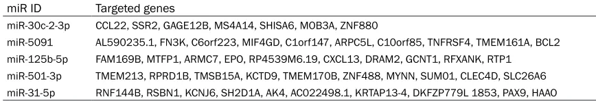
After the investigation of these five known miRNAs that were differentially expressed between CHD patients and healthy individuals, we then predicted the targeted genes that could potentially be targeted by these miRNAs using Targetscan. After prediction of the miRNA targeted genes, we found that five predicted targeted genes of differentially expressed miRNAs appeared to be involved in a broad range of biological processes (Table 5). Most of the targets related to metabolic pro-cess (e.g., macromolecule metabolic propro-cess, primary metabolic process and cellular meta-bolic process), cellular component organization or biogenesis (e.g., cell junction assembly, cell morphogenesis, multicellular organismal pro-cess), cellular process (e.g., cellular protein modification process, cell differentiation, cell maturation), developmental process (e.g., car-diac muscle tissue development, muscle tissue morphogenesis, mesoderm formation), cell proliferation, and reproduction (Figure 2). Discussion
[image:5.612.88.522.98.172.2]Both genetic and environmental factors cumu-latively contribute to CHD risk in human popula-tions. Recent studies have leveraged some genetic variations to identify multiple sites of CHD susceptibility; however, the causal mecha-nisms largely remain elusive [10]. MiRNA, first described in 1993, regulate gene translation by binding at complementary mRNA sequences, Table 5. GO analysis for predicted targets of differentially expressed plasma miRNA between CHD patients and healthy individuals
miR ID Targeted genes
miR-30c-2-3p CCL22, SSR2, GAGE12B, MS4A14, SHISA6, MOB3A, ZNF880
miR-5091 AL590235.1, FN3K, C6orf223, MIF4GD, C1orf147, ARPC5L, C10orf85, TNFRSF4, TMEM161A, BCL2
miR-125b-5p FAM169B, MTFP1, ARMC7, EPO, RP4539M6.19, CXCL13, DRAM2, GCNT1, RFXANK, RTP1
miR-501-3p TMEM213, RPRD1B, TMSB15A, KCTD9, TMEM170B, ZNF488, MYNN, SUM01, CLEC4D, SLC26A6
then initiating mRNA cleavage or obstructing mRNA incorporation [11]. Studies have found that miRNA modulates a wide range of patho-physiological processes such as tumorigene-sis, cardiovascular diseases, pain, viral infec-tions, cellular proliferation, cellular differentia-tion, and apoptosis during the last decade [12]. Our study from the plasma miRNA expression profile of CHD patients found that miRNAs were differentially expressed between plasma from CHD and healthy group. Among differentially expressed miRNAs, 30c-2-3p and miR-5091 were upregulated. MiR-30 family mem-bers have been implicated in cancer, myocar-dial matrix remodeling, cellular senescence, osteoblast differentiation, adipogenesis, and epithelial-to-mesenchymal transition [13]. In vitro, overexpression miR-30 promotes angio-genic sprouting in epithelial cells. MiR-30 is among the most highly expressed miRNAs in cardiac myocytes [14]. A previous study showed that miR-30 downregulate connective tissue growth factor, a key profibrotic protein, thereby establish an important role for it in the control of structural changes in the extracellular matrix of the myocardium [15]. In our study, the level of plasma miR-30c-2-3p was 132 folds upregu-lation, it suggests that dysregulation of miR-30c-2-3p in the heart may contribute to myo-cardial matrix remodeling after myomyo-cardial in- farction for CHD patients. Molecular markers have the potential to help in the disease diag-nosis and treatment selection. Previous stud-ies have identified a few serum markers, such as cardiac troponins, myoglobin, heart fatty acid binding protein, and glycogen phosphory-lase BB are used to help in establishing the diagnosis of myocardial infarction [16]. New markers of early detection of myocardial dam-age are is still on. Our study identified that plas-ma miR-30c-2-3p was high upregulated betwe- en CHD patients and healthy controls, thereby it points to the possibility of miR-30c-2-3p as a circulating molecular marker in the diagnosis of CHD. Therefore, more studies from larger CHD patient groups will be required, which will all- ow to determine the cutoff values to classify the plasma miR-30c-2-3p expression level as “high” or “low”.
This study showed that plasma miR-5091 was 3.78-fold upregulated in CHD patients. The miR-5091 targeted genes predicted by Targ-
etscan contain FN3K, MIF4GD, TNFRSF4, and BCL2, and so on. The FN3K gene encodes an enzyme which catalyzes the fructosamines phosphorylation which may result in deglyca-tion [17]. The MIF4GD protein interacts with the N-terminus of the stem-loop binding protein and the 3’ end of histone mRNA, thereby, this interaction facilitates the activation of histone mRNA translation [18]. The TNFRSF4 protein is a member of the TNF-receptor superfamily sh- own to activate NF-kappaB signaling pathway [19]. The BCL2 gene encodes an integral outer mitochondrial membrane protein that blocks the apoptotic death of lymphocytes [20]. How the miR-5091 regulates the FN3K, MIF4GD, TNFRSF4, and BCL2 genes, thereby leading to CHD keeps unknown.
MiRNA can regulate cardiac and skeletal mus-cle function in both development and disease. It is noteworthy that miR-125b-5p have been shown to be functionally involved in cardiovas-cular diseases such as acute myocardial infarc-tion, heart failure, and hypertrophy [21]. MiR-125b-5p is an abundant miRNA present in diac valve, and has been shown to protect car-diac muscle from ischemia/reperfusion injury by reducing infarct size by 60%. In addition, miR-125b-5p has been associated with athero-sclerosis and CHD development [22]. The plas-ma level of miR-5091 was significantly lower in patients with CHD when compared with healthy controls in this study. Our study is consistent with previous studies [23]. It suggests that miR-5091 may be involved in the pathogenesis of CHD, but not be released from the myocardi- um.
both of miR-31-5p and miR-501-3p may be closely involved in the pathogenesis of CHD. Acknowledgements
This study was supported by research grants from Science and Technology Innovation Com- mission of Shenzhen Municipality (No. JCYJ- 20140414154847277).
Disclosure of conflict of interest
None.
Address correspondence to: Shunchang Sun, De- partment of Laboratory Medicine, Ruijin Hospital North, School of Medicine, Shanghai Jiao Tong Uni- versity, 888 Shuangding Road, Jiading, Shanghai 201801, China. Tel: 8613636359946; E-mail: [email protected]
References
[1] Colantonio LD, Bittner V, Reynolds K, Levitan EB, Rosenson RS, Banach M, Kent ST, Derose SF, Zhou H, Safford MM, Muntner P. Association of serum lipids and coronary heart disease in contemporary observational studies. Circula- tion 2016; 133: 256-264.
[2] Laugsand LE, Ix JH, Bartz TM, Djousse L, Kizer JR, Tracy RP, Dehghan A, Rexrode K, Lopez OL, Rimm EB, Siscovick DS, O’Donnell CJ, Newman A, Mukamal KJ, Jensen MK. Fetuin-A and risk of coronary heart disease: a Mendelian ran-domization analysis and a pooled analysis of AHSG genetic variants in 7 prospective stud-ies. Atherosclerosis 2015; 243: 44-52. [3] Wei S, Wang Y, Xu H, Kuang Y. Screening of
po-tential biomarkers for chemoresistant ovarian carcinoma with miRNA expression profiling data by bioinformatics approach. Oncol Lett 2015; 10: 2427-2431.
[4] Zhu J, Wang S, Zhang W, Qiu J, Shan Y, Yang D, Shen B. Screening key microRNAs for castra-tion-resistant prostate cancer based on miR-NA/mRNA functional synergistic network. On- cotarget 2015; 6: 43819-43830.
[5] Rachagani S, Macha MA, Menning MS, Dey P, Pai P, Smith LM, Mo YY, Batra SK. Changes in microRNA (miRNA) expression during pan- creatic cancer development and progression in a genetically engineered KrasG12D;Pdx1-Cre mouse (KC) model. Oncotarget 2015; 6: 40295-40309.
[6] Debernardi S, Massat NJ, Radon TP, Sangara- lingam A, Banissi A, Ennis DP, Dowe T, Chelala C, Pereira SP, Kocher HM, Young BD, Bond-Smith G, Hutchins R, Crnogorac-Jurcevic T.
Noninvasive urinary miRNA biomarkers for early detection of pancreatic adenocarcinoma. Am J Cancer Res 2015; 5: 3455-3466. [7] Imhof A, Koenig W, Jaensch A, Mons U, Brenner
H, Rothenbacher D. Long-term prognostic val-ue of IgM antibodies against phosphorylcho-line for adverse cardiovascular events in pa-tients with stable coronary heart disease. Ath- erosclerosis 2015; 243: 414-420.
[8] Gong J, Wu Y, Zhang X, Liao Y, Sibanda VL, Liu W, Guo AY. Comprehensive analysis of human small RNA sequencing data provides insights into expression profiles and miRNA editing. RNA Biol 2014; 11: 1375-1385.
[9] Cheng WC, Chung IF, Tsai CF, Huang TS, Chen CY, Wang SC, Chang TY, Sun HJ, Chao JY, Cheng CC, Wu CW, Wang HW. YM500v2: a small RNA sequencing (smRNA-seq) database for human cancer miRNome research. Nucleic Acids Res 2015; 43: D862-867.
[10] Shi Y, Zhou L, Huang LH, Lian YT, Zhang XM, Guo H, Wu TC, Cheng LX, He MA. Plasma ferri-tin levels, genetic variations in HFE gene, and coronary heart disease in Chinese: a case-con-trol study. Atherosclerosis 2011; 218: 386-390.
[11] Wang J, Lei ZJ, Guo Y, Wang T, Qin ZY, Xiao HL, Fan LL, Chen DF, Bian XW, Liu J, Wang B. miR-NA-regulated delivery of lincRNA-p21 suppre- sses β-catenin signaling and tumorigenicity of colorectal cancer stem cells. Oncotarget 2015; 6: 37852-37870.
[12] Balzano F, Deiana M, Dei Giudici S, Oggiano A, Baralla A, Pasella S, Mannu A, Pescatori M, Porcu B, Fanciulli G, Zinellu A, Carru C, Deiana L. miRNA stability in frozen plasma samples. Molecules 2015; 20: 19030-19040.
[13] Zhang T, Tian F, Wang J, Jing J, Zhou SS, Chen YD. Endothelial cell autophagy in atherosclero-sis is regulated by miR-30-mediated transla-tional control of ATG6. Cell Physiol Biochem 2015; 37: 1369-1378.
[14] Qi F, He T, Jia L, Song N, Guo L, Ma X, Wang C, Xu M, Fu Y, Li L, Luo Y. The miR-30 family inhib-its pulmonary vascular hyperpermeability in the premetastatic phase by direct targeting of Skp2. Clin Cancer Res 2015; 21: 3071-3080. [15] Guess MG, Barthel KK, Harrison BC, Leinwand
LA. miR-30 family microRNAs regulate myo-genic differentiation and provide negative feedback on the microRNA pathway. PLoS One 2015; 10: e0118229.
[17] Hellwig A, Scherber A, Koehler C, Hanefeld M, Henle T. A new HPLC-based assay for the mea-surement of fructosamine-3-kinase (FN3K) and FN3K-related protein activity in human erythrocytes. Clin Chem Lab Med 2014; 52: 93-101.
[18] Wan C, Hou S, Ni R, Lv L, Ding Z, Huang X, Hang Q, He S, Wang Y, Cheng C, Gu XX, Xu G, Shen A. MIF4G domain containing protein regulates cell cycle and hepatic carcinogenesis by an-tagonizing CDK2-dependent p27 stability. On- cogene 2015; 34: 237-245.
[19] Schreiber TH, Wolf D, Bodero M, Gonzalez L, Podack ER. T cell costimulation by TNFR super-family (TNFRSF)4 and TNFRSF25 in the con-text of vaccination. J Immunol 2012; 189: 3311-3318.
[20] Zhu P, Zhao MY, Li XH, Fu Q, Zhou ZF, Huang CF, Zhang XS, Huang HL, Tan Y, Li JX, Li JN, Huang S, Ashraf M, Lu C, Chen JM, Zhuang J, Guo HM. Effect of low temperatures on BAX and BCL2 proteins in rats with spinal cord isch-emia reperfusion injury. Genet Mol Res 2015; 14: 10490-10499.
[21] Nagpal V, Rai R, Place AT, Murphy SB, Verma SK, Ghosh AK, Vaughan DE. MiR-125b is criti-cal for fibroblast-to-myofibroblast transition and cardiac fibrosis. Circulation 2016; 133: 291-301.
[22] Yang L, Ma Y, Han W, Li W, Cui L, Zhao X, Tian Y, Zhou Z, Wang W, Wang H. Proteinase-activated receptor 2 promotes cancer cell mi-gration through RNA methylation-mediated re-pression of miR-125b. J Biol Chem 2015; 290: 26627-26637.
[23] Kozomara A, Griffiths-Jones S. miRBase: inte-grating microRNA annotation and deep-se-quencing data. Nucleic Acids Res 2011; 39: D152-D157.
[24] Yamagishi M, Nakano K, Miyake A, Yamochi T, Kagami Y, Tsutsumi A, Matsuda Y, Sato-Otsubo A, Muto S, Utsunomiya A, Yamaguchi K, Uchimaru K, Ogawa S, Watanabe T. Polycomb-mediated loss of miR-31 activates NIK-dependent NF-κB pathway in adult T cell leuke-mia and other cancers. Cancer Cell 2012; 21: 121-135.
[25] Mlcochova J, Faltejskova-Vychytilova P, Ferr- acin M, Zagatti B, Radova L, Svoboda M, Nemecek R, John S, Kiss I, Vyzula R, Negrini M, Slaby O. MicroRNA expression profiling identi-fies miR-31-5p/3p as associated with time to progression in wild-type RAS metastatic color- ectal cancer treated with cetuximab. Oncot- arget 2015; 6: 38695-38704.
[26] Magni S, BuoliComani G, Elli L, Vanessi S, Ballarini E, Nicolini G, Rusconi M, Castoldi M, Meneveri R, Muckenthaler MU, Bardella MT, Barisani D. miRNAs affect the expression of in-nate and adaptive immunity proteins in celiac disease. Am J Gastroenterol 2014; 109: 1662-1674.