Original Article
The protetive role of aspirin in patients with
acute ischemic stroke
Yanhai Ren1*, Chen Zhao1*, Zhirui Sun2, Guangyao Liu1
1Department of Neurosurgery, Hong Qi Hospital, Mudanjiang Medical University, No. 5, Aimin District, Tong Xiang Road, Mudanjiang, Heilongjiang Province, P. R. China; 2The Third Affiliated Hospital of Qiqihar Medical University, P. R. China. *Equal contributors.
Received February 17, 2016; Accepted May 15, 2016; Epub July 15, 2016; Published July 30, 2016
Abstract: Acute ischemic stroke is regarded as the second leading cause of death. As a primary antiplatelet agent, aspirin has been widely used for the prevention of ischemic stroke. In this study, we aimed to explore the mecha-nism of aspirin to improve ischemic stroke. The rats were divided into four groups, sham group, MCAO group, MCAO+saline group and Aspririn+MCAO group. After aspirin treatment, the neurological deficits, cerebral Infarct Size and Reactive Oxygen Species (ROS) Production were significantly improved in comparison with saline group. Administration of aspirin significantly reduced the protein level of NOD2. Further study demonstrated the down-stream signaling such as NF-κB, and JNK activation was obviously suppressed. Meanwhile, enhanced anti-apoptotic protein, Bcl-2, and decreased pro-apoptotic protein, Bax, can be detected. Consistent with the above data, the levels of TNFα, IL-1β and IL-6 were significantly suppressed in the aspirin group compared with saline group. To conclude, through suppressing NOD2-induced inflammatory responses and ROS production, aspirin treatment could alleviate the deterioration of cerebral function after acute ischemic stroke.
Keywords: Aspirin, acute ischemic stroke, NOD2, inflammation, ROS
Introduction
Acute ischemic stroke is regarded as the sec-ond leading cause of death and is often consid-ered as the most common cause of permanent disability among adults in the world [1, 2]. Cerebral ischemia is one of the most important reasons that contributes to stroke [3]. It is reported that the balance between the pro-inflammatory and anti-pro-inflammatory responses can be broken [4]. Further studies reveal that inhibition of inflammatory responses could reduce brain injury and enhance neurological outcome [5, 6]. Thus, prevention of inflamma-tory responses is especially important for the treatment of acute ischemic stroke.
Innate immunity plays a key role in the first line of defense against infection and many inflam-matory responses, including cerebral injury [7, 8]. (NOD)-like receptors (NLRs) are key mem-bers that induce inflammatory responses in innate immunity defense [9]. Recent studies indicated the formation of inflammasome
com-plex after focal cerebral ischemia and inhibition of NLRP1 decreased the detrimental injury of post ischemic inflammation [10, 11]. Moreover, deficiency of NLRP3 could improve neurovascu-lar damage after ischemic stroke [12]. Fur- thermore, the two members of NLR family, NOD1 and NOD2, activates NF-κB signaling through the caspase recruitment domain (CARD) thereby triggering the inflammatory responses [13]. NOD2 was found to be upregu-lated in microglia and astrocytes after bacteria infectious [14]. However, the specific role of NOD1 and NOD2 has not fully explored after cerebral ischemic injury (CIR).
Asprin protects rats from acute ischemic stroke
this study, we first determined that aspirin ther-apy could decrease the expression of NOD2 and reduce NF-κB signaling activation, thereby improving cerebral ischemic injury.
Materials and methods
Animals
Adult male Sprague-Dawley rats weighing 250-270 g were purchased from the Animal Breeding Center of Spfanimals Company (Beijing, China). All animals were fed a standard laboratory diet or high fat diet for 10 weeks in a temperature-controlled (20-24°C) and humidity-temperature-controlled (45-55%) environment. A 12 h/12 h light/dark cycle was maintained. The animals had free access to food and water. All the experimental procedures were approved by the Animal Care Center of MuDanJiang Medical University. All animal experiments were performed in accor-dance with the institutional guidelines and ethics.
Animal models and experimental protocol
After 24 h of acclimatization, the rats were anesthetized with chloral hydrate at a dose of 400 mg·kg-1 (i.p.). The rectal temperature was maintained at 37 ± 0.5°C throughout the surgi-cal procedure. The MCAO operation by the intraluminal filament method was performed as previously description with some modifications [18]. In brief, 4-0 monofllament nylon suture with a round tip was inserted from the left external carotid artery into the lumen of the internal carotid artery to occlude the origin of the MCA. Aspirin was fed by gavage to rats in experimental group at doses of 2 mg/kg/day for 4 weeks. For control group, water was given only. The rats were randomly divided into the following 4 groups (n = 10/group): sham group; MCAO group; MCAO+saline group; Aspri- rin+MCAO group. The sham and vehicle-treated rats were injected with physiological saline. Neurological defects were determined at 24 h after the MCAO followed by an examination of the brain infarct volume. The entire brain or the cortex was then removed and processed to detect cerebral infarct size.
Assessment of neurological defects
The neurological defects were detected by a single researcher after the MCAO. The research-er was blinded to the expresearch-erimental treatment
groups. The neurological behaviors were scored on 5-point scale as described previously [19].
Cerebral infarct size
The cerebral infract volumes were determined using TTC staining. After treatment, the brains were quickly removed and sliced into 6 coronal sections 40 μm thick. The brain slices were treated with 2% TTC saline solution and incu-bated at 37.5°C for 30 min, followed by 10% formalin fixation overnight according to a previ-ously described method [20].
Measurement of reactive oxygen species (ROS) production
Reactive oxygen species (ROS) production in rat brain mitochondria was monitored by the fluorescent probe DCFH-DA promptly after mitochondria were prepared. Intracellular ROS can oxidize DCFH-DA to dichlorofluorescein (DCF), an intensely fluorescent chemical. Mi- tochondria isolated from different groups (0.5 mg protein) were incubated with 10 μM DCFA at 37°C for 60 min, and the fluorescence intensity of DCF was measured at an excitation wave-length of 488 nm and emission wavewave-length of 527 nm in the microplate reader.
Western blot
The protein bands were visualized using the ECL Western blotting detection kit (Life Technologies). The relative intensities of the bands were quantified by densitometric analysis.
Reverse transcription-PCR
[image:3.612.92.522.69.307.2]RNA was extracted with RNAvizol reagent (Vigorous, Beijing, China) and reverse tran-scribed with the EasyScript One-Step gDNA
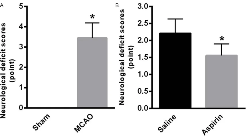
Figure 1. Aspirin improves neurological defects. A. After 24 h of MCAO, the rats of MCAO group (3.45 ± 0.74) demon-strated higher neurological defects compared with sham group. B. After aspirin treatment, the neurological deficits were significantly improved (1.56 ± 0.34) in comparison with saline group. Data were presented as mean ± SE from 3 independent experiments. *P < 0.05, **P < 0.01.
[image:3.612.94.521.380.576.2]Asprin protects rats from acute ischemic stroke
Removal and cDNA Synthesis SuperMix (AE311-03, Transgene, Beijing, China). The PCR pro-gram was described as follows: 48°C for 45 min; 96°C for 2 min; 25 repeats of 94°C for 30 s, 55°C for 45 s, and 68°C for 80 s; followed by a final extension at 65°C for 6 min. All PCRs were run in triplicate and gene expression, rela-tive to GAPDH, was calculated by the compara-tive ΔΔCt method.
Statistical analyses
The data are represented as means ± standard error (SE) of the indicated number of measure-ments. The statistical significance of differenc-es between groups was determined by one-way analysis of variance (ANOVA). p value < 0.05 was considered statistically significant.
Results
Aspirin improves neurological defects
After 24 h of MCAO, the rats of MCAO group (3.45 ± 0.74) demonstrated higher neurological defects compared with sham group (Figure 1A). After aspirin treatment, the neurological deficits were significantly improved in compari-son with saline group (P < 0.05).
Aspirin improves cerebral infarct size
As shown in Figure 2A, the cerebral infarct size was significantly increased in the serial coronal brain sections of the MCAO rats. Administration
of aspirin significantly decreased the infarct volume compared with the saline group (Figure 2A and 2B).
Aspirin suppresses the mitochondrial ROS production
Mitochondria are the major sources of ROS duction in cells. As shown in Figure 3, ROS pro-duction was significantly increased in the rats of MCAO group. In comparison, treatment with aspirin significantly improved ROS production in comparison with the saline group (Figure 3).
Aspirin resulted in the deficiency of NOD2 after cerebral IR injury
It is reported that TLR signaling plays a key in inflammatory responses [21]. NOD2 was recently found to activate ROS production after cerebral I/R injury [22]. We found the expres-sion of NOD2 was significantly increased in the rats of MCAO group compared with sham group (Figure 4A). In comparison, administration of aspirin significantly reduced the protein level of NOD2 (Figure 4B). Further study demonstrated the downstream signaling, NF-κB, and JNK acti-vation, was obviously suppressed (Figure 4B). Meanwhile, enhanced anti-apoptotic protein, Bcl-2, and decreased pro-apoptotic protein, Bax, can be detected (Figure 4B). These data suggested that aspirin resulted in the deficien-cy of NOD2 after cerebral IR injury.
Aspirin decreased the I/R induced inflamma-tion
The NF-κB and JNK signaling are key contribu-tors to the inflammatory responses. Then, we found that the levels of TNFα, IL-1β and IL-6 were obviously increased in the MCAO group compared with sham group (Figure 5A). Since decreased NF-κB and JNK activation was iden-tified in the aspirin treatment group, we deter-mined the level of inflammatory factors. The data suggested that the levels of TNFα, IL-1β and IL-6 were significantly suppressed in the aspirin group compared with saline group (Figure 5B), suggesting the anti-inflammatory role of aspirin after I/R injury.
Discussion
In this study, we determined that aspirin exerts a protective role on the acute ischemic stroke. After cerebral ischemic, obvious brain injury,
Figure 4. Aspirin resulted in the deficiency of NOD2 after cerebral IR injury. A. The expression of NOD2 was signifi-cantly increased in the rats of MCAO group compared with sham group. B. Administration of aspirin signifisignifi-cantly reduced the protein level of NOD2 and the downstream signaling, NF-κB, and JNK activation. Data were presented as mean ± SE from 3 independent experiments. *P < 0.05, **P < 0.01.
[image:5.612.91.524.483.654.2]Asprin protects rats from acute ischemic stroke
including neurology defects and enhanced cerebral infarct, can be detected. Our results indicated that aspirin could significantly improve brain injury after CIR in the MCAO rat models. Here, we demonstrated that aspirin treatment could obviously decrease NF-κB and JNK activation, thereby suppressing inflamma-tory responses and cell apoptosis. More impor-tantly, we found that aspirin could decrease the expression of NOD2, thereby alleviating inflam-matory damage and ROS production after CIR. After cerebral ischemic injury, an acute inflam-mation phase appears with production of large amounts of pro-inflammatory factors [23]. Growing evidences have indicated the impor-tant role of NOD2 in the inflammatory signaling after cerebral ischemic injury [24, 25]. In this study, we showed the enhanced expression of NOD2 after CIR. However, whether aspirin could suppress the expression of NOD2 has not been fully studied. Here, we clearly showed that administration with aspirin could markedly down-regulate the protein level of NOD2 in the ischemic brain.
To elucidate the specific mechanism in which NOD2 regulate ischemic injury, we compared the protein expression in the ischemic rat brains compared with the sham group. We found that upregulation of NOD2 could obvi-ously enhance the activation of NF-κB and JNK signaling. The activation of NF-κB and JNK sig-naling strongly enhance the transcription of pro-inflammatory factors, such as TNFα, IL-1β and IL-6 [26]. In comparison, administration of aspirin could inhibit the activation of NF-κB and JNK signaling. Consistent with decreased NF-κB and JNK activation, aspirin treatment downregulate the expression of TNFα, IL-1β and IL-6.
NOD-derived ROS production is the major ori-gin of cerebral ischemia [24]. Increasing evei-dence indicates that enhanced ROS production prompts innate immunity responses and enhances cell apoptosis [27, 28]. It has been shown that NOD2 contributes to ROS-induced cell apoptosis. Therefore, it is necessary to determine the levels of ROS production [28]. Obviously, ROS production was enhanced in rats of MCAO group. More importantly, our results demonstrated that aspirin treatment significantly decreased ROS generation and cell apoptosis. Our results showed that aspirin
treatment could suppress NOD2-derived ROS production and inflammation.
In summary, this study for the first time showed that aspirin could decrease NOD2 expression in the rats of acute ischemic stroke. Through suppressing NOD2-induced inflammatory res- ponses and ROS production, aspirin treatment could alleviate the deterioration of cerebral function after acute ischemic stroke.
Disclosure of conflict of interest None.
Address correspondence to: Dr. Guangyao Liu, De- partment of Neurosurgery, Hong Qi Hospital, Mudanjiang Medical University, No. 5, Aimin District, Tong Xiang Road, Mudanjiang 157000, Heilongjiang Province, P. R. China. E-mail: renhaiyan151216@163. com
References
[1] Flynn RW, MacWalter RS and Doney AS. The cost of cerebral ischaemia. Neuropharmacolo-gy 2008; 55: 250-256.
[2] Poisson SN, Schardt TQ, Dingman A and Ber-nard TJ. Etiology and treatment of arterial isch-emic stroke in children and young adults. Curr Treat Options Neurol 2014; 16: 315.
[3] Muir KW, Tyrrell P, Sattar N and Warburton E. Inflammation and ischaemic stroke. Curr Opin Neurol 2007; 20: 334-342.
[4] Chamorro A and Hallenbeck J. The harms and benefits of inflammatory and immune respons-es in vascular disease. Stroke 2006; 37: 291-293.
[5] Emsley HC and Hopkins SJ. Acute ischaemic stroke and infection: recent and emerging con-cepts. Lancet Neurol 2008; 7: 341-353. [6] Wang X. Investigational anti-inflammatory
agents for the treatment of ischaemic brain in-jury. Expert Opin Investig Drugs 2005; 14: 393-409.
[7] Nguyen MD, Julien JP and Rivest S. Innate im-munity: the missing link in neuroprotection and neurodegeneration? Nat Rev Neurosci 2002; 3: 216-227.
[8] Kim JY, Kawabori M and Yenari MA. Innate in-flammatory responses in stroke: mechanisms and potential therapeutic targets. Curr Med Chem 2014; 21: 2076-2097.
[9] Inohara, Chamaillard, McDonald C and Nunez G. NOD-LRR proteins: role in host-microbial in-teractions and inflammatory disease. Annu Rev Biochem 2005; 74: 355-383.
innate immunity and mediators of inflamma-tory diseases. J Leukoc Biol 2008; 83: 13-30. [11] Abulafia DP, de Rivero Vaccari JP, Lozano JD,
Lotocki G, Keane RW and Dietrich WD. Inhibi-tion of the inflammasome complex reduces the inflammatory response after thromboem-bolic stroke in mice. J Cereb Blood Flow Metab 2009; 29: 534-544.
[12] Yang F, Wang Z, Wei X, Han H, Meng X, Zhang Y, Shi W, Li F, Xin T, Pang Q and Yi F. NLRP3 deficiency ameliorates neurovascular damage in experimental ischemic stroke. J Cereb Blood Flow Metab 2014; 34: 660-667.
[13] Kufer TA. Signal transduction pathways used by NLR-type innate immune receptors. Mol Bio Syst 2008; 4: 380-386.
[14] Ting JP, Duncan JA and Lei Y. How the nonin-flammasome NLRs function in the innate im-mune system. Science 2010; 327: 286-290. [15] Sneader W. The discovery of aspirin: a
reap-praisal. BMJ 2000; 321: 1591-1594.
[16] Johnson ES, Lanes SF, Wentworth CE 3rd, Sat-terfield MH, Abebe BL and Dicker LW. A metare-gression analysis of the dose-response effect of aspirin on stroke. Arch Intern Med 1999; 159: 1248-1253.
[17] Chen WH, Cheng X, Lee PY, Ng W, Kwok JY, Tse HF and Lau CP. Aspirin resistance and adverse clinical events in patients with coronary artery disease. Am J Med 2007; 120: 631-635. [18] Jiang WL, Zhang SP, Zhu HB, Jian H and Tian
JW. Cornin ameliorates cerebral infarction in rats by antioxidant action and stabilization of mitochondrial function. Phytother Res 2010; 24: 547-552.
[19] Longa EZ, Weinstein PR, Carlson S and Cum-mins R. Reversible middle cerebral artery oc-clusion without craniectomy in rats. Stroke 1989; 20: 84-91.
[20] Saraf MK, Prabhakar S and Anand A. Neuro-protective effect of Bacopa monniera on isch-emia induced brain injury. Pharmacol Biochem Behav 2010; 97: 192-197.
[21] Yang CS, Kim JJ, Lee SJ, Hwang JH, Lee CH, Lee MS and Jo EK. TLR3-triggered reactive oxy-gen species contribute to inflammatory re-sponses by activating signal transducer and activator of transcription-1. J Immunol 2013; 190: 6368-6377.
[22] Liu HQ, Zhang XY, Edfeldt K, Nijhuis MO, Idborg H, Back M, Roy J, Hedin U, Jakobsson PJ, La-man JD, de Kleijn DP, Pasterkamp G, Hansson GK and Yan ZQ. NOD2-mediated innate im-mune signaling regulates the eicosanoids in atherosclerosis. Arterioscler Thromb Vasc Biol 2013; 33: 2193-2201.
[23] Jin R, Yang G and Li G. Inflammatory mecha-nisms in ischemic stroke: role of inflammatory cells. J Leukoc Biol 2010; 87: 779-789. [24] Farina C, Aloisi F and Meinl E. Astrocytes are
active players in cerebral innate immunity. Trends Immunol 2007; 28: 138-145.
[25] Stephenson D, Yin T, Smalstig EB, Hsu MA, Pa-netta J, Little S and Clemens J. Transcription factor nuclear factor-kappa B is activated in neurons after focal cerebral ischemia. J Cereb Blood Flow Metab 2000; 20: 592-603. [26] Murata Y, Fujiwara N, Seo JH, Yan F, Liu X,
Tera-saki Y, Luo Y, Arai K, Ji X and Lo EH. Delayed inhibition of c-Jun N-terminal kinase worsens outcomes after focal cerebral ischemia. J Neu-rosci 2012; 32: 8112-8115.
[27] Kahles T, Luedike P, Endres M, Galla HJ, Stein-metz H, Busse R, Neumann-Haefelin T and Brandes RP. NADPH oxidase plays a central role in blood-brain barrier damage in experi-mental stroke. Stroke 2007; 38: 3000-3006. [28] Chen H, Song YS and Chan PH. Inhibition of