Paper
12
Ultrasonographic scoring system for superficial digital
3flexor tendon injuries in horses: Intra- and inter-rater
4variability
5R. Alzola, C.M. Riggs, D.S. Gardner and S.L. Freeman
67 8
R. Alzola, BVMS, MSc, Cert AVP (EP, ESST, ESO, VDI) MRCVS, 9
Oakham Veterinary Hospital, Ashwell Road, Oakham, Rutland, UK. 10
C.M. Riggs, BVSc, PhD, DEO, DipECVS, MRCVS, 11
Veterinary Clinical Services, Hong Kong Jockey Club, Sha Tin Racecourse, New Territories, 12
Hong Kong. 13
D.S. Gardner, BSc, PhD, DSc, 14
S.L. Freeman, BVetMed, PhD, CertVA, CertVR, CertES, DipECVS, FHEA, FRCVS, 15
School of Veterinary Medicine and Science, University of Nottingham, College Road, Sutton 16
Bonington, Loughborough, Leicestershire, UK. 17
18
E-mail for correspondence: rafael.alzola@oakhamvethospital.co.uk 19
20 21
Superficial digital flexor tendon (SDFT) tendinopathy is an important musculoskeletal 22
problem in horses. The study objective was to validate an ultrasonographic scoring 23
system for SDFT injuries. Ultrasonographic images from fourteen Thoroughbred 24
racehorses with SDFT lesions (seven core; seven diffuse) and two controls were blindly 25
assessed by five clinicians on two occasions. Ultrasonographic parameters evaluated 26
were: type and extent of the injury, location, echogenicity, cross-sectional area and 27
longitudinal fibre pattern of the maximal injury zone (MIZ). Inter-rater variability and 28
intra-rater reliability were assessed using Kendall's coefficient of concordance (KC) 29
and Lin´s concordance correlation coefficient (LC), respectively. Type of injury (core 30
vs. diffuse) had perfect inter/intra-rater agreement. Cases with core lesions had very 31
strong inter-rater agreement (KC ≥0.74, p<0.001) and intra-rater reliability (LC ≥0.73) for
32
all parameters apart from echogenicity. Cases with diffuse lesions, had strong inter-33
rater agreement (KC ≥0.62) for all parameters, but weak agreement for echogenicity (KC
34
=0.22); intra-rater reliability was excellent for MIZ location and fibre pattern (LC ≥0.82),
35
and moderate (LC ≥0.58) for cross sectional area and number of zones affected. This
36
scoring system was reliable and repeatable for all parameters, except for echogenicity. 37
A validated scoring system will facilitate reliable recording of SDFT injuries, and inter-38
study meta-analyses. 39
Introduction
43Superficial digital flexor tendinopathy is a common injury in equine athletes; it 44
frequently occurs in racehorses during normal activity, following undefined periods of 45
accumulation of exercise and age-related microdamage without any preceding clinical 46
symptoms. Its prevalence in Thoroughbred racehorses varies significantly between 47
different disciplines ranging from 24% (Avella and others 2009) to 43% (Pickersgill 48
2000) in National Hunt horses and from 3.4% (Rossdale and others 1985) to 11.1% 49
(Kasashima and others 2004) in Flat-racing Thoroughbred horses. However, there are 50
limited data concerning other disciplines (Palmer and others 1994; van den Belt and 51
others 1994; Dyson 1998). Although complete tendon healing is a long and often 52
frustrating process (Goodship and others 1994; Smith and Schramme 2003) that 53
usually takes between 6-18 months, re-injury rates can be as high as 56% (Marr and 54
others 1993; Dyson 2004). Therefore, tendinopathy remains a significant cause of 55
wastage in Thoroughbred racehorses and a major health and welfare concern, as it is 56
a debilitating and potentially career-ending injury (Dowling and others 2000, Williams 57
and others 2001, Oikawa and Kasashima 2002, Perkins and others 2005). 58
There are many imaging modalities used to evaluate this condition, including 59
radiography (Verschooten and De Moor 1978), scintigraphy (Martinelli and Chambers 60
1995), thermography (Denoix and Audigie 2004), ultrasonography (Smith 2008), 61
ultrasound tissue characterisation (UTC) (Van Schie and others 2001) and magnetic 62
resonance imaging (MRI) (Karlin and others 2011). All of these imaging modalities are 63
useful, as each of them assists differently in the diagnosis and differentiation of 64
superficial digital flexor tendinopathy. Objective, accurate and repeatable imaging of 65
the SDFT is difficult, with MRI, UTC and ultrasonography possibly being the most 66
reliable methods. Ultrasonography, as opposed to MRI and UTC, is practical, cost 67
effective and a readily accessible imaging technique that allows real time evaluation 68
of the soft tissues. As a result, it is considered the diagnostic method of choice for 69
assessing equine tendon injuries (Smith 2008) in order to reach a diagnosis or to 70
determine readiness for return to exercise/competition (Palmer and others 1994). In 71
addition, with further assessments, ultrasonography can also be helpful when 72
monitoring recovery and response to treatment. Nevertheless, ultrasonography has a 73
limited field of view and image acquisition depends on the operator, the angle of 74
incidence, the equipment and the physical and physiological status of the tissue 75
(Pickersgill, 2000). Ultrasonographic images have been traditionally assessed using 76
both subjective and objective scales to evaluate the severity of injuries (Genovese and 77
others 1986). Objective measurements are repeatable values which can be measured 78
independently of the operator´s experience, such as percentage of cross-sectional 79
area affected. On the other hand, subjective measurements refer to measures that 80
could vary depending on operator´s experience and opinion, such as echogenicity. 81
Ultrasonographic scoring systems have been described before, but there are currently 82
no published studies which describe repeatability and reliability. 83
84
The objectives of this study were to: 1) develop a robust, reliable and repeatable 85
ultrasonographic scoring system for superficial digital flexor tendinopathy using 86
objective and subjective measurements of ultrasonographic parameters and 2) 87
determine inter-rater variability and intra-rater reliability for a panel of subjectively 88
90
Materials and Methods
9192
Participants and ultrasonographic data: Five experienced equine orthopaedic 93
clinicians, including three ECVS diplomates and two RCVS certificate holders, working 94
in specialist centres, reviewed and scored the ultrasonographic images using the 95
predefined SDFT scoring system. Fourteen ultrasonographic studies from 96
Thoroughbred racehorses with only forelimb SDFT lesions, including seven cases of 97
SDFT tendonitis with a core lesion and seven cases of diffuse SDFT tendonitis 98
(without a core lesion), were non-randomly selected from a large hospital database. 99
Cases were selected based on having a set of images of diagnostic quality with both 100
transverse and longitudinal views of regions of interest, and were selected to represent 101
a range of lesions with differing severity. In addition, two Thoroughbred racehorses 102
with a complete set of normal ultrasonographic images of the SDFT were also included 103
(control/no-injury cases). Each ultrasonographic study was obtained using a high-104
frequency linear ultrasonic transducer (5-13MHz), an acoustic stand-off pad and 105
acoustic gel. Transverse (zones 1A - 3C) and longitudinal (L1-L3) images of the SDFT 106
were obtained from the carpal bone down to ergot in the palmar metacarpal region. 107
DICOM (Digital Imaging and Communication in Medicine) data was used to store all 108
the ultrasonographic images in a web-shared folder to allow free access to the 109
participants. All the images were independently reviewed on two occasions, four to six 110
weeks apart using a dedicated DICOM viewer, and scored by completing an online 111
questionnaire (SurveyMonkey; https://www.surveymonkey.co.uk/) with objective and 112
subjective measurements for each case. On each occasion, the ultrasonographic 113
studies were presented to the participant in a computer-generated random order. 114
Throughout the study, participants were blinded to any case information and 115
outcomes. 116
117
Predefined scoring system (Fig.1): The ultrasonographic images of each case were 118
initially assessed qualitatively for the presence of an SDFT lesion (scored as 1 = SDFT 119
tendonitis with core lesion, 2 = diffuse SDFT tendonitis without core lesion or 3 = 120
normal SDFT). In cases where lesions were found, two further categories were 121
assessed qualitatively (using case logic on the survey tool to exclude these 122
assessments in cases considered to be normal). These two categories were the 123
number of zones affected (from 1 zone to ≥5 zones), and the location of the maximal
124
injury zone (MIZ; seven different sites on the leg: zones 1A - 3C (Rantanen and others 125
2011). Three semi-quantitative ultrasonographic criteria were also defined for the MIZ: 126
a) lesion echogenicity (MIZ-echogenicity, scored as 1 = anechoic, 2 = hypoechoic or 127
3 = hyperechoic compared to normal tendon tissue), b) estimated lesion cross-section 128
area (MIZ-CSA (%), scored as 1 = <25%, 2 = ≥25-50%, 3 = ≥50-75% or 4 = ≥75% of
129
the lesion cross-sectional area affected) and c) estimated lesion longitudinal fibre 130
pattern (MIZ-LFP (%), scored as 0 = normal, 1 = <25%, 2 = ≥25-50%, 3 = ≥50-75% or
131
4 = ≥75% of the lesion longitudinal fibre pattern affected) (Fig. 2). Grey-scale digital
132
images for the different transverse zones (1A-3C) and for the criteria to be used for 133
each category were provided as examples. The scoring system for diffuse SDFT 134
tendonitis without a core lesion was also clarified following initial feedback from the 135
participants. Specifically, scores for the percentage of affected cross sectional area 136
and/or longitudinal fibre pattern of the MIZ related only to the maximum seen in the 137
scores were also provided for these parameters in diffuse lesions. This clarification 139
was only provided for injuries without a core lesion and related to the diffuse nature of 140
these injuries. 141
142
Statistical analysis:All data was analysed using Genstat v16 (VSNi, Rothampsted, 143
UK). The ability of each rater to reproduce the same score for each category on two 144
occasions (i.e. intra-rater reliability) was evaluated using Lin’s Concordance 145
correlation coefficient, which quantifies the agreement between two independent 146
scores of the same parameter (0 = no agreement, 1 = perfect agreement). A value 147
≥0.75 is considered as very strong agreement and 95% confidence intervals are used 148
to represent the experimental variability around each score. Kendall´s coefficient of 149
concordance was used to measure the degree of agreement/consensus between 150
participants for each SDFT parameter scored (i.e. the inter-rater variability, where a 151
score of 0 = no agreement and 1 = perfect agreement). Statistical significance was 152
considered at p<0.05, with p<0.001 indicating a highly statistically significant effect. 153
Results
154All participants successfully (Kendall´s and Lin´s Coefficient = 1) distinguished the type 155
of SDFT injury (core vs. diffuse) for all cases (Table 1). 156
157
Reliability of the SDFT scoring system (Intra-rater agreement): For the seven 158
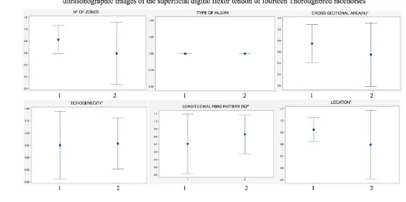
cases of SDFT tendonitis with a core lesion, the intra-rater reliability was very good 159
(Lin´s Coefficient [LC] = ≥0.73; Fig. 3) for the majority of ultrasonographic parameters,
160
including: number of zones (LC = 0.84), maximal injury zone (MIZ) location (LC = 161
0.93), MIZ-cross-section area (MIZ-CSA (%); LC = 0.77) and MIZ-longitudinal fibre 162
pattern (MIZ-LFP (%); LC = 0.73). For the seven cases with a diffuse SDFT injury 163
(without a core lesion), the intra-rater reliability was excellent (LC ≥0.86) for
MIZ-164
location (LC = 0.82) and MIZ-LFP (%) (LC = 0.85) but only moderate (LC = 0.41-0.60) 165
for the number of zones (LC = 0.62) and MIZ-CSA (%) (LC = 0.58). In contrast, the 166
intra-rater agreement for MIZ-echogenicity for SDFT lesions with a core lesion was 167
weak (LC = 0.31 [-0.05,0.50] 95% confidence interval). Similarly, the Lin´s Coefficient 168
for the cases with diffuse SDFT tendonitis (without a core lesion) was also weak (LC 169
= 0.30 [0.07,0.49] 95% confidence interval). 170
171
Variability of the SDFT scoring system (Inter-rater agreement): For cases of SDFT 172
tendonitis with a core lesion, the inter-rater agreement was very strong (Kendall´s 173
Coefficient [KC] ≥0.74, P<0.001; Fig. 4) for almost all ultrasonographic parameters
174
including the number of zones (KC = 0.76), MIZ-location (KC = 0.80), MIZ-CSA (%) 175
(KC = 0.84) and MIZ-LFP (%) (KC = 0.74). For cases of diffuse SDFT tendonitis 176
(without a core lesion), the inter-rater agreement was strong (KC = ≥0.62 - <0.69) for
177
the following ultrasonographic parameters: number of zones (KC = 0.64), MIZ-location 178
(KC = 0.62) and MIZ-CSA (%) (KC = 0.69) and very strong for the MIZ-LFP (%) (KC = 179
0.87). The inter-rater agreement for MIZ-echogenicity for both SDFT lesions with or 180
without core lesions was weak (KC = 0.31, χ2 26.8, P=0.01) and (KC = 0.30, P=0.36)
181
respectively. 182
Discussion
185186
At present, MRI is the most sensitive imaging modality for the evaluation of tendon 187
injury (Karlin and others 2011). However, ultrasonography is widely available, 188
portable, cheap and safe and recent improvements in US technology make it the most 189
commonly used imaging modality for equine practitioners to evaluate SDFT injuries. 190
Several ultrasonographic scoring scales to evaluate injured tendons have been 191
developed over the last 30 years in veterinary practice (Genovese and others 1986, 192
Reef and others 1993, Van den Belt and others 1993, Saini and others 2002, Geburek 193
and others 2016), but there is no internationally agreed protocol for reporting SDFT 194
injuries, making it difficult to compare datasets. In an attempt to provide a semi-195
quantitative evaluation, each of these scoring systems focuses on different 196
parameters: Cross-sectional area and echogenicity (Genovese and others 1986 and 197
Van den Belt and others 1993); length of the lesion and percentage of the cross-198
sectional area affected (Reef and others 1993) or echogenicity only (Saini and others 199
2002). A fundamentally more powerful method of ultrasonographic diagnosis is 200
ultrasound tissue characterization (UTC) which quantifies tendon integrity based on a 201
computerized analysis of the stability of echo-patterns in contiguous ultrasound 202
images (Geburek and others 2016). Although this technique has great potential for the 203
future, at present it is mainly being applied in a research environment. With the 204
exception of UTC, the reliability and repeatability of the ultrasonographic parameters 205
included in each system should be investigated. Ideally only parameters with high 206
reliability and repeatability should be included. 207
208
This is the first study which describes the reliability and repeatability of an ultrasound 209
scoring system for SDFT injuries. Scoring systems (i.e. qualitative, semi-quantitative 210
and quantitative) are widely used in human medicine to provide a framework for 211
standardization of clinical management, benchmarking outcomes and planning or 212
analysing research. The ultrasonographic scoring system developed in this study, 213
obtained by categorizing type and extent of SDFT injury together with location and 214
ultrasonographic characteristics of the maximal injury zone (MIZ), will allow equine 215
practitioners to apply these criteria in veterinary medicine. In comparison with 216
previously described scoring systems, we have included more ultrasonographic 217
parameters with higher reliability and repeatability which allow for a more detailed 218
characterization of the injury. Two of the previously proposed ultrasonographic 219
systems (Genovese and others 1986 and Van den Belt and others 1993, Saini and 220
others 2002) rely heavily on echogenicity which in our study had weak intra/inter rater 221
agreement. Contrary to the scoring system proposed by Reef and others (1993), this 222
ultrasonographic scoring system also required subjective visual assessment of the 223
area of tendon damaged to assess the echogenicity. 224
225
This study presents a simple, repeatable and thus reliable scoring system for tendon
226
injury evaluation using ultrasonographic features of the MIZ as a representative part 227
of the injury. Contrary to previously described ultrasonographic scoring scales 228
(Genovese and others 1986), our system described here is quick (taking on average 229
5 to 10 minutes) and simple to complete, requiring only minimal training which will
230
facilitate its incorporation into routine practice. However, it still relies on subjective 231
ultrasonographic parameters, some of which have poor reliability and repeatability. 232
This scoring system could allow standardization of the SDFT evaluations in clinical 233
colleagues, and enabling practices to monitor and audit clinical cases by comparing 235
and contrasting findings and responses to treatment between different cases. We 236
acknowledge that scoring diffuse SDF tendonitis without a core lesion is more 237
subjective and difficult than SDF tendonitis with a core lesion. In this study both 238
Kendall´s and Lins coefficients were lower for the majority of the categories without a 239
core lesion (with wider confidence intervals as expected), but the tendency was similar 240
in both groups (see Fig. 4). This fact was also highlighted by our study: in order to 241
significantly improve the initial inter-rater agreement of clinicians assessing tendonitis 242
without a core lesion, a detailed explanation and images of all the categories had to 243
be provided to each of the participants prior to assessment. 244
245
Limitations of the study:The main limitation of this study is that ultrasound images
246
were retrospectively reviewed. The images were also obtained by multiple clinicians
247
with different ultrasonographic equipment which could alter image quality. Although all
248
the images were of diagnostic quality, no attempt was made to assess or compare the
249
quality of the images which could have affected some categories of the scoring
250
system. In addition, lack of ultrasonographic images of the contralateral limb for
251
comparison is a weakness. However, in our study images of two control horses with 252
no-injury were reliably interpreted by all practitioners. Nevertheless, we acknowledge 253
that having images of the contralateral limb could have significantly improved our 254
scores. 255
256 257
With regard to echogenicity, which showed poor reliability and agreement, the test 258
conditions could have influenced results to some extent; for example, the brightness 259
in the room, the type of screen or the dedicated DICOM viewer used by the participants 260
were not recorded but could have influenced echogenicity score of the cases. Some 261
of the participants changed the test conditions between part one and two of the study, 262
by using different screens and DICOM viewers to score the cases. Echogenicity is 263
highly dependent on the positioning of the probe and angle of the ultrasound beam in 264
comparison with the longitudinal axis of the tendon fibres. Assessment of the 265
echogenicity in real time by the operator would have led to a better evaluation of the 266
echogenicity score. Nevertheless, echogenicity is an ultrasonographic parameter 267
commonly used to characterize tendon injury in horses and whilst this study 268
highlighted low intra- and inter-rater agreement, all cases were acute injuries that were 269
either scored hypoechoic or anechoic by all participants. 270
In summary, this study describes a scoring system which uses both qualitative and 271
semi-quantitative measures that can be simply and consistently applied by equine 272
practitioners and researchers. The development of a validated scoring system is 273
important to enable standardised clinical recording of SDTF injuries for equine 274
practitioners both for repeated assessments within the same patient, and also for 275
comparison of lesions between different patients. It will also enable inter-study 276
comparisons and meta-analysis of future SDFT research projects by minimizing 277
variation between different operators and/or different studies. 278
279
Word count: 2592 280
283
Ethics statement: This study was reviewed and approved by the ethical committee 284
of the School of Veterinary Medicine and Science, University of Nottingham. 285
286
Competing interests: No declared. 287
288
Funding: No sources of funding. 289
290
Acknowledgments: The authors would like to extend their thanks to Dr Neal Ashton, 291
Dr James Doles and Dr Heidi Janicke for their assistance with scoring 292
ultrasonographic images. 293
References:
297298
AVELLA, C. S., ELY E. R., VERHEYEN K. L. P., PRICE J. S., WOOD J. L. N. and 299
SMITH R. K. W. (2009) Ultrasonographic assessment of the superficial digital flexor 300
tendons of National Hunt racehorses in training over two racing seasons. Equine Vet. 301
J. 41, 449-454. 302
DENOIX, J. M., AUDIGIE, F. (2004) Imaging of the musculoskeletal system in horses. 303
In: Equine Sports Medicine and Surgery. Ed: Elsevier Saunders, Philadelphia, USA. 304
pp 161-187. 305
DOWLING, B. A., DART, A. J., HODGSON, D. R. and SMITH, R. K. W. (2000) 306
Superficial digital flexor tendonitis in the horse. Equine Vet. J. 32, 369-378. 307
DYSON, S. J. (1998) Superficial digital flexor tendonitis: a comparison of treatment 308
methods and rehabilitation programmes. In: Proceedings of the Conference on Equine 309
Sports Medicine, Ed: A. Lindner, Wageningen Pers. pp 111-118. 310
DYSON, S. J. (2004) Medical management of superficial digital flexor tendonitis: a 311
comparative study in 219 horses (1992-2000). Equine Vet. J. 36, 415-419. 312
GEBUREK, F., GAUS, M., VAN SCHIE, H. T. M., ROHN, K., & STADLER, P. M. 313
(2016). Effect of intralesional platelet-rich plasma (PRP) treatment on clinical and 314
ultrasonographic parameters in equine naturally occurring superficial digital flexor 315
tendinopathies – a randomized prospective controlled clinical trial. BMC Veterinary 316
Research, 1-16 317
GENOVESE, R. L., RANTANEN, N. W., HAUSER, M. L., SIMPSON, B. S. (1986) 318
Diagnostic ultrasonography of equine limbs. Vet Clin. North Am. Equine Pract. 2, 145-319
226. 320
GOODSHIP, A. E., BIRCH, H. L., WILSON, A. M. (1994): The patho- biology and 321
repair of tendon and ligament injury. Vet Clin North Am: Equine Pract. 10, 323–349. 322
KARLIN, W. M., STEWART, A. A., DURGAM, S. S., NAUGHTON, J. F., O’DELL-323
ANDERSON, K. J., STEWART, M. C. (2011) Evaluation of experimentally induced 324
injury to the superficial digital flexor tendon in horses by use of low-field magnetic 325
resonance imaging and ultrasonography. J. Am. Vet. Radiol., 72, 791-798. 326
KASASHIMA, Y., TAKAHASHI, T., SMITH, R. K., GOODSHIP, A. E., KUWANO, A., 327
UENO, T. and HIRANO, S. (2004) Prevalence of superficial digital flexor tendonitis 328
and suspensory desmitis in Japanese Thoroughbred flat racehorses in 1999. Equine 329
Vet. J. 36, 346-350. 330
MARR, C. M., LOVE, S., BOYD, J. S. and MCKELLAR, Q. (1993) Factors affecting 331
the clinical outcome of injuries to the superficial digital flexor tendon in National Hunt 332
and point-to-point racehorses. Vet. Rec. 132, 476-479. 333
MARTINELLI, M. J. and CHAMBERS, M. D. (1995) Scintigraphy Series: Equine 334
nuclear bone scintigraphy: physiological principles and clinical application. Equine Vet. 335
Educ. 7, 281-287. 336
OIKAWA, M. and KASASHIMA, Y. (2002) The Japanese experience with tendonitis in 337
PALMER, S. E., GENOVESE, R. L., LONGO, K. L., et al. (1994) Practical 339
management of superficial digital flexor tendinitis in the performance horse. Vet. Clin. 340
North Am. Equine Pract. 110, 425-481. 341
PERKINS, N. R., REID, S. W. J. and MORRIS, R. S. (2005) Risk factors for injury to 342
the superficial digital flexor tendon and suspensory apparatus in Thoroughbred 343
racehorses in New Zealand. N. Z. Vet. J. 53, 184-192. 344
PICKERSGILL, C. H. (2000) Epidemiological Studies into Orthopaedic Conditions of 345
the Equine Athlete. MVM thesis, University of Glasgow, Glasgow. 346
RANTANEN, N. W., JORGENSEN, J.S. and GENOVESE, R.L. (2011) 347
Ultrasonographic evaluation of the equine limb: technique. In, Ross M.W., Dyson S. 348
(eds) Diagnosis and Management of Lameness in the Horse, 2nd edition. Ed: Elsevier 349
Saunders, St. Louis, Missouri, USA. pp191-192. 350
REEF, V., MARTIN, B., and ELSER, A. (1993) Types of tendon and Ligament injuries 351
detected with diagnostic ultrasound: detection and follow-up. Proc. Am. Ass. Equine 352
Practitioners. 34, 245. 353
ROSSDALE, P. D., HOPES, R., DIGBY, N. J. and OFFORD, K. (1985) 354
Epidemiological study of wastage among racehorses 1982 and 1983. Vet. Rec. 116, 355
66-69. 356
SAINI, N. S., ROY, K. S., BANSAL, P. S., SINGH, B., SIMRAN, P. S. (2002) A 357
preliminary study on the effect of ultrasound therapy on the healing of surgically 358
severed achilles tendons in five dogs. J. Vet. Med. A. Physiol. Pathol. Clin. Med. 49, 359
321-328. 360
SMITH, R. K. (2008) Tendon and ligament injury. Proc. 54th Ann. Conv. Am. Assoc. 361
Eq. Pract. 54, 475–501. 362
SMITH, R. K., SCHRAMME, M. (2003): Tendon injury in the horse: current theories 363
and therapies. In Practice 25, 529–539. 364
VAN DEN BELT, A. J., BECKER, C. K., DIK, K. J. (1993) Desmitis of the accessory 365
ligament of the deep digital flexor tendon in the horse: clinical and ultrasonographic 366
features. A report of 24 cases. J. Vet. Med. A. 40, 492-500. 367
VAN DEN BELT, A. J., DIK, K. and BARNEVELD, A. (1994) Ultrasonographic 368
evaluation and long-term follow-up of flexor tendonitis/desmitis of the 369
metacarpal/metatarsal region in Dutch Warmblood and Standardbred racehorses. Vet. 370
Quart. 16, Suppl. 2, S76-S80. 371
VAN SCHIE, H. T. M., BAKKER, E. M., JONKER, A. M., VAN DE LEST, C. H. A., VAN 372
WEEREN, P. R. (2001). Efficacy of computerized discrimination between structure-373
related and non-structure-related echoes in ultrasonographic images for the 374
quantitative evaluation of the structural integrity of superficial digital flexor tendons in 375
horses. American Journal of Veterinary Research 62 (7), 1159-1166. 376
VERSCHOOTEN, F., DE MOOR, A. (1978) Tendinitis in the horse: its radiographic 377
diagnosis with air-tendograms. J. Am. Vet. Radiol. Soc. 19, 23-30. 378
WILLIAMS, R. B., HARKINS, L. S., HAMMOND, C. J. and WOOD, J. L. N. (2001) 379
Racehorse injuries, clinical problems and fatalities recorded on British racecourses 380
from flat racing and National Hunt racing during 1996, 1997 and 1998. Equine Vet. J. 381
Figure 1: Flowchart summarising the predefined scoring system used in this study. 383
384
Table 1: Summary of Kendalls and Lin’s coefficients of concordance
SDFT tendonitis with core lesion Diffuse SDFT tendonitis without core
lesion
Parameter assessed Lin’s coeff. Kendal’s
coeff.
P-value Lin’s coeff.
Kendal’s
coeff. P-value
Type of Injury (Core vs. Diffuse) 1 1 <0.001 1 1 <0.001
Nº of zones 0.84 (0.72-0.91) 0.76 <0.001 0.62 (0.37-0.79) 0.64 <0.001
MIZ: Location 0.93 (0.88-0.96) 0.80 <0.001 0.82 (0.67-0.90) 0.62 <0.001
MIZ: CSA (%) 0.77 (0.60-0.87) 0.84 <0.001 0.58 (0.31-0.76) 0.69 <0.001
MIZ: Echogenicity 0.31*(-0.05-0.50) 0.34* 0.013 0.30*(0.07-0.49) 0.22* 0.36
MIZ: LFP (%) 0.73 (0.54-0.85) 0.74 <0.001 0.85 (0.72-0.92) 0.87 <0.001
Figure2: Example of the semi-quantitative ultrasonographic criteria (echogenicity, cross-section area and longitudinal fibre pattern) used to score the lesion at the maximal injury zone (MIZ) in equine cases with superficial digital flexor tendon injuries. Transverse and longitudinal ultrasonographic images of the MIZ of SDFT injury:
a) SDFT tendonitis with a core lesion; lesion echogenicity (MIZ-echogenicity) scored as 1 = anechoic, lesion cross-section area (MIZ-CSA (%) scored as 1 = < 25% and lesion longitudinal fibre pattern (MIZ-LFP (%) scored as 3 = 50-75%.
b) SDFT tendonitis without a core lesion; lesion echogenicity (MIZ-echogenicity) scored as 2 = hypoechoic, lesion cross-section area (MIZ-CSA (%) scored as 4 = ≥75% and lesion longitudinal fibre pattern (MIZ-LFP (%) scored as 3 = 50-75%.
a) b)