Expression of polycomb protein BMI-1 maintains the
plasticity of basal bronchial epithelial cells
Elizabeth Torr1, Meg Heath2, Maureen Mee3, Dominick Shaw1, Tyson V. Sharp4& Ian Sayers1
1 Division of Respiratory Medicine, Queens Medical Centre, University of Nottingham, Nottingham, United Kingdom 2 Cytogenetics Unit, Nottingham City Hospital, Hucknall Road, Nottingham, United Kingdom
3 School of Life Sciences, Queens Medical Centre, University of Nottingham, Nottingham, United Kingdom
4 Centre for Molecular Oncology, Barts Cancer Institute, Queen Mary, University of London, London, United Kingdom
Keywords
BMI-1, bronchial epithelial cells, lifespan, plasticity.
Correspondence
Ian Sayers, Division of Respiratory Medicine, Queen’s Medical Centre, University of Nottingham, Nottingham, NG7 2UH, United Kingdom.
Tel: 0115 82 31066 Fax: 0115 82 31059
E-mail: ian.sayers@nottingham.ac.uk
Funding Information
This work was supported by Asthma UK Grant 10/006.
Received: 7 April 2016; Revised: 7 June 2016; Accepted: 7 June 2016
doi: 10.14814/phy2.12847
Physiol Rep, 4 (16), 2016, e12847, doi: 10.14814/phy2.12847
Abstract
The airway epithelium is altered in respiratory disease and is thought to con-tribute to disease etiology. A caveat to disease research is that the technique of isolation of bronchial epithelial cells from patients is invasive and cells have a limited lifespan. The aim of this study was to extensively characterize the plas-ticity of primary human bronchial epithelial cells that have been engineered to delay cell senescence including the ability of these cells to differentiate. Cells were engineered to express BMI-1 or hTERT using viral vector systems. Cells were characterized at passage (p) early (p5), mid (p10), and late (p15) stage for: BMI-1, p16, and CK14 protein expression, viability and the ability to dif-ferentiate at air–liquid interface (ALI), using a range of techniques including immunohistochemistry (IHC), immunofluorescence (IF), transepithelial elec-trical resistance (TEER), scanning electron microscopy (SEM), MUC5AC and beta tubulin (BTUB) staining. BMI-1-expressing cells maintained elevated levels of the BMI-1 protein and the epithelial marker CK14 and showed a suppression of p16. BMI-1-expressing cells had a viability advantage, differen-tiated at ALI, and had a normal karyotype. In contrast, hTERT-expressing cells had a reduced viability, showed limited differentiation, and had an abnormal karyotype. We therefore provide extensive characterization of the plasticity of BMI-1 expressing cells in the context of the ALI model. These cells retain properties of wild-type cells and may be useful to characterize res-piratory disease mechanisms in vitro over sustained periods.
Introduction
The airway epithelium acts as the critical interface between the environment and organ physiology. It acts as a barrier to potential pathogens and extraneous particles and helps regulate host defense mechanisms, including the inflammation process. Under normal conditions, the bronchial epithelium is composed of ciliated columnar, mucus-secreting goblet and Clara cells that secrete surfac-tant. There is accumulating evidence that the airway epithelium is intrinsically different in airway diseases such as asthma and chronic obstructive pulmonary disease (COPD) if compared with the normal type. For example,
airway epithelium contributes to disease etiology. There-fore, the airway epithelium represents an attractive thera-peutic target in asthma. However, more research tools and cellular assay systems are needed to rapidly move this area of important research forward.
The most common approach to study bronchial epithe-lial cells from healthy controls and patients with respira-tory disease is to isolate cells using bronchoscopic brush technique (Kelsen et al. 1992) and then culture the cells as basal cell monolayers or using air–liquid interface (ALI) differentiation (Stewart et al. 2012a,b). ALI cells form an epithelial barrier that closely resembles the in vivo architec-ture of the airway epithelium and is composed of basal, goblet, and ciliated cells allowing data to be generated in a more physiological context. However, the bronchoscopic procedure is invasive with risk to the individual, especially in people with severe asthma. Moreover, adequate numbers of cells are hardly collected. Similarly, once isolated and grown in vitro, the primary airway epithelial cells have a limited lifespan (i.e., the point at which the cells stop pro-liferating, typically 4–5 passages). This is often too short to obtain key experimental data. Importantly, after p4 bron-chial epithelial cells begin to deviate in their morphology/ phenotype, lose their plasticity required for the differentia-tion at ALI. These factors make the identificadifferentia-tion and care-ful characterization of a method to delay cell senescence while maintaining the plasticity of the cells highly desir-able. One approach to delay cell senescence is the suppres-sion of cyclin-dependent kinase inhibitor, p16(Ink4a), a tumor suppressor that induces a G1 cell cycle arrest. B-cell-specific Moloney murine leukemia virus integration site 1 (BMI-1) is a polycomb protein thought to repress p16(Ink4a) expression and has previously been shown to delay cell senescence (Vonlanthen et al. 2001).
In this study, we set out to engineer primary human bronchial epithelial cells to express different levels of BMI-1 via alternative promoter constructs and extensively characterize the cells for effects on viability and plasticity such as the ability to differentiate at ALI. This was assessed using a range of techniques including immuno-histochemistry (IHC), immunofluorescence (IF), transep-ithelial electrical resistance (TEER), and scanning electron microscopy (SEM). Importantly, we also investigated gen-ome stability/integrity through effects on karyotype. Both wild-type early passage cells and cells engineered to express hTERT (as a commonly used immortalization technique) were included in these analyses. We demon-strate the BMI-1-engineered cells assessed at early (p5), mid (p10), and late (p15) passage produce elevated levels of BMI-1 expression with associated suppression of p16. These cells retain both viability and differentiation potential of wild-type cells while importantly not demon-strating the changes in cell karyotype. These features were
apparent across all donor cell populations engineered to express the elevated levels of BMI-1. These data are in contrast to hTERT cells that were no longer viable and did not differentiate at mid passage and demonstrated gross chromosomal abnormalities. Therefore, BMI-1-engi-neered bronchial epithelial cells delayed cell senescence and retained their plasticity including the ability to differ-entiate at ALI in a comparative manner to low passage unmodified cells.
Materials and Methods
BMI-1 plasmid construction
Human BMI-1 full-length cDNA was originally obtained from GeneserviceTM
Mammalian Gene Collection (MGC) Clone, amplified and cloned into pLVX-Puro (Clontech) using XhoI/BamHI restriction sites and into pFLRu-FH using MluI/BamHI restriction sites, using standard molec-ular biology techniques. The retroviral vector pWZL-BLAST-FLAG-HA-hTERT and corresponding empty vector backbone were purchased from Addgene (Maida et al. 2009). All vectors were sequence verified.
Packaging of lentivirus and retrovirus
HEK293T cells were seeded at 29106 cells per 10-cm diameter dish and were cultured in DMEM, 10% FCS, 1% penicillin/streptomycin overnight. For lentiviral produc-tion, the cells were transfected with a mix containing an 8:1 ratio of packaging: envelope vectors, that is, 4.4 lg of pCMV delta R8.9, 0.6lg of pCMV-VSV-G (McKay et al. 2011), and 5 lg of the appropriate lentiviral vector (empty or BMI-1 insert) with 36 lL of TransIT-LT1 (Geneflow). For retroviral production, the cells were transfected with 5lg of pKAT (Addgene) and 5lg of retroviral vector (empty or with hTERT insert) and 36lL of TransIT-LT1 (Geneflow). Cells were grown for 48–72 h and the super-natant containing packaged virus was harvested and ultra-centrifuged (48,384g for 3.5 h at 4°C) to produce a concentrated viral stock which was stored at -80°C.
Primary bronchial epithelial cell culture and air liquid interface
published methods (Stewart et al. 2012a,b). BEDM is composed of 50:50 Dulbecco’s Modified Eagle’s Medium (DMEM, Sigma-Aldrich, Dorset, UK):BEBM with Lonza singlequots, excluding triiodo-L-thyronine and retinoic acid, but including GA-1000 (Gentamicin and Ampho-tericin-B). BEDM is supplemented with 50 nmol/L reti-noic acid, added at time of use. All cells were cultured on 6.5-mm polyester Transwell inserts with a pore size of 0.4lm (Corning Life Sciences, Amsterdam, The Nether-lands). Cells were plated at 30,000 cells per insert in an appropriate medium. When confluent (~3 days), cells were raised to ALI. Medium was replaced and the apical face washed with media every 48 h. Cells were fixed for immunostaining, histology sectioning, and scanning elec-tron microscopy after 28 days at ALI.
Generation of genetically modified NHBEC
Passage 2 NHBEC were plated in a 6-well plate at 59104 cells per well and grown overnight. These were purchased heterogeneous NHBEC and not clonal cells. The media was replaced with 800lL of BEGM with 2lg/mL of polybrene (Hexadimethrine bromide H9268 Sigma) and 6.25 lL of lentivirus or 12.5lL of retrovirus stock solutions (minimal concentrations to give >90% transfection determined empirically). The plates were incubated at 37°C for 6.5 h with gentle rocking of the plate every 30 min for the first 2 h. The viral mixture was removed and replaced with BEGM. After 72 h, selection media was added to the wells with 300 ng/mL puromycin for the lentiviral-infected cells and 600 ng/mL blasticidin for the retroviral cells. Cells with antibiotic resistance after 6 days of selection were considered to have been stably infected with the appropri-ate virus expressing BMI-1 or hTERT.
Western blotting
A total of 2 9105 cells were lysed in SDS loading buffer, heat denatured, and proteins were separated by elec-trophoresis using 12% SDS resolving gels. The proteins were transferred to nitrocellulose and probed for BMI-1, p16, and bactin expression using mouse anti-p16 (Milli-pore MAB4133; Watford, UK) at 1 in 1000 dilution (1 lg/mL), mouse anti-BMI-1 (Millipore Clone F6 Cat No 05-637) primary antibody at 1 in 1000 dilution, and rabbit polyclonal anti-bactin (Abcam ab8227, lot 712923, 0.65 mg/mL) primary antibody at 1 in 5000 dilution. Sec-ondary antibodies were used at 1 in 10000 dilutions and consisted of goat anti-mouse HRP (Jackson Immuno 115-035-062) and goat polyclonal anti-rabbit HRP (Sigma A0545). ECL reagent was used to visualize proteins as directed by the manufacturer (GE Healthcare RPN2209; GE Healthcare, Amersham, UK).
Immunofluorescence
ALI-cultured cells were fixed in situ on inserts and trans-ferred to the glass slides for visualization. Cells were fixed using 4% formaldehyde and blocked/permeabilized with PBS, 10% goat serum, 1% BSA, and 0.15% Triton-X. Cells were incubated with appropriate primary antibodies at 4°C overnight, and FITC labeled secondary for 1 h at room temperature before mounting in HardSet DAPI (Vector Labs). These methods were as previously described (Stewart et al. 2012b) with the addition of BMI-1 (Millipore Clone F6) and CK14 (Chemicon MAB3232) antibodies to the panel. Cells were visualized using the Zeiss spinning disk confocal microscope and Volocity software (PerkinElmer, Cambridge, UK).
Cell viability
A total of 2.5 9103 cells were plated in quadruplicate wells of a 96-well plate in 200 lL BEGM +GA and incu-bated for 96–120 h before analysis. 3-(4,5-dimethylthia-zol-2-yl)-2,5-diphenyltetrazolium bromide (MTT) at 500 lg/mL in BEGM was added to the wells for 4 h before termination of the experiment. The media was then replaced with 200 lL isopropanol and incubated for 10 min to lyse the cells. The plate was read at 570 nm (with a background subtraction of 690 nm) using a Flexs-tation (Molecular Devices, Wokingham, UK).
Transepithelial electrical resistance
Transepithelial electrical resistance (TEER) was measured in differentiating cells using an EVOM2 epithelial volt-ohm meter (World precision Instruments UK, Stevenage) to indicate development of tight junctions as described previously (Stewart et al. 2012b). Briefly, medium was aspirated and replaced with 1 mL growth medium in the basolateral and 0.5 mL growth medium in the apical compartment. Cultures were equilibrated in the incubator for 30 min before TEER measurement. Apical medium was then aspirated to restore ALI. TEER values of insert and medium alone wells was subtracted from the mea-sured TEER and Ocm2 was calculated by multiplying by the insert area. For comparison across cell lines and pas-sage, we calculated area under curve using TEER data from days 1–28 using GraphPad Prism 6.02 for Windows (GraphPad Software, San Diego, CA).
Karyotyping
and fixing in methanol/glacial acetic acid. The cells were fixed further two times before slides were produced and aged, then banded using trypsin and Giemsa staining before imaging and analyses (completed by Cytogenetics Unit, City Hospital, Nottingham, UK).
Histological analyses
Fixed transwells were paraffin embedded, sectioned, and then stained with hematoxylin and eosin (H & E) or alcian blue using standard approaches.
Scanning electron microscopy
Transwells at day 28 ALI were washed with BEGM media to remove any mucous and then fixed with 3% glutaralde-hyde in 0.1 mol/L sodium cacodylate buffer for a mini-mum of 24 h. The wells were washed three times with 0.1 mol/L phosphate/cacodylate buffer and post fixed for 1 h at room temperature with 1% aqueous osmium tetrox-ide. The samples were washed with distilled water and then dehydrated using a graded series of ethanol washes (50, 70, 90, and 100%, respectively). Samples remained submerged in 100% ethanol, processed when they were dried, and coated with gold particles. The samples were then imaged at low and high magnification using a scanning electron microscope (model JSM-35; JOEL).
Telomerase activity assay
TRAPeze XL Telomerase detection kit was used as directed by the manufacturer to determine hTERT activity (Milli-pore S7707). A total of 19105 cells were suspended in 200 lL of CHAPS lysis buffer and incubated on ice for 30 min. The positive cells (19106, provided by the kit) were treated in the same way. The samples were then cen-trifuged at 120,000g for 20 min at 4°C. PCR tubes contain-ing 10lL 5X TRAPeze XL reaction mix, 0.4lL Taq polymerase (5units/lL), 37.6 lL dH2O, and 2lL of cell
extract, positive template or buffer for the control samples were run at 30°C for 30 mins followed by 36 cycles of 94°C for 30 sec, 59°C for 30 sec, and 72°C for 1 min. This was followed by 72°C for 3 min, 55°C for 25 min, and 4°C hold. The samples were then diluted in 150lL of buffer (10 mmol/L Tris-HCl pH 7.4, 0.15 mol/L NaCl, 2 mmol/L MgCl2) and fluorescent signals were read at excitation
485 nm and emission 535 nm and excitation 585 nm and emission 620 nm on the Flexstation (Molecular Devices).
Statistical analyses
GraphPad Prism 6.02 for Windows was used for statistical analyses. Quantitative data were analyzed using Student’s
t test or ANOVA with Dunnett’s multiple comparison test. AP<0.05 was considered significant.
Results
Cell generation
We specifically chose two lentiviral vectors with differing promoters: Cytomegalovirus (CMV) in pLVX-Puro or human ubiquitin C (UbiC)) in pFLRu-FH to express BMI-1 in human bronchial epithelial cells with the hypothesis that the CMV promoter may lead to greater BMI-1 overexpression than the UbiC promoter (Qin et al. 2010). Lentiviruses were used to transduce passage 2 NHBECs from two donors. This protocol therefore gener-ated four independent cell populations expressing recom-binant BMI-1 to evaluate the effect of BMI-1 on cell plasticity. The transduction efficiency was >95%, as indi-cated by limited cell death following antibiotic selection and the pWZL-based constructs gave similar findings (data not shown). Cell populations generated were fol-lowed up for~12 months with focused analyses presented for (1) Early passage (infection passage 3/actual passage 6), (2) Mid passage (infection passage 5–7/actual passage 8–10), and (3) Late passage (infection passage 8–12/actual passage 11–15). Finally, a subset of analyses was com-pleted using extended passage cells (infection passage 17/ actual passage 20). It is important to note that we arbi-trarily designated these early, mid, and late passage defini-tions based on passage 20 being the maximum achieved. NHBECs transduced with lentivirus containing plasmid vector that did not contain the BMI-1 expression cassette, that is, empty vectors, stopped growing at mid passage. Similarly, for the cells containing the pWZL constructs including hTERT insert cells were not viable at mid pas-sage, although there were donor differences in these cell senescence times.
NHBEC-BMI-1 cell populations maintain elevated levels of BMI-1 expression and feature p16 suppression during prolonged culture
expression in engineered late passage cells for both donors demonstrating robust elevated BMI-1 through to late pas-sage (Fig. 1C). Early and mid paspas-sage cells showed similar elevated BMI-1 expression as determined by immunofluo-rescence (data not shown). In order to confirm that genetic modification of cells to overexpress hTERT trans-lated to increased hTERT enzymatic activity, we deter-mined telomerase activity in wild-type cells, cells transfected with empty vector, and those cells engineered to express hTERT. hTERT activity was elevated in the hTERT-expressing cell populations in both donors across early-mid-late passages as anticipated (Fig. 1D).
NHBEC-BMI-1 cell populations have extended viability
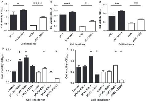
In order to investigate the effect of the genetic modifica-tions on cell viability, we used the MTT assay as described (Portelli et al. 2014). Early passage cells con-taining either BMI-1 vector or pWZL-hTERT showed increased viability in both donors over 96 or 120 h (Fig. 2A, B and C, P<0.05). Similarly, mid passage cells showed alterations in viability compared to wild-type cells over 96 h (empty vector controls were no longer viable at this stage). For donor 1, both BMI-1 cell populations increased viability compared to wild-type cells (ANOVA, post hoc P<0.05), whereas hTERT-engineered cells did
not show altered viability compared to wild-type control. For donor 2, pFLRu-BMI-1 significantly increased cell viability and hTERT reduced viability when compared to wild-type control (ANOVA, post hoc P<0.05) with pLVX-BMI-1 cell viability showing no difference (Fig. 2D). In a similar analysis of late passage cells (assayed over 120 h), both donors showed pLVX-BMI-1 significantly increased viability, whereas hTERT cells had a decreased viability (Fig. 2E, ANOVA, post hoc P<0.05). Late passage pFLRu-BMI-1 cells showed no differences in viability compared to wild-type control cells. These data suggest that late passage NHBECs trans-duced with the pLVX-BMI-1 lentivirus are the only cell populations to maintain high viability.
NHBEC-BMI-1 cell populations maintain epithelial cell marker expression over prolonged lifespan
In order to begin to characterize the cell phenotype in the genetically modified cells, we examined the expression of the epithelial lineage marker, cytokeratin (CK) 14. For both donors, a very similar level and distribution of CK14 was observed in wild-type cells at passage 3 and late passage cells for both BMI-1 vectors using immunofluo-rescence (Fig. 3). Similarly, donor 1 hTERT-expressing cells showed comparable expression, however, for donor 2
Cell line
Telomerase activity
(Relative fluorescence)
Positive controlControl cellspWZL(—)- earlyhTERT - early
hTERT- midhTERT - lateControl cellspWZL(—) - earlyhTERT - earlyhTERT - mid
0 10 20 30 40
BMI-1 cDNA
— + — +
Anti BMI-1
Anti B-actin 50 kDa
37 KDa
37 KDa
A
C
BMI-1 cDNA
— + — +
pFLRu pLVX pFLRu pLVX
Anti p16
Anti B-actin 20 kDa
16 KDa
37 KDa
B
Control pFLRu-BMI-1 pLVX-BMI-1
(i) (ii) (iii)
(iv) (v) (vi)
[image:5.612.146.458.72.276.2]D
pWZL-hTERT, the cells did not reach late passage and infection passage 8 (passage 10) was the maximum achieved with limited CK14 expression observed.
ALI differentiated NHBEC-BMI-1 cell
populations show comparative morphology to nonmodified wild-type cells
To compare the morphology of genetically modified cells to low passage (p3) wild-type cells grown at air–liquid interface, we examined sections stained with H & E and alcian blue to detect mucins. Wild-type NHBECs gener-ated a pseudostratifed epithelial layer upon ALI showing similarities to that observed in situ including mucin stain-ing at the apical side and the presence of cilia (Fig. 4). Significantly, early, mid, and late passage BMI-1-expres-sing cells from both lenti-vector constructs maintained the ability to differentiate and showed the classical
pseudostratified epithelial layer in this model, however, late passage BMI-1 cells had more diffuse mucin staining (Fig. 4). In contrast, hTERT-expressing cells failed to show a differentiated phenotype even at early passage and in mid and late passage, it was not possible to generate sections as the cells did not grow and adhere to the tran-swell.
NHBEC-BMI-1 cell populations retain the ability to differentiate into multiple cell types at air liquid interface
In order to further characterize the differentiated pheno-type of wild-pheno-type and genetically modified cells, we used immunofluorescence for MUC5AC which is a marker of goblet cells and beta tubulin, which is a marker of ciliated cells. These data show that early, mid, and late passage BMI-1-expressing cells maintain differentiation potential
pFL Ru
pFLRu
BMI-1 pFLRu
pFLR uBM
I-1
A B C
*
****
***
*
**
**
D E
[image:6.612.72.546.70.400.2]* *
*
*
*
*
*
*
Figure 2. BMI-1 epithelial cell populations have an extended lifespan with viability increased compared to wild-type bronchial epithelial cells. Viability (MTT assay) was determined for early passage cells at (A) pFLRu/96 h, (B) pLVX/96 h, and (C) pWZL/120 h demonstrating that the insertion of BMI1 or hTERT leads to increased viability in Donor 1 (solid bars) and Donor 2 (open bars),*P<0.05,**P<0.005,***P<0.0005,
and generate a differentiated layer composed of multiple cell types comparable to wild-type control cells from the same experiments (Fig. 5). As anticipated in the z-stack
images, both the beta tubulin and MUC5AC staining is particularly apical. hTERT-expressing cells maintain these features until cell senescence for this donor.
Control pFLRu-BMI-1 pLVX-BMI-1 pWZL-hTERT Isotype
(i) (ii) (iii) (iv)
(vi) (vii) (viii) (ix)
(v)
[image:7.612.146.457.70.234.2](x)
Figure 3. BMI-1 epithelial cell populations retain epithelial cell marker expression over extended passage. Immunofluorescence for cytokeratin 14 confirmed epithelial lineage in wild-type cells (Control) and for late passage engineered cells for Donor 1 (i–iv) and Donor 2 (vi–ix). Note pWZL-hTERT in donor 2, mid passage was the maximum passage achieved.
Wild-type (p3) pFLRu-BMI-1 pLVX-BMI-1 pWZL-hTERT
Late
Mi
d
E
arl
y
[image:7.612.70.538.304.565.2]NHBEC-BMI-1 cell populations retain the ability to differentiate and form an epithelial barrier
One of the hallmarks of the ALI model is the develop-ment of tight junctions between the epithelial cells form-ing a barrier. A surrogate measure of this is the development of TEER. Therefore, we examined this out-come in both wild-type and genetically modified cells. Both donors demonstrated the capacity to develop TEER as shown by the wild-type cells (passage 3) time course data (Fig. 6A and B). In Figure 6A, a decrease in TEER was observed between days 14–21 which we have observed for other primary human cell donors. Similar plots were generated for early, mid, and late passage-engineered cells and to facilitate multiple comparisons area under the curve values were generated. At early passage donor 1 cells, all developed a TEER except for the pWZL empty vector cell population (Fig. 6C). The level of TEER was variable with pLVX-BMI-1 showing the most comparable to wild-type control levels (Fig. 6C). Donor 2 early passage data demonstrated that only the BMI-1-containing cells were capable of develop-ing a TEER and in the case of the pLVX-BMI-1, this was higher than the wild-type control. All empty vector
controls and the pWZL-hTERT did not develop a mea-surable TEER at passage 6 (Fig. 6D). At mid passage, the empty vector controls were no longer viable and comparison of the engineered cells showed that in both donors, the BMI-1-expressing cells demonstrated a mea-surable TEER, albeit lower than wild-type, whereas the hTERT-engineered cells did not (Fig. 6E and F). At late passage, again the BMI-1-expressing cells demonstrated a TEER but at a reduced level compared to wild-type cells (Fig. 6G and H). Across the passage stages, there was a gradual loss of TEER development in the BMI-1 cell lines when compared to the wild-type cells (100%) at each stage, for example, Donor 1 pFLRu-BMI-1: 56.5%, 28.1%, and 29.1% (of wild-type control, 100%) across early, mid, and late passages. Similarly, Donor 1 pLVX-BMI-1: 89.5%, 43.5%, 42.1% (of wild-type control, 100%) across early, mid, and late passages. Donor 2 data were more heterogeneous but showed a similar trend. These data demonstrated that the BMI-1-expres-sing cells maintain plasticity, differentiate at ALI, and develop TEER. However, due to our low number of technical replicates, we were not able to provide more quantitative comparisons of the absolute levels of differ-entiation, for example, TEER achieved across cell popu-lations and/or passages with certainty.
Wild-type (p3) pFLRu-BMI-1 pLVX-BMI-1 pWZL-hTERT
Late
Mi
d
E
arly
[image:8.612.76.541.69.310.2]Beta tubulin MUC5AC Beta tubulin MUC5AC Beta tubulin MUC5AC Beta tubulin MUC5AC
0 500 1000 1500 Contr ol
pFLRu pLV X pWZL pFLRu BMI pLVX BMI pWZ L hTE
RT
Cont rol
pFLR u BMI
pLVX BMI pWZL hTER T Cont rol pFLR u BMI
pLVX BMI pWZL hTER T Contr ol pFLR u BM I pLVX BMI pWZL hTERT Cont rol
pFLRu pLV X pWZL pFLRu BMI pLVX BMI pWZ L hTE
RT
Cont rol
pFLR u BMI
pLVX BMI pWZL hTER T N.D A 2000 20,000 15,000 10,000 5000 0 20,000 15,000 10,000 5000 0 20,000 15,000 10,000 5000 0 20,000 15,000 10,000 5000 0 20,000 15,000 10,000 5000 0 20,000 15,000 10,000 5000 0 2000 1500 1000 500 0
3 5 7 10 12
Days at ALI
TEER ( Ω cm 2) TEER ( Ω cm 2) AUC (TEER, Ω cm 2) AUC (TEER, Ω cm 2) AUC (TEER, Ω cm 2) AUC (TEER, Ω cm 2) AUC (TEER, Ω cm 2) AUC (TEER, Ω cm 2) Cell line Cell line Cell line Cell line Cell line Cell line
14 17 19 21 24 28 3 5 7 10 12
Days at ALI
14 17 19 21 24 28
C E G B D F H
Donor 1 – wild-type (p3) Donor 2 – wild-type (p3)
Donor 1 – early passage Donor 2 – early passage
Donor 1 – mid passage Donor 2 – mid passage
[image:9.612.138.467.69.619.2]Donor 1 – late passage Donor 2 – late passage
NHBEC-BMI-1 cell populations retain the ability to differentiate forming ciliated cells at the apical surface
To further characterize the differentiated epithelial layer observed in the ALI model, we used scanning electron microscopy to visualize the apical surface and examine cilia. These images clearly show cilia projections on the surface of wild-type cells as we have observed previously (Fig. 7). For genetically engineered cells, cilia were also present including the hTERT genetically modified cells and there was a large degree of heterogeneity. For mid passage pLVX-BMI-1 cells, there was potentially a reduced pattern of cilia compared to wild-type control. Importantly, there was evidence for late passage BMI-1 cells, particularly the pLVX-BMI-1 cells to have ciliated surfaces (Fig. 7).
NHBEC-BMI-1 cell populations maintain a stable genome over extended passage
As many approaches to prevent cell senescence have unwanted effects on the genetic information of the cells,
we completed karyotyping of wild-type and genetically modified cells at each passage stage to identify gross changes in chromosomes (>5 megabase rearrangements). For all BMI-1-expressing cells assayed at early, mid, and late passage, normal karyotypes were present. In contrast, an abnormal karyotype was observed for donor 2 hTERT-expressing cells at mid passage suggesting gross alterations on chromosomes 6, 13, and 16 and chromosomal frag-ments that could not be mapped (Fig. 8).
Discussion
In this study, we set out to evaluate methods to delay cell senescence while maintaining plasticity/differentiation potential in primary human bronchial epithelial cells. This is important as these cells have a key role to play in respi-ratory disease warranting further research. However, such primary cells are isolated from patients by invasive proce-dures and have limited lifespan ex vivo. We have devel-oped two lentiviral systems that enabled us to elevate the expression of polycomb protein BMI-1 and importantly suppress p16 levels in cells containing these genetic modi-fications. Extensive characterization over 12 months
Control cells (p3)
pFLRu-BMI-1
pLVX-BMI-1
pWZL-hTERT
Late
Mi
d
E
[image:10.612.77.543.364.639.2]arly
A
B
[image:11.612.157.442.71.647.2]C
demonstrated that cells expressing higher levels of BMI-1 have (1) increased viability, (2) an extended number of cell divisions, (3) maintain basal epithelial morphology, (4) maintain plasticity, that is, the ability to form a differ-entiated pseudostratified air–liquid interface model, and (5) importantly maintain a normal karyotype. These data suggest that the cell populations generated using BMI-1 induction maintain cell plasticity over extended periods in the laboratory and may be useful for primary human bronchial epithelial cell research.
To date, either viral oncogenes have been used to pre-vent cell senescence in human bronchial epithelial cells (Willey et al. 1991; Zeitlin et al. 1991; Viallet et al. 1994) or the introduction of human telomerase reverse tran-scriptase (hTERT) (Zabner et al. 2003; Piao et al. 2005). These studies have shown some success, however, a major limitation is alterations in karyotype (Piao et al. 2005). Therefore, viral oncogene independent approaches for prevention of cell senescence have been sought, the most favored approach being one with an underlying mecha-nism that decreases the levels of cyclin-dependent kinase inhibitor, p16(Ink4a), a tumor suppressor that induces a G1 cell cycle arrest.
BMI-1 is a polycomb transcriptional repressor protein and is part of the polycomb repressive complex-1 (PRC1) complex. This complex regulates transcriptional activity at several loci including the Ink4a locus which encodes the tumor suppressor proteins p16(Ink4a) and p14(Arf). BMI-1 has been extensively characterized in the context of numerous cancers, with both protein and mRNA levels correlating with disease prognosis (Cao et al. 2011). The contribution of BMI-1 to multiple cancers is thought to be due to its role in self-renewal and differentiation of stem cells (Lessard and Sauvageau 2003). With these unique properties of regulating cell senescence, BMI-1 has been the focus of attention particularly in the delay of cell senescence in many cell types, for example, stem cell fee-der cells (McKay et al. 2011). Recently, transduction of HBEC with combinations of hTERT and BMI-1 has gen-erated some promising data (Fulcher et al. 2009). The resultant hTERT/BMI-1 cell populations continued to divide longer than nonmodified cells, and importantly, at passage 14 and 15 had a diploid karyotype and formed differentiated pseudostratified morphology following ALI culture. Interestingly, BMI-1 alone was also introduced to bronchial epithelial cells in the study of Fulcher and col-leagues and delayed cell senescence, however, these BMI-1 cell populations were not extensively characterized for plasticity, a hallmark of low passage basal epithelial cells.
In this study, we set out to significantly extend these studies and focus away from the use of hTERT to the use of BMI-1 alone to delay cell senescence in bronchial epithelial cells and provide more definitive
characterization of the plasticity of cells particularly in the context of ALI models. To this end, we developed two new lentiviral systems engineered to express elevated BMI-1 under the control of CMV or UbiC promoter and a control retroviral system expressing hTERT for compar-ison. These systems were used to transduce passage 2 nor-mal bronchial epithelial cells from two donors. These genetically engineered cells showed sustained elevation of BMI-1 as determined by western blot and immunofluo-rescence and similarly elevated telomerase activity for the hTERT-engineered cells. As anticipated, the BMI-1-expressing cells showed consequent suppression of p16 as observed by several groups (Meng et al. 2010). It is important to note that BMI-1 is a master regulator of a very large number of genes and gene families of relevance to cell senescence, for example, see a recent study using microarray analyses in BMI-1 knockout mice cells (Zacharek et al. 2011). In this study, a large number of genes were differentially expressed (elevated) when BMI-1 was absent which included multiple homeodomain genes and Cdkn2a (p16/p19) and Cdkn2b (p15). In a more recent study using BMI-1 / mouse brain tissue in con-junction with RNA-seq,>500 genes were shown to be up regulated due to a loss of BMI-1 including identifying a role for BMI-1 in TGF-b/BMP-ER stress pathways (Gar-giulo et al. 2013). Therefore, while our p16 suppression data confirms that our genetic manipulation of BMI-1 has downstream effects on BMI-1 responsive networks, it is highly likely that multiple pathways underlie the effects we have observed, for example, p14Arf suppression by BMI-1 may be highly relevant for apoptosis mechanisms through the mdm2/TP53 pathway.
data is in contrast to a recent study that engineered pri-mary airway epithelial cells to express hTERT and reported that these cells grew beyond 40 passages, how-ever, it is difficult to make conclusions as this study only used cells from one donor (Walters et al. 2013). Over extended passage the BMI-1-expressing cells maintained epithelial marker expression (CK14), whereas this was more variable with the hTERT-engineered cells. To date, no other study has evaluated changes in CK14 marker expression post 1 engineering, however, both BMI-1/hTERT- and hTERT-engineered cells demonstrated maintenance of an epithelial phenotype based on addi-tional markers including KRT5 in hTERT cell populations (Walters et al. 2013).
An important finding of our current study was that BMI-1-expressing cells (early-mid-late up to passage 15) maintain cell plasticity and the ability to differentiate at air–liquid interface generating a pseudostratified layer com-posed of basal, goblet, and ciliated cells as observed by sec-tioning transwells. These data are novel and significantly extend data on BMI-1/hTERT- or hTERT-engineered cells that can also maintain this capacity (Fulcher et al. 2009; Walters et al. 2013). However, the hTERT cell populations generated in this study did not maintain this plasticity and showed limited differentiation in contrast to a recent report (Walters et al. 2013). These data question the utility of hTERT alone and suggest BMI-1 alone to be the preferred genetic tool to maintain plasticity in our study. Of note, several groups are developing combination approaches to prevent cell senescence in primary epithelial cells using hTERT, for example, hTERT/BMI-1 can lead to cells that reach at least 38 population doublings (Fulcher et al. 2009) and hTERT/cyclin-dependent kinase 4 (Cdk4) epithelial cells have shown >100 population doublings (Roig et al. 2010). However, in this study, we focused to the use of BMI-1 alone to reduce transformation of the cells away from a diploid karyotype and to maintain plasticity.
In agreement with the potential utility of BMI-1 cell populations, further measures of differentiation in the context of the air–liquid interface model, mainly goblet cell (MUC5AC), ciliated cell (beta tubulin) staining, development of TEER, and SEM showing apical cilia were all maintained in the late passage BMI-1 cell populations but not in the hTERT cell populations.
Overall, these data show that for the first time BMI-1-expressing cells have an extended lifespan, albeit not exten-sive and suggest that passage 15 cells show very similar plasticity properties to wild-type cells for these key out-comes. Of critical importance, compared to other methods that incorporate the use of hTERT, we did not observe any alterations in karyotype in BMI-1-expressing cells, whereas the hTERT cells (p8) had chromosomal abnormalities. These findings are in complete agreement to the recent
study of hTERT-engineered bronchial epithelial cell popu-lations which identified chromosomal abnormalities as early as passage 9 (Walters et al. 2013).Interestingly, anal-yses of passage 14/15 BMI-1/hTERT failed to identify chro-mosomal abnormalities suggesting that BMI-1 may be protective in this context (Fulcher et al. 2009).
This study has several strengths over previous reports such as including the evaluation of multiple BMI-1 and hTERT vectors in parallel in multiple primary human donors. Similarly, the extensive characterization of these cells for cell plasticity in the context of the ALI model and for karyotype is novel. These data from four geneti-cally modified cell populations show comparable effects of BMI-1 elevation. However, we acknowledge that greater numbers of donors and technical replicates are required to provide more quantitative characterization of cell differentiation, for example, TEER. This represents a limitation of this study. We have previously identified a large degree of heterogeneity in both intra- and inter-donor variation in the air–liquid interface model (Stewart et al. 2012b), even in the absence of genetic modification suggesting a substantial study is required involving large numbers of donors over many years to further define quantitative changes in cell plasticity using this model.
In conclusion, we provide an in-depth characterization of the plasticity of human bronchial epithelial cells that have been engineered to express BMI-1. These cells retain the plasticity observed in wild-type low passage cells including the ability to differentiate at ALI that can be used to model the lining of the airways. These findings suggest these cells may show utility in many aspects of respiratory research including in defining airway epithelial abnormalities in diseases such as asthma, which has the potential to lead to therapeutic opportunities.
Acknowledgments
We thank Professor Greg Longmore (Washington Univer-sity) for providing the pFLRu-FH plasmid, the Advanced Microscopy Unit (Life Sciences, University of Notting-ham) for their help with SEM, and Dr John G. Foster for critical reading of this manuscript.
Conflicts of Interest
No conflicts of interest, financial or otherwise, are declared by the authors.
References
Fulcher, M. L., S. E. Gabriel, J. C. Olsen, J. R. Tatreau, M. Gentzsch, E. Livanos, et al. 2009. Novel human bronchial epithelial cell lines for cystic fibrosis research. Am. J. Physiol. Lung Cell. Mol. Physiol. 296:L82–L91. Gargiulo, G., M. Cesaroni, M. Serresi, N. de Vries, D.
Hulsman, S. W. Bruggeman, et al. 2013. In vivo RNAi screen for BMI1 targets identifies TGF-beta/BMP-ER stress pathways as key regulators of neural- and malignant glioma-stem cell homeostasis. Cancer Cell 23:660–676.
Gras, D., A. Bourdin, I. Vachier, L. de Senneville, C. Bonnans, and P. Chanez. 2012. An ex vivo model of severe asthma using reconstituted human bronchial epithelium. J. Allergy Clin. Immunol. 129:1259–1266e1.
Kelsen, S. G., I. A. Mardini, S. Zhou, J. L. Benovic, and N. C. Higgins. 1992. A technique to harvest viable
tracheobronchial epithelial cells from living human donors. Am. J. Respir. Cell Mol. Biol. 7:66–72.
Kicic, A., T. S. Hallstrand, E. N. Sutanto, P. T. Stevens, M. S. Kobor, C. Taplin, et al. 2010. Decreased fibronectin production significantly contributes to dysregulated repair of asthmatic epithelium. Am. J. Respir. Crit. Care Med. 181:889–898.
Lessard, J., and G. Sauvageau. 2003. Bmi-1 determines the proliferative capacity of normal and leukaemic stem cells. Nature 423:255–260.
Lopez-Guisa, J. M., C. Powers, D. File, E. Cochrane, N. Jimenez, and J. S. Debley. 2012. Airway epithelial cells from asthmatic children differentially express proremodeling factors. J. Allergy Clin. Immunol. 129:990–997e Maida, Y., M. Yasukawa, M. Furuuchi, T. Lassmann, R.
Possemato, N. Okamoto, et al. 2009. An RNA-dependent RNA polymerase formed by TERT and the RMRP RNA. Nature 461:230–235.
McKay, T. R., M. V. Camarasa, B. Iskender, J. Ye, N. Bates, D. Miller, et al. 2011. Human feeder cell line for derivation and culture of hESc/hiPSc. Stem Cell Res. 7:154–162.
Meng, S., M. Luo, H. Sun, X. Yu, M. Shen, Q. Zhang, et al. 2010. Identification and characterization of Bmi-1-responding element within the human p16 promoter. J. Biol. Chem. 285:33219–33229.
Parker, J., S. Sarlang, S. Thavagnanam, G. Williamson, D. O’Donoghue, R. Villenave, et al. 2010. A 3-D well-differentiated model of pediatric bronchial epithelium demonstrates unstimulated morphological differences between asthmatic and nonasthmatic cells. Pediatr. Res. 67:17–22.
Piao, C. Q., L. Liu, Y. L. Zhao, A. S. Balajee, M. Suzuki, and T. K. Hei. 2005. Immortalization of human small airway epithelial cells by ectopic expression of telomerase. Carcinogenesis 26:725–731.
Portelli, M. A., M. Siedlinski, C. E. Stewart, D. S. Postma, M. A. Nieuwenhuis, J. M. Vonk, et al. 2014. Genome-wide
protein QTL mapping identifies human plasma kallikrein as a post-translational regulator of serum uPAR levels. FASEB J 28:923–934.
Qin, J. Y., L. Zhang, K. L. Clift, I. Hulur, A. P. Xiang, B. Z. Ren, et al. 2010. Systematic comparison of constitutive promoters and the doxycycline-inducible promoter. PLoS ONE 5:e10611.
Roig, A. I., U. Eskiocak, S. K. Hight, S. B. Kim, O. Delgado, R. F. Souza, et al. 2010. Immortalized epithelial cells derived from human colon biopsies express stem cell markers and differentiate in vitro. Gastroenterology 138:1012–1021. e15
Stewart, C. E., E. E. Torr, N. H. Mohd Jamili, C. Bosquillon, and I. Sayers. 2012a. Evaluation of differentiated human bronchial epithelial cell culture systems for asthma research. J. Allergy (Cairo) 2012:943982.
Stewart, C. E., H. S. Nijmeh, C. E. Brightling, and I. Sayers. 2012b. uPAR regulates bronchial epithelial repair in vitro and is elevated in asthmatic epithelium. Thorax 67:477–487. Viallet, J., C. Liu, J. Emond, and M. S. Tsao. 1994.
Characterization of human bronchial epithelial cells immortalized by the E6 and E7 genes of human papillomavirus type 16. Exp. Cell Res. 212:36–41. Vonlanthen, S., J. Heighway, H. J. Altermatt, M. Gugger, A.
Kappeler, M. M. Borner, et al. 2001. The bmi-1 oncoprotein is differentially expressed in non-small cell lung cancer and correlates with INK4A-ARF locus expression. Br. J. Cancer 84:1372–1376.
Walters, M. S., K. Gomi, B. Ashbridge, M. A. Moore, V. Arbelaez, J. Heldrich, et al. 2013. Generation of a human airway epithelium derived basal cell line with multipotent differentiation capacity. Respir. Res. 14:135.
Willey, J. C., A. Broussoud, A. Sleemi, W. P. Bennett, P. Cerutti, and C. C. Harris. 1991. Immortalization of normal human bronchial epithelial cells by human papillomaviruses 16 or 18. Cancer Res. 51:5370–5377.
Xiao, C., S. M. Puddicombe, S. Field, J. Haywood, V. Broughton-Head, I. Puxeddu, et al. 2011. Defective epithelial barrier function in asthma. J. Allergy Clin. Immunol. 128:549–556. e1–12.
Zabner, J., P. Karp, M. Seiler, S. L. Phillips, C. J. Mitchell, M. Saavedra, et al. 2003. Development of cystic fibrosis and noncystic fibrosis airway cell lines. Am. J. Physiol. Lung Cell. Mol. Physiol. 284:L844–L854.
Zacharek, S. J., C. M. Fillmore, A. N. Lau, D. W. Gludish, A. Chou, J. W. Ho, et al. 2011. Lung stem cell self-renewal relies on BMI1-dependent control of expression at imprinted loci. Cell Stem Cell 9:272–281.