www.wjpr.net Vol 4, Issue 1, 2015. 1012
CYTOTOXIC EFFECT ON HUMAN OSTEOSARCOMA CELL LINE
MG-63 AND ANTIMICROBIAL ACTIVITY ON CLINICAL
PATHOGENS BY PLANT MEDIATED SILVER NANOPARTICLES
Ritika Chauhan, Mohammed Ibrahim, Shankar Narayanan and Jayanthi Abraham*
Microbial Biotechnology Laboratory, School of Biosciences and Technology, VIT University, Vellore-632014, Tamil Nadu.
ABSTRACT
The present investigation reveals the green synthesis of silver nanoparticles using Solanum nigrum and Cardiospermum halicacabum. The silver nanoparticles were synthesized and characterized by UV-Vis spectrophotometer (UV-Vis), Fourier transfer infrared spectrophotometer (FT-IR), X-ray diffraction (XRD), atomic force mass spectroscopy (AFM) and particle shape of the synthesized nanoparticles were determined by scanning electron microscopy (SEM). Furthermore, synthesized nanoparticles were evaluated for antimicrobial effect against various pathogens and cytotoxic effect on human osteosarcoma (MG-63) cell line. The plant mediated silver nanoparticles exhibited good antimicrobial activity against clinical pathogens and cytotoxic effect against bone cancer cells. Plant mediated silver nanoparticles possess therapeutic properties which may be useful in pharmaceutical applications.
KEYWORDS: Plant extracts; Cytotoxicity effect; Green synthesis.
INTRODUCTION
The green synthesis of metal nanoparticles has emerged as an alternative approach towards novel therapeutics against many ailments.[1] The present research attracts synthesis of green nanoscale particles for the development of therapeutic agents.[2,3] The synthesis of metal nanoparticles employs the use of non-biodegradable toxic chemicals as reducing agents which pose a potential threat to environment and biological system. The biological and green synthesis approach has been suggested as valuable alternative to chemical and physical
Volume 4, Issue 01, 1012-1027. Research Article ISSN 2277– 7105
Article Received on 28 October 2014,
Revised on 18 Nov 2014, Accepted on 10 Dec 2014
*Correspondence for
Author
Dr. Jayanthi Abraham,
www.wjpr.net Vol 4, Issue 1, 2015. 1013 methods.[4] For ecofriendly synthesis of silver nanoparticles, the use of micro-organisms or the extracts of medicinal plants holds unique attention among researchers.[5] Silver nanoparticles are well known for its antimicrobial effect in medicinal field[6,7] and it also possess wide applications in physics, material science and chemistry.[8] Silver nanoparticle follows unique mode of action leading to the development of a new generation of antibiotics to overcome drug resistance[9], antitumor[10], antiproliferative[11] and antiangiogenic agent.[12] AgNPs also play an effective role in tumor control via their cytotoxic effects,[13] in treatment of diseases including retinal neovascularization [14,15] and acquired immunodeficiency syndrome caused by human immunodeficiency virus (HIV)[16,17] The increasing interest in the development of nanomedicine using silver nanoparticles has led to treatment of various infectious diseases including hepatitis B,[18] respiratory syncytial virus, herpes simplex virus type 1,[19] and monkey pox virus. [20]
Plant products have been used from ancient times for its bioactivity and for discovering new drugs. The bioactive compounds thus, extracted are highly efficient against microbial infections and various pharmaceutical properties. Millions of species are known to have medicinal value and several studies have reported the antimicrobial activity of plants.[21] Solanum nigrum and Cardiospermum halicacabum are important sources of medicinal properties. These plants possess antiseptic, antidysentric, antidiuretic and are recommended in ayurveda for the management of gastric ulcers.[22] By considering the medicinal aspects of both the plants and nanomedicine the present investigation was carried out to study comparative analysis of bioactivity and synthesis of nanoparticles and evaluate their efficiency. To our knowledge not much of the work has been reported regarding cytotoxicity of silver nanoparticles against bone cancer cells, most of the cytotoxicity effect have been carried out on cervical (HeLa) and breast (MCF-7) cancer cells. Therefore the present work was aimed at cytotoxicity of plant mediated silver nanoparticles against bone cancer cells (MG-63).
MATERIALS AND METHODS Sample collection
www.wjpr.net Vol 4, Issue 1, 2015. 1014 Extraction of phytochemical compounds
30 g of finely ground powder of Solanum nigrum and Cardiospermum halicacabum was extracted with n-hexane, ethyl acetate and methanol subsequently in soxhlet extractor not exceeding the boiling point of the solvent. The extracts obtained were filtered through Whattman No.1 filter paper and then concentrated under reduced atmospheric pressure. The dry extracts were stored at 4C for further biological analysis.
Phytochemical screening
The preliminary phytochemical analysis was performed by following the protocol of Trease and Evans.[23] Test for Alkaloids: 0.5 g of the extracts was dissolved in 5 ml of 1% HCL and it was kept in a boiling water bath. 1 ml of filtrate was treated with a drop of Meyer’s reagent. Turbidity or the change in precipitate was taken as an indicator for the presence of alkaloids. Test for Tannins: 0.5 ml of each sample was mixed with 10 ml of boiling water and filtered. Few drops of 6% FeCl3 were added to the filtrate and appearance of deep green color confirmed the presence of tannins.
Test for Flavanoids: 0.2 ml of the plant extracts was dissolved in CH3OH and heated. A chip of Mg metal was added to the mixture followed by the addition of few drops of HCL. The appearance of reddish orange indicates the presence of flavonoids.
Test for Steroids: 0.5 ml of the extract was dissolved in 3 ml of CHCl3 and filtered. To the filtrate concentrated H2SO4 was added, which forms a lower layer. A reddish brown color indicates the presence of steroids.
Antimicrobial assay
The antimicrobial activity of plant extracts were estimated by Kirby-Bauer method against pathogenic microorganisms. The pathogens were swabbed onto Muller-Hinton agar plates and four wells were punctured. The plant extracts were introduced into the wells with different concentration of 25, 50, 75, 100 µl. The plates were incubated at 37 C for 24 h. The diameter of zone of clearance around the wells after the incubation period confirmed the antimicrobial activity of the respective extract.
Synthesis of silver nanoparticles
www.wjpr.net Vol 4, Issue 1, 2015. 1015 leaf extract was allowed to cool and was filtered using Whattman No.1 filter paper. The green synthesis of AgNPs was carried out by adding 1 mM silver nitrate (AgNO3) to 90 ml of deionized water which was mixed with 10 ml of Solanum nigrum and Cardiospermum halicacabum aqueous leaf extract. The conical flask containing silver nanoparticles were incubated for 24-72 h at room temperature.
Characterization of synthesized nanoparticle
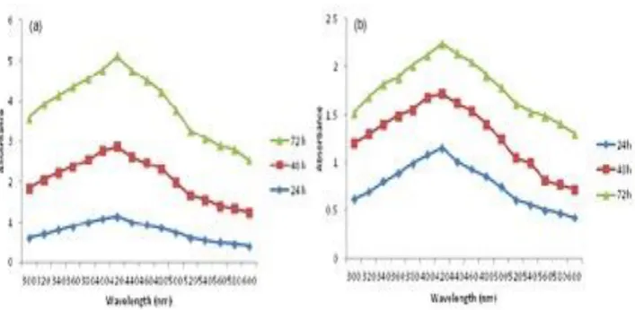
The reduction of pure Ag+ ions was monitored by measuring UV-visible spectrum of the reaction medium. The progress of the reaction between metal ions and the leaf extracts was monitored by UV-visible spectra of Ag nanoparticles in aqueous solution with different reaction times of 24, 48 and 72 h. UV-visible spectral analysis was carried out by using Perkin-Elmer Lambda 25 spectrophotometer with a resolution of 1 nm between 300 and 600 nm.
The Ag nanoparticles solution (100 ml) was centrifuged at 5000 rpm for 20 min. The pellet was washed three times with 10 ml of de-ionized water to get rid of the free proteins/enzymes. The samples were dried and ground with potassium bromide (KBr) pellets and analyzed on a JASCO FT/IR-5300 model in the diffuse reflectance mode operating at a resolution of 4000 cm-1.
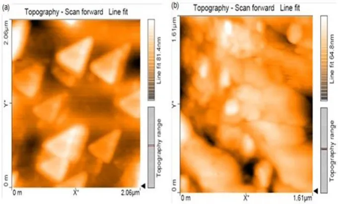
The topography and particle size of synthesized silver nanoparticles was studied using AFM (Model-Nanosurf easyscan 2 AFM, made in Switzerland) working in the contact mode. The size of the nano objects was measured with atomic resolution from the thin film of the sample which was prepared on a glass slide by dropping 100 μL of the sample on the slide. The topographical images were obtained in non-contact mode using silicon nitride tips at a resonance frequency of 218 kHz.
www.wjpr.net Vol 4, Issue 1, 2015. 1016 Antimicrobial activity of synthesized silver nanoparticles
The silver nanoparticles (AgNPs) synthesized from the leaf extracts were tested for their antimicrobial activity by well diffusion method against pathogenic bacteria such as Escherichia coli, Pseudomonas aeruginosa, Staphylococcus aureus, Shigella sp., Enterococci sp., Solmonella typhi, Klebsiella pneumonia, Proteus mirabilis and Serratia marsenences. The pathogenic cultures were swabbed onto sterile Muller Hinton agar plates. The antimicrobial activity was determined by well-diffusion assay, wells of 6 mm were punctured onto Muller Hinton agar plates by using sterilized well borer. 100 µl of the sample of synthesized nanoparticles was introduced into the wells and was incubated for 24 h at 37 C, the diameter zone of inhibition was measured.
Antioxidant Assay
DPPH radical-scavenging activity
The free radical scavenging activity of synthesized silver nanoparticles was analyzed by 2, 2-diphenyl-1 picrylhydrazyl (DPPH). 300 µl of 1 mM synthesized nanoparticles was mixed with 2.7 ml of methanolic solution containing DPPH radicals of 0.05 mM.[24] The mixture was shaken vigorously and allowed to stand for 30 min in dark. The reduction of the DPPH radical was determined by measuring the absorption at 517 nm. The radical scavenging activity (RSA) was calculated as a percentage of DPPH discoloration using the equation % RSA = [(ADPPH-AS)/ADPPH]/100, where AS is the absorbance of the solution when the sample extract has been added at a particular level, and ADPPH is the absorbance of the DPPH solution. [25]
Ferric Reducing Antioxidant Power Assay
The ferric reducing antioxidant power assay (FRAP) method is based on the reduction of a ferric 2, 4, 6-tripyridyl-s-triazine complex (Fe3+-TPTZ) to the ferrous form (Fe2+- TPTZ). The FRAP assay was carried out by the method of Benzie and Strain.[26] 1 mM of synthesized silver nanoparticles was analyzed for reducing assay, 20 µl of the sample was added to 3 mL of FRAP reagent and the increase in the reagent was measured at 593 nm after 30 min of incubation.
Cytotoxic assay
www.wjpr.net Vol 4, Issue 1, 2015. 1017 containing 10% fetal bovine serum (FBS). All cells were maintained at 37°C, 5% CO2, 95% air and 100% relative humidity. The monolayer cells were detached with trypsin-ethylenediaminetetraacetic acid (EDTA) to make single cell suspensions and viable cells were counted using a hemocytometer and diluted with medium containing 5% FBS to give final density of 1x10⁵ cells/ml. 100 µl per well of cell suspension were seeded into 96-well plates at plating density of 10,000 cells/well and incubated to allow cell attachment at 37°C, 5% CO2, 95% air and 100% relative humidity. [27]
After 48h of incubation, 15 µl of MTT (5 mg/ml) in phosphate buffered saline (PBS) was added to each well and incubated at 37°C for 4 h. The medium with MTT was separated and the formazan formed crystals were solubilized in 100 µl of DMSO and then measured the absorbance at 570 nm using micro plate reader. The percentage cell inhibition was determined using the following formula: Percentage of cell inhibition = 100- Abs (sample)/Abs (control) x100. [28] Nonlinear regression graph was plotted between % Cell inhibition and Log concentration and IC50 was determined using GraphPad Prism software.
RESULTS AND DISCUSSION Phytochemical analysis
www.wjpr.net Vol 4, Issue 1, 2015. 1018 Table 1 Antimicrobial activity of hexane, ethyl acetate and methanol extracts of
Solanum nigrum against various pathogens
S.
No. Test Organisms
Zone of inhibition (mm) Hexane extract (µl)
Zone of inhibition (mm) Ethyl acetate extract (µl)
Zone of Inhibition (mm) Methanol extract (µl)
25 50 75 100 25 50 75 100 25 50 75 100
1 E. coli - 5 10 10 - 9 - 20 - - 10 25 2 P. aeruginosa - - 20 22 - - - 10 1 - 20 25 3 S. aureus - - 10 10 - - 5 10 2 - 5 10 4 Shigella sp. - - 10 10 - - 5 12 5 - 10 10 5 Enterococci sp. - - - 20 - - - - 6 Salmonella sp. - 10 10 20 - - - 10 - - - - 7 Klebsiella. - - - 10 - - 10 15 8 P. mirabilis - - - 10 10 - - - - 9 S. marcescences - - - 5 20 - - - -
Table2. Antimicrobial activity of hexane, ethyl acetate and methanol extracts of Cardiospermum halicacabum against various pathogens
S.
No. Test Organisms
Zone of inhibition (mm) Hexane extract (µl)
Zone of inhibition (mm) Ethyl acetate extract (µl)
Zone of inhibition (mm) Methanol extract (µl)
25 50 75 100 25 50 75 100 25 50 75 100
1 E. coli - 10 15 15 - - - - 2 P. aeruginosa - - - - 3 S. aureus - - - - 4 Shigella sp. - - 10 25 - - - - 5 Enterococci sp. - - - - 6 Salmonella sp. - - - - 7 Klebsiella sp. - - - - 8 P. mirabilis - - 15 30 - - - - 9 S. marcescences - - 10 20 - - - -
Synthesis and Characterization of silver nanoparticles
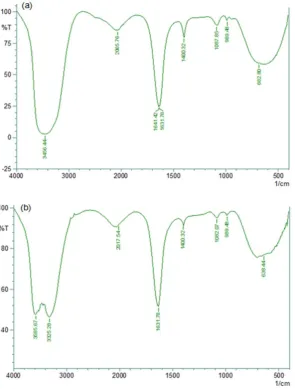
www.wjpr.net Vol 4, Issue 1, 2015. 1019 silver nanoparticles produced by Solanum nigrum showed bands at 3456.44 cm-1, which falls near 3280 cm-1 was considered as the stretching vibrations of primary amines while the corresponding bending vibrations were observed in 1641.42 cm-1 and 1631.78 cm-1 as shown in Figure 2(a). Silver nanoparticles produced by Cardiospermum halicacabum showed bands in the range of 3325.28 cm-1, which is closely related to the band 3280 cm-1 corresponds to the stretching vibrations of primary amines. The strong peak at 3585.67 cm-1 corresponds to the stretching of alcohols and phenolic groups. The strong band observed at 1631.78 cm-1 corresponds to the stretching vibrations of C=C as shown in Figure 2(b). The vibrational bands corresponding to bonds flavonoids and terpenoids in plant extracts. Hence, it may be assumed that these biomolecules are responsible for capping and stabilization. The bands observed in FTIR of bio synthesized AgNPs using plant leaf extracts confirms the presence of proteins in the corresponding plant extracts.
Fig. 1(a) UV-visible spectrum analysis of plant mediated silver nanoparticles synthesized by Solanum nigrum. (b) UV-visible spectrum analysis of plant mediated silver nanoparticles synthesized by Cardiospermum halicacabum.
[image:8.595.117.471.332.505.2]www.wjpr.net Vol 4, Issue 1, 2015. 1020 Fig. 2(a) FT-IR spectra of the biofunctional peaks for the synthesized silver nanoparticles by Solanum nigrum. (b) FT-IR spectra of the biofunctional peaks for the synthesized silver nanoparticles by Cardiospermum halicacabum.
The scanning electron micrographs of synthesized nanoparticles represent irregular shape by Solanum nigrum as shown in Figure 5(a) whereas Cardiospermum halicacabum also showed irregular morphology of synthesized nanoparticles as shown in Figure 5(b). Furthermore, EDAX was performed to confirm the synthesis of silver nanoparticles as shown in Figure 6(a) for Solanum nigrum and Figure 6(b) for Cardiospermum halicacabum.
[image:9.595.152.448.77.467.2]www.wjpr.net Vol 4, Issue 1, 2015. 1021 showed moderate antioxidant effect with very light color change when analyzed by DPPH and FRAP assay.
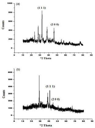
Fig. 3(a) XRD pattern peaks of silver nanoparticles synthesized by Solanum nigrum.
(b) XRD pattern peaks of silver nanoparticles synthesized by Cardiospermum halicacabum.
Cytotoxic effect
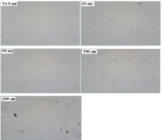
[image:10.595.143.454.128.543.2]www.wjpr.net Vol 4, Issue 1, 2015. 1022 mortality and for Solanum nigrum IC 50 it was found to be 538.3 µg/ml which exhibits dose dependent relation on cell death. Jacob[29] reported that silver nanoparticles may induce reactive oxygen species and cause damage to cellular components leading to cell death.
Fig. 4(a) Atomic force microscopy of silver nanoparticles synthesized by Solanum nigrum. (b) Atomic force microscopy of silver nanoparticles synthesized by
Cardiospermum halicacabum.
Fig. 5(a) Scanning electron micrographs of silver nanoparticles synthesized by Solanum nigrum. (b) Scanning electron micrographs of silver nanoparticles synthesized by
Cardiospermum halicacabum.
[image:11.595.125.466.149.355.2] [image:11.595.132.463.443.627.2]www.wjpr.net Vol 4, Issue 1, 2015. 1023 reported the anti-proliferative effects of carvacrol isolated from O. vulgare against human prostate cancer cell line, LNCap. Figure 7 and Figure 8 represents the cytotoxicity effect of Solanum nigrum and Cardiospermum halicacabum against bone cancer cell line (MG-63) respectively.
Fig. 6(a) EDAX micrograph of silver nanoparticles synthesized by Solanum nigrum. (b)EDAX micrograph of silver nanoparticles synthesized by Cardiospermum halicacabum.
[image:12.595.127.470.156.322.2] [image:12.595.127.464.405.695.2]www.wjpr.net Vol 4, Issue 1, 2015. 1024 Fig. 8 Cytotoxicity analysis of plant mediated silver nanoparticles by Cardiospermum halicacabum against bone cancer cells (MG-63).
But studies related to green synthesis of Solanum nigrum and Cardiospermum halicacabum with anticancer studies in regard to osteosarcoma is lacking. Most of the researchers mostly focus on women oriented cancer studies such as cervical cancer cells (HeLa cell lines) or breast cancer cells (MCf-7). In our study we explore anticancer effect of plant mediated silver nanoparticles against bone cancer cell (MG 63) as osteosarcoma is regarded as disseminated disease at the time of diagnosis and it is found to be radioresistant and also resistant to most of the cytostatic agents.
Declaration of conflicts of interest There is no conflict of interest to declare.
REFERENCES
[image:13.595.128.467.70.394.2]www.wjpr.net Vol 4, Issue 1, 2015. 1025 2. Shanmugasundaram T, Radhakrishnan M, Gopikrishnan V, Pazhanimurugan R, Balagurunathan R. A study of the bactericidal, anti-biofouling, cytotoxic and antioxidant properties of actinobacterially synthesized silver nanoparticles. Colloid Surf B, 2013; 111: 680- 687.
3. Dubey M, Bhadauria S, Kushwah B. Green synthesis of nanosilver particles from extract of eucalyptus hybrid leaf. Dig J Nanomater Biostruct, 2009; 4: 537-543.
4. Mohanpuria P, Rana NK, Yadav SK. Biosynthesis of nanoparticles: technological concepts and future applications. J Nanopart Res, 2008; 10: 507-517.
5. Reddy JN, Vali D N, Rani M, Rani SS. Evaluation of antioxidant, antibacterial and cytotoxic effects of green synthesized silver nanoparticles by Piper longum fruit. Mater Sci Eng, 2014; 34: 115-122.
6. Castellano JJ, Shafii SM, Ko F, Donate G, Wright TE, Mannari RJ. Comparative evaluation of silver-containing antimicrobial dressings and drugs. Int Wound J, 2007; 4: 114-22.
7. Richard JW, BA Spencer, McCoy LF, Carina E, Washington J, Edgar P (2002). Acticoat versus silverlon: the truth. J Burns Surg Wound Care, 2002; 1: 11-20.
8. Yang P, Wei W, Tao C. Determination of trace thiocyanate with nano-silver coated multi-walled carbon nanotubes modified glassy carbon electrode. Anal Chim. Acta, 2007; 585-331.
9. Singh M, Singh S,. Prasad S, Gambhir IS. Interaction and nanotoxic effect of ZnO and Ag nanoparticles on mesophilic and halophilic bacterial cells. Dig J Nanomater Bios, 2008; 3; 115.
10.Ahamed M, Karns M, Goodson M (2008). DNA damage response to different surface chemistry of silver nanoparticles in mammalian cells. Toxicol Applied Pharma, 2008; 233; 404-410.
11.AshaRani PV, Mun GLK, Hande MP, Valiyaveettil S. Cytotoxicity and genotoxicity of silver nanoparticles in human cells. ACS Nano. 2009; 3; 279–290.
12.Gurunathan S, Lee KJ, Kalishwaralal K, Sheikpranbabu S, Vaidyanathan R, Eom SH. Antiangiogenic properties of silver nanoparticles. Biomaterials, 2009; 30:6341.
www.wjpr.net Vol 4, Issue 1, 2015. 1026 14.Bhattacharya K, Davoren M, Boertz J, . Schins R P F, Hoffmann E, Dopp E. Titanium dioxide nanoparticles induce oxidative stress and adduct formation but not DNA-breakage in human lung cells. Part. Fibre Toxicol, 2009; 6: 17.
15.Kalishwaralal K, Deepak V, Ramkumarpndian S, Nellaiah H, Sangiliyandi G (2008) Extracellular biosynthesis of silver nanoparticles by the culture supernatant of Bacillus licheniformis. Mater. Letters, 2008; 62: 4411-4413.
16.Sun L, Singh AK, Vig K, Pillai SR, Singh SR. Silver nanoparticles inhibit replication of respiratory syncytial virus. J Biomed Biotechnol, 2008;4: 149.
17.Lara HH, Trevino ENG., Turrent LI, Singh DK. Silver nanoparticles are broad-spectrum bactericidal and virucidal compounds. Nanobiotechnology,2011; 9: 30
18.Lu L, Sun RW, Chen R, Hui CK, Ho CM, Luk JM, Lau GK, Che CM. Silver nanoparticles inhibit hepatitis B virus replication. Antivir Ther, 2008; 13: 253-62.
19.Baram-Pinto D, Shukla S, Perkas N, Gedanken A, Sarid R. Inhibition of herpes simplex virus Type 1 infection by silver nanoparticles capped with mercaptoethane sulfonate. Bioconjugate Chem 2009; 40: 1497.
20.Rogers JV, Parkinson CV, Choi YW, Speshock JL, Hussain SM. A preliminary assessment of silver nanoparticle inhibition of Monkeypox virus plaque formation. Nanoscale Res Lett, 2008; 129.
21.Okemo WFP, Ansorg R. Antibacterial activity of east African medicinal plants. J Ethnopharmacol 1999; 60: 79-84.
22.Kavitha R, Kamalakamnan P, Deepa T, Elamathi R, Shridhar S, Kumar SJ. In vitro antimicrobial activity and phytochemical analysis of Indian medicinal plant Couroupita guianensis aubl. J Chem Phar Res, 2011; 3: 115-121.
23.Trease GE, Evans WC, A textbook of Pharmcognosy, London: Bacilliere Tinall Ltd, 1989; 19-21.
24.Barros L, Ferreira M J, Queiro B, Ferreira CFR, Baptista P (2007). Total phenols, ascorbic acid, β-carotene and lycopene in Portugese wild edible mushrooms and their antioxidant activities. Food Chem; 2007; 103: 413-419.
25.Hatano T, Kagawa H, Yasuhara T, Okuda T. Two new flavonoids and other constituents in licorice root: Their relative astringency and radical scavenging effects. Chem Pharm Bull, 1988; 36: 2090–2097.
www.wjpr.net Vol 4, Issue 1, 2015. 1027 27.Mosmann T. Rapid colorimetric assay for cellular growth and survival: application to
proliferation and cytotoxicity assays. J. Immunol. Methods, 1983; 65: 55-63.
28.Monks A, Scudiero D, Skehan P, Shoemaker R, Paull K. Feasibility of high flux anticancer drug screen using a diverse panel of cultured human tumour cell lines. J Natl Cancer Inst 1991: 83: 757-766.
29.Jacob SJP, Finub JS, Narayanan A. Synthesis of silver nanoparticles using Piper longum leaf extracts and its cytotoxic activity against Hep-2 cell line. Colloid Surf B, 2012; 91: 212.