MELPHALAN SPANLASTICS FOR ORAL ADMINISTRATION –
FORMULATION AND DEVELOPMENT
Dr. Prathima Srinivas* and Swathi Bharatha
Department of Pharmaceutics, Osmania University, Hyderabad-500081, India.
ABSTRACT
The main aim of the present study was to optimize the composition of
spanlastics containing Melphalan for oral administration with a view to
achieve sustained release. Melphalan loaded spanlastics were prepared
by Ethanol Injection Method using non-ionic surfactants (Span 40 &
Span 60) and edge activators (Tween 20, Tween 60, Tween 80,
Sodium taurocholate and Sodium deoxycholate) in different ratios like
85:15, 80:20, 75:25, 70:30, 60:40 and 50:50. Spanlastics were
characterized for their particle size, surface morphology, zeta potential,
entrapment efficiency, in-vitro diffusion, drug compatibility studies
and stability studies. Spanlastics were prepared using Span 60 as non-
ionic surfactants and Sodium taurocholate(STC), Sodium deoxycholate(SDC) as edge
activators. This combination 80:20 w/w was found to be the optimized formulation. Scanning
electron microscopy revealed spherical shape. The entrapment efficiency and percentage drug
release of optimized formulation were found to be 78% and 80% respectively.
KEYWORDS: Spanlastics, Melphalan, Ethanol injection method, Non ionic surfactant,
Edge activator.
1. INTRODUCTION
Melphalan, 4-[bis(2-chloroethyl)amine]-L-phenylalanine is an antineoplastic agent, used in
the treatment of multiple myeloma, advanced ovarian adenocarcinoma, breast cancer,
childhood neuroblastoma. The clinical use of Melphalan has practical disadvantages mainly
due to its poor water solubility and poor bioavailability. Therefore, the development of novel
carrier for Melphalan was attempted to achieve sustained release.
Volume 5, Issue 12, 430-440. Research Article ISSN 2277– 7105
*Corresponding Author
Dr. Prathima Srinivas
Department of
Pharmaceutics, Osmania
University,
Hyderabad-500081, India. Article Received on 09 Oct. 2016,
Revised on 29 Oct. 2016, Accepted on 19 Nov. 2016
Cevc and Blume (1992) introduced the first generation of highly deformable elastic
liposomes, referred to as transferosomes.[1] Touitou et al. (1997) developed ethosomes, soft
vesicular carriers mainly consisting of phospholipids and ethanol.[2] A second generation of
elastic vesicles mainly consisting of nonionic surfactant (NIS) was introduced by Van den
Bergh in 1999.[3] Recently, a novel surfactant-based nanovesicular elastic carrier system
named spanlastics was developed. Spanlastics are advanced drug delivery systems. These are
elastic non-ionic surfactant vesicles, composed of spans as vesicle forming nonionic
surfactant (NIS) and edge activator (EA).[4]
In the present study Span 60 and Span 40 are used as non ionic surfactants. Elasticity is
attributed to the use of edge activators. Edge activators are the surfactant molecules that
provide flexibility to the vesicles and improve the solubility of water insoluble drugs. Tween
20, Tween 60, Tween 80, Sodium taurocholate and Sodium deoxycholate are used as edge
activators.
The main aim of the current study was to enhance the sustained release of Melphalan. In
order to achieve this goal, Melphalan loaded spanlastics were prepared by ethanol injection
method, using different edge activators.
2. MATERIALS AND METHODS
2.1MATERIALS
Melphalan was a kind gift from Aurobindo Pharma Limited, Hyderabad, India. Span 40,
Span 60, Tween 20 and NaoH were purchased from SD fine chemical limited, Mumbai,
India. Tween 60, Tween 80, Sodium taurocholate and Sodium deoxycholate were purchased
from Oxford laboratory, Mumbai, India.
2.2 EXPERIMENTAL
2.2.1 Preparation of Melphalan spanlastics
Melphalan spanlastics were prepared by Ethanol Injection Method. In this method, the chosen
edge activator was dissolved in 10ml of water and was injected slowly through a 16-guage
needle in to 10ml of organic phase containing Span and drug previously dissolved in ethanol.
The resultant solution was stirred on magnetic stirrer at 60°C. The prepared spanlastics
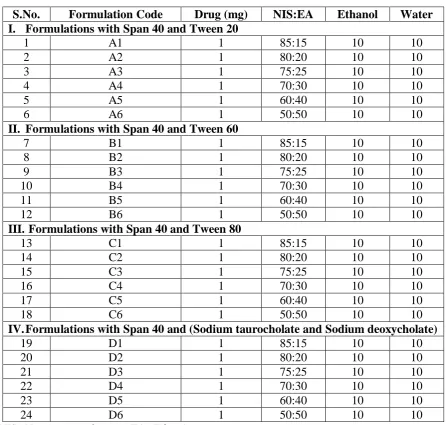
Table 1: Various Span40-based formulations of Spanlastics
S.No. Formulation Code Drug (mg) NIS:EA Ethanol Water
I. Formulations with Span 40 and Tween 20
1 A1 1 85:15 10 10
2 A2 1 80:20 10 10
3 A3 1 75:25 10 10
4 A4 1 70:30 10 10
5 A5 1 60:40 10 10
6 A6 1 50:50 10 10
II. Formulations with Span 40 and Tween 60
7 B1 1 85:15 10 10
8 B2 1 80:20 10 10
9 B3 1 75:25 10 10
10 B4 1 70:30 10 10
11 B5 1 60:40 10 10
12 B6 1 50:50 10 10
III. Formulations with Span 40 and Tween 80
13 C1 1 85:15 10 10
14 C2 1 80:20 10 10
15 C3 1 75:25 10 10
16 C4 1 70:30 10 10
17 C5 1 60:40 10 10
18 C6 1 50:50 10 10
IV.Formulations with Span 40 and (Sodium taurocholate and Sodium deoxycholate)
19 D1 1 85:15 10 10
20 D2 1 80:20 10 10
21 D3 1 75:25 10 10
22 D4 1 70:30 10 10
23 D5 1 60:40 10 10
24 D6 1 50:50 10 10
NIS=Non ionic surfactant; EA=Edge Activator.
Table 2: Various Span 60-based formulations of Spanlastics
S.No. Formulation Code Drug (mg) NIS :EA Ethanol Water
V. Formulations with Span 60 and Tween 20
1 E1 1 85:15 10 10
2 E2 1 80:20 10 10
3 E3 1 75:25 10 10
4 E4 1 70:30 10 10
5 E5 1 60:40 10 10
6 E6 1 50:50 10 10
VI.Formulations with Span 60 and Tween 60
7 F1 1 85:15 10 10
8 F2 1 80:20 10 10
9 F3 1 75:25 10 10
10 F4 1 70:30 10 10
[image:3.595.65.532.560.768.2]12 F6 1 50:50 10 10
VII. Formulations with Span 60 and Tween 80
13 G1 1 85:15 10 10
14 G2 1 80:20 10 10
15 G3 1 75:25 10 10
16 G4 1 70:30 10 10
17 G5 1 60:40 10 10
18 G6 1 50:50 10 10
VIII. Formulations with Span 60 and (Sodium taurocholate and Sodium deoxycholate)
19 H1 1 85:15 10 10
20 H2 1 80:20 10 10
21 H3 1 75:25 10 10
22 H4 1 70:30 10 10
23 H5 1 60:40 10 10
24 H6 1 50:50 10 10
3. EVALUATION OF SPANLASTICS
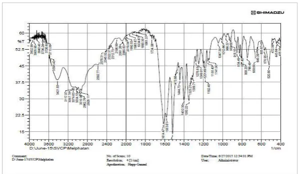
3.1 Fourier Transform Infrared (FTIR) analysis
The drug excipient compatibility study was determined by FTIR (Fourier Transform Infrared
Spectroscopy). Samples were scanned in the range from 400- 4000cm-1. The IR spectrum of
the pure drug was compared with IR spectrum of combination of drug and excipients to
check the interactions.
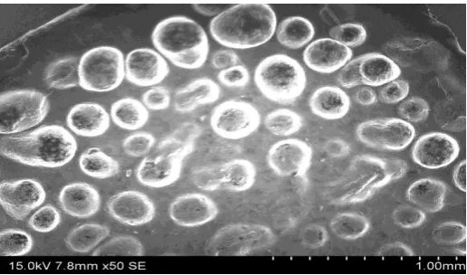
3.2 Surface morphology
The morphology of spanlastics was determined by scanning electron microscopy (SEM)
using HITACHI S-3700N Instrument. SEM gives a three dimensional image of the globules.
The samples were examined at suitable accelerating voltage 15kV.
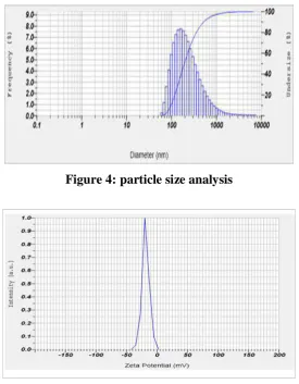
3.3 Particle size measurement and Zeta potential
The particle size and zeta potential of Melphalan spanlastics were measured by Particle size
analyzer and Zeta sizer (HORIBA SZ 100). For the measurement, 100μl of formulation was
diluted with appropriate volume of PBS pH 7.4. The vesicle diameter and zeta potential were
determined.
3.4 Entrapment efficiency
For the determination of Entrapment efficiency, the unentrapped drug was first separated by
centrifugation at 1500RPM for 60min and supernatant was collected. The collected
supernatant was estimated using UV Visible Spectrophotometer at 259nm.
3.5 In-vitrodiffusion Study
In-vitro diffusion studies were carried out using Franz-diffusion cell. The membrane was
soaked for 24h in 0.1N HCl. The donor compartment was covered with dialysis membrane.
The receptor compartment was filled with 7.4 pH phosphate buffer. The prepared formulation
was introduced into dialysis membrane in the donor compartment. The whole assembly was
maintained at 37°C and the speed of stirring was controlled. 3ml aliquot of drug sample was
withdrawn from receptor compartment at specified time intervals 1, 2, 3, 4, 5, 6, 7 and 8h and
replaced with fresh medium to maintain sink condition. The samples were analyzed
spectrophotometrically at 259nm.
3.6 Stability Studies
Stability studies were conducted on optimized formulation according to ICH guidelines. The
spanlastics suspension was kept in sealed ampoule and stored at 25°C±2°C/60±5% RH and
40°C±2°C/75±5% RH for 3months. Samples were withdrawn periodically (intervals 30, 60,
90 days) and analyzed for physical appearance and entrapment efficiency.
4. RESULTS AND DISCUSSION
4.1. Fourier Transform Infrared (FTIR) analysis
The results showed that there was no chemical interaction or changes between Melphalan
spanlastics and melphalan pure drug. Melphalan spanlastics showed same characteristic
[image:5.595.148.447.504.677.2]absorption bands as that of melphalan.
Figure 2: FTIR image of melphalan spanlastics suspension
4.2Surface Morphology
The three dimensional image of globules was shown in Fig. 3. The image indicates that the
obtained spanlastics have a smooth surface and are spherical in shape.
Figure 3: SEM photograph of spanlastic formulation
4.3Particle size measurement and Zeta potential
The particle size distribution of spanlastics was characterized. The particle size of optimized
spanlastics formulation was found to be 234nm. The particle size was found to depend on the
HLB value of the non ionic surfactants used. The formulations prepared using STC and SDC
showed higher particle size compared to formulations using Tween 80 and Tween 60. With
the increase in the HLB value, the vesicle size also was found to increase as shown in Table
3. Zeta potential of optimized formulation was found to be -18.4mv as shown in Fig. 5. This
[image:6.595.169.427.361.513.2]Figure 4: particle size analysis
Figure 5: zeta potential
Table 3: Particle size of Spanlastics based on HLB value
Edge activator HLB value Particle size (nm)
Sodium tauro cholate and Sodium deoxy cholate 16 and 26 234
Tween 20 16.7 210
Tween 60 14.9 195
Tween 80 15 200
HLB= Hydrophilic Lipophilic Balance.
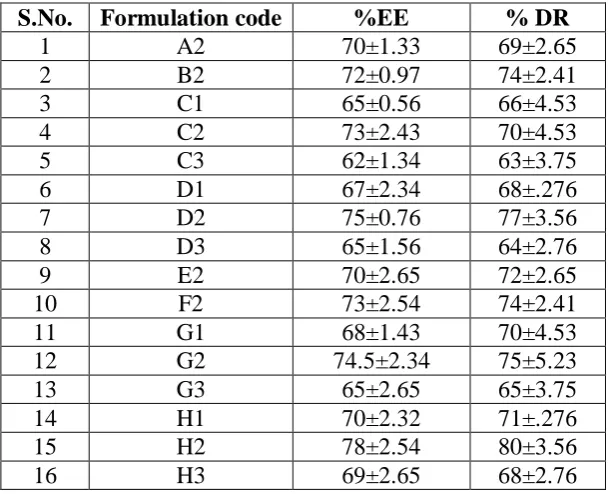
4.4Entrapment efficiency
Entrapment efficiency of selected formulations was in the range of 65 to 78%. Highest
entrapment efficiency (78%) was found in H2 formulation. H2 formulation contains a
Span 60 and combination of Sodium taurocholate and Sodium deoxycholate in the ratio of
80:20. High entrapment efficiency of Span 60 may be due to the solid nature, hydrophobicity
and high-Phase transition temperature. The entrapment efficiency was found to increase when
the Non ionic surfactant: Edge activator ratio was increased to 80:20. When the proportion of
[image:7.595.165.434.70.215.2]efficiency decreased indicating that beyond an optimum concentration, the NIS showed drug
[image:8.595.162.437.124.274.2]leakage and aggregate formation.
[image:8.595.146.451.337.584.2]Figure 6: entrapment efficiency of selected formulations
Table 4: Entrapment efficiency and percent drug release of selected formulations
EE=Entrapment Efficiency, %DR= percentage drug release.
4.5In-vitro diffusion Studies
The percentage drug release of span 40-based spanlastics was in the range of 63 to 77%,
while that of span 60-based formulations was in the range of 65 to 80%. H2 formulation
showed highest % drug release of 80% in 8h. The Rate of drug release was influenced by
the concentration of non ionic surfactant and edge activator concentration as shown in Fig. 8,
Fig. 9 and Fig. 10. It was observed that drug release was increased with an increasing the
S.No. Formulation code %EE % DR
1 A2 70±1.33 69±2.65
2 B2 72±0.97 74±2.41
3 C1 65±0.56 66±4.53
4 C2 73±2.43 70±4.53
5 C3 62±1.34 63±3.75
6 D1 67±2.34 68±.276
7 D2 75±0.76 77±3.56
8 D3 65±1.56 64±2.76
9 E2 70±2.65 72±2.65
10 F2 73±2.54 74±2.41
11 G1 68±1.43 70±4.53
12 G2 74.5±2.34 75±5.23
13 G3 65±2.65 65±3.75
14 H1 70±2.32 71±.276
15 H2 78±2.54 80±3.56
amount of non ionic surfactant. Concentration of edge activator also affects drug release from
[image:9.595.158.437.124.276.2]spanlastics.
[image:9.595.162.435.314.454.2]Figure 7: Cumulative percent drug release of selected span 40 formulations
Figure 8: Comparative percent drug release of selected span 60 formulations
Figure 9: Comparative drug release of span 40 and span 60 formulations
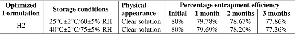
4.6 Stability Studies
It was observed that there was no change in the physical appearance of the formulation. The
[image:9.595.157.438.493.661.2]at different temperatures as shown in Table 5. Spanlastics suspension retained good stability
[image:10.595.46.553.147.216.2]throughout the study.
Table 5. Stability studies
Optimized
Formulation Storage conditions
Physical appearance
Percentage entrapment efficiency
Initial 1 month 2 months 3 months
H2 25°C±2°C/60±5% RH
40°C±2°C/75±5% RH Clear solution Clear solution 80% 80% 79.78% 79.69% 78.67% 78.20% 77.86% 77.36% 5. CONCLUSION
Sustained release spanlastics were successfully formulated using Span 40, Span 60 as non
ionic surfactants and Tween 20, Tween 60, Tween 80, Sodium taurocholate and Sodium
deoxycholate as edge activators. Prepared spanlastics showed good entrapment efficiency and
sustained release up to 8h. The stability studies indicate that the optimized formulation was
stable without physical and chemical degradation. Melphalan loaded spanlastics could be a
promising dosage form to improve sustained release of Melphalan by oral route.
6. REFERENCES
1. Cevc G, Blume G. Lipid vesicles penetrate into intact skin owing to the transdermal
osmotic gradients and hydration force. Biochem Biophys Acta, 1992; 1104(1): 226-32.
2. Touitou E, Alkabes M, Dayan N. Ethosomes: novel lipid vesicular system for enhanced
delivery. Pharm Res, 1997; S14: 305-6.
3. Van den Bergh BA. Elasticity of vesicles affects hairless mouse skin structure and
permeability. J Control Release, 1999; 62(3): 367-79.
4. Kakkar S, Kaur IP. Spanlastics-a novel nanovesicular carrier system for ocular delivery.
Int J Pharm, 2011; 413(1-2): 202-10.
5. Abdulaziz M, Mahallawi, Omneya MK, Shoukri RA.Ciprofloxacin loaded spanlastics for
ototopical non-invasive delivery to the middle ear: in-vitro and ex-vivo studies. Inventi
Impact: NDDS, 2014; 2014(3): 174-79.
6. Kaur IP. Development and evaluation of novel surfactant based elastic vesicular system
for ocular delivery of fluconazole. J Ocul Pharmacol Therap, 2012; 28(5): 484-96.
7. Basha M. Design and optimization of surfactant-based nanovesicles for ocular delivery of
clotrimazole. J Liposome Res, 2013; 23(3): 203-10.
8. EI Zaafarany GM. Role of edge activators and surface charge in developing
9. Hao Y. Studies on a high encapsulation of colchicine by a niosome system. Int J Pharm,
2002; 244(1-2): 73-80.
10.Irfan MD, Verma S, Ram A. Preparation and characterization of ibuprofen loaded
transferosome as a novel carrier for transdermal drug delivery system. Asian J Pharm Clin
Res, 2012; 5(3): 162-65.
11.Abdallah MH. Transfersomes as a transdermal drug delivery system for enhancement the
antifungal activity of nystatin. Int J Pharm Pharm Sci, 2013; 5(4): 560-67.
12.Prakash G, Manjunatha, Hiremath D, Santhosh F. Perindopril erbumine loaded
ethosomes: design and in vitro characterization. Sch Acad J Pharm, 2015; 4(2): 138-44.
13.Saahil A, Singh HL, Tiwari R. Dermal delivery of drugs using different vesicular carriers:
a comparative review. Asian J Pharm, 2012; 6(2): 237-44.
14.Suresh DK, Chaudhari YS, Priyanka B, Pallavi D, Nikita B, Khushbu S, Pankit Shah,
Mishra P. Transfersomes: a promising approach for transdermal drug delivery system.
Asian J Pharm Sci Res, 2013; 3(5): 1-17.
15.Touitou E. Ethosomes – novel vesicular carriers for enhanced delivery: characterization
and skin penetration properties. J Control Release. 2000; 65(3): 403-18.
16.Prem Kumar Y, Vinod Kumar K, Raja Shekar R, Ravi M, Sai Kishore V. Formulation
and evaluation of econazole niosomes. Sch Acad J Pharm, 2013; 2(4): 315-18.
17.Kushal M, Monali M, Mishra D, Mittal P, Umesh S, Pragna S. Oral controlled release
drug delivery system: an overview. Int Res J Pharm, 2013; 4(3): 70-76.
18.Bansal S, Kashyap CP, Agarwal G, Harikumar SL. A comparative review on vesicular
drug delivery system and stability issues. Int J Res Pharm Chem, 2012; 2(3): 704-13.
19.Rokade VS, Kerunath KP. Formulation and evaluation of novel anti-bacterial
ciprofloxacin loaded niosomal cream. Int Res J Pharm, 2015; 6(8): 519-27.
20.Srikanth K, Rama Mohan Gupta V, Devanna N. Formulation and evaluation of nystatin