The development of specialisation in diagnostic
radiography : concepts, contexts and implications
FERRIS, Christine Margaret
Available from Sheffield Hallam University Research Archive (SHURA) at:
http://shura.shu.ac.uk/3193/
This document is the author deposited version. You are advised to consult the
publisher's version if you wish to cite from it.
Published version
FERRIS, Christine Margaret (2005). The development of specialisation in diagnostic
radiography : concepts, contexts and implications. Doctoral, Sheffield Hallam
University.
Copyright and re-use policy
See
http://shura.shu.ac.uk/information.html
SHEFFIELD HALLAM UNIVERSITY LEARNING CENTRE COLLEGIATE CRESCENT
SHEFFIELD S10 2BP
ProQuest Number: 10700846
All rights reserved
INFORMATION TO ALL USERS
The quality of this reproduction is dependent upon the quality of the copy submitted.
In the unlikely event that the author did not send a com plete manuscript and there are missing pages, these will be noted. Also, if material had to be removed,
a note will indicate the deletion.
uest
ProQuest 10700846
Published by ProQuest LLC(2017). Copyright of the Dissertation is held by the Author.
All rights reserved.
This work is protected against unauthorized copying under Title 17, United States C ode Microform Edition © ProQuest LLC.
ProQuest LLC.
789 East Eisenhower Parkway P.O. Box 1346
The Development of Specialisation in Diagnostic Radiography: Concepts, Contexts and Implications
Christine Margaret Ferris
A thesis submitted in partial fulfilment of the requirements of Sheffield Hallam University
Abstract
The aim of this project is to examine how specialisation in diagnostic radiography has occurred. In particular, this research aims to examine the contemporary ethos of specialism in diagnostic radiography; how the higher status of technology has developed over patient centredness; the impact of the working relationship between radiology and diagnostic radiography on this development; the relationship between gender and the nature of occupation in the development of diagnostic radiography.
Qualitative data was collected using 31 semi-structured interviews that took the form of oral histories where possible. The time-span covered is 1932-2001.
Findings show that a paradigm shift is required. Defining specialism and expert practice is difficult, as both are negotiated constructs that tend to have local meaning. Not all specialisms in diagnostic radiography increase professional autonomy and management has a key role to play in the development of radiography as a profession to enable full engagement with consultant status. To a great extent, diagnostic radiography and its associated specialisms is governed by an emphasis on technology rather than the patient. This emphasis has roots in power difference between radiologists and radiographers and within the hierarchy of radiography. Radiologists have controlled radiographers to provide a service to radiology rather than the patients, and, although this is still evident in some hospitals, it is changing. Task offloading from radiologists to radiographers confuses the notions of specialism and radiography is in danger of seeking professional development through emulating radiology rather than using radiographic caring skills and expanding practice with a humanistic and patient focused emphasis. Radiography emerged as a pioneering, elite profession that could originally be regarded as a specialism of nursing. Technology and medicine gradually reduced radiographic practice to have a technological and quantitative focus. There is now a demand to once again broaden radiographic practice to actively contribute to a patient-centred service in an autonomous, more qualitative and self-directed manner. A collection of oral histories has been a direct result of this research. Specialisms are a broad area of diagnostic
Acknowledgements
If I honestly reflect on my studies I find I owe a debt to others that spans my own history. People unknown to me have made my life easier so that I can study when not in my paid employment. It is important to acknowledge all these unknown persons; but it is also necessary to acknowledge those people who I know have directly shaped my life and my work. Adversity is a good teacher. Even those people that have created difficulties for me are owed my respect for what I have learned. I thank all for their role in this part of my life.
My husband Bryan has urged me on with consistent support and seemingly unlimited belief in me, thank you for keeping me going through the times when I lost my self-belief. Thank you to my children David, Wendy and Gavin for being their magical selves. There are some from behind the scenes that know this journey and have encouraged and supported my work. I wish to thank them, my “godmothers”, Dr Frances Gordon, Dr. Gail Mountain and Dr. Anne Hollows.
There are special mentors that I must acknowledge due to their importance in my work. In particular Professor Anne Parry for her unstinting searches for the meaning in my thoughts and writing when this project was in its infancy and beyond. Anne, along with Professor Mike Worboys, helped me shape and mould a complex concept into a feasible research project. Of course, like any student, I am indebted always to those people that do their best to improve on my best. Thanks especially to Professor Chas Critcher for stepping in at the last moment to assess the continuity of my work.
The radiographers and nurses who were the respondents for this study were generous to a fault with their time, perspectives and stories, not to mention their chocolate biscuits. I thank them for their honesty, good wishes, their energy and their ability to recount aspects of our professional history that may otherwise go unrecorded.
Contents
Page
Abstract i
Acknowledgements ii
Contents iii
Chapter One Introduction 1 1.1 Purpose of research 1 1.2 Aims and objectives 2 1.3 Background and rationale 3 1.4 The boundaries of the project 4 1.5 Potential importance 4 1.6 Clarification of roles 5 1.7 Current specialisms in diagnostic 7
radiography (applicable to UK only)
1.8 Concepts of specialism 11 1.9 Structure of the thesis 12
Summary 15
Chapter Two Development of the radiographic 16 profession
Introduction 16
2.1 Professionalisation 17 2.2 The medical context 20 2.3 The service context 24 2.4 The educational dimension 25 2.5 Steering forces in the evolution of 31
diagnostic radiographic practice
Summary 32
Chapter Three The context of medical specialism 34
Introduction 34
3.1 Influential social forces 38
Page
Chapter Three The context of medical specialism continued
3.2 Social forces - development of the 3 8 city
3.3 Social forces - social attitudes - 39 gender
3.4 Social forces - institutions 41 3.5 Current influences 44
Summary 46
Chapter Four Models of specialism 47
Introduction 47
4.1 Anatomical model of specialism 48 4.2 Technological model of specialism 49 4.3 Procedural model of specialism 53 4.4 Age-related model of specialism 54
Summary 57
Chapter Five A model of expert practice 58
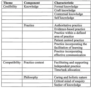
Introduction 58 5.1 Expert practice 61 5.2 Emerging themes on expert 62
practice
5.3 Credibility 64 5.4 Credible practice 70 5.5 Compatibility 74
Summary 79
Chapter Six Methodology 80
Introduction 80
6.1 Theoretical perspective 80 6.2 Methodology 81 6.3 Sources and forms of evidence 83
6.4 Sample 85
6.5 Trustworthiness and rigour 88
Page
Chapter Six Methodology continued
6.6 Data analysis 91 6.7 Design issues 92
Summary 95
Chapter Seven Findings - the contemporary ethos of 96 specialism in diagnostic radiography
Introduction 96 7.1 Discussion 98 7.2 Credibility 98 7.3 Credible knowledge 98 7.4 Credible practice 103 7.5 Compatibility 112 7.6 Compatible philosophy 116 7.7 National clinical priorities 118 7.8 Areas for consultant 118
appointments
Summary 120
Chapter Eight Findings - how the higher status of 121 technology has developed over
patient-centredness
Introduction 121 8.1 Radiography as a specialism 122 8.2 Aspirations and opportunities 124 8.3 Characteristics of diagnostic 127
radiography specialisms
8.4 Expert practice 138
Summary 141
Page
Chapter Nine Findings - the impact of radiology and 142 gender on the nature of occupation in the
development of diagnostic radiography
Introduction 142
9.1 Radiology 143
9.2 Referring clinicians 154 9.3 The influence of gender/city 158
Summary 161
Chapter Ten Incidental findings - some hidden 162 history of radiography
Introduction 162 10.1 Some general incidental findings 162 10.2 Some specific incidental findings 163 10.3 Challenges of practice 164 10.4 Social forces - times of conflict - 173
its impact on practitioners
10.5 Role of the radiographer 176
Summary 180
Chapter Eleven Reflexive account 182
Introduction 182 11.1 Radiographer educationist 183 11.2 Impact of being female 185 11.3 My concept of specialism 186 11.4 The sample 186 11.5 The sampling strategy 187 11.6 Ethical issues 188 11.7 Data analysis 189 11.8 Meta-synthesis 189
11.9 Learning 189
Summary 191
Page
Chapter Twelve Conclusions, implications and 192 recommendations
Introduction 192 12.1 The contemporary ethos of 193
specialism in diagnostic radiography
12.2 Technology vs patient-centredness 194 12.3 The impact of the working 195
relationship between radiology and .diagnostic radiography on the
development of specialisation in diagnostic radiography
12.4 The relationship between gender 196 and the nature of occupation in the
development of diagnostic radiography
12.5 Implications and recommendations 196
References 199
Appendices 218
Appendix A Tables from meta-synthesis 219 Appendix B Tables of respondents 226 Appendix C Interview schedules 229 Appendix D Lists of codes 235 Appendix E Interview summaries 240
Tables
Table 1 Summary of findings 64 Table 2 Types of knowledge 70 Table 3 Relationship of interview questions 88 and project aims
Table 4 Questions related to the first aim 96 Table 5 Model of expert practice 97 Table 6 Questions related to the second aim 121 Table 7 Recognition of characteristics by 129 respondents
Table 8 Questions related to third and fourth 142 aims
Table 9 The “norm” past and present 180 Table 10 Methodological characteristics of the 220 qualitative studies included in the meta
synthesis
Table 11: Characteristics of participants and 221 individual studies used in the meta-synthesis
Table 12 Summary of metaphors, themes or 222 concepts included in the meta-synthesis of the
studies.
Table 13 Examples of demonstration of fit 225 Table 14 Table of respondents 227 Table 15 Gender, practice years and 228 practitioner/manager role
Table 16 A start list of codes 236 Table 17 Final list of codes used to analyse 238 the 31 interviews
Table 18 List of codes used to analyse the 10 239 leading voice interviews to identify
characteristics of expert practice
CHAPTER ONE
Introduction
Generally, the work of an imaging department is described using technical and procedural jargon and terminology. Consequently staff are described in similar terms, e.g. there may be a
Computed Tomography (CT) Superintendent Radiographer or a Magnetic Resonance Imaging (MRI) Senior 1 Radiographer. Patient groups that clearly have identifiable and significant needs are referred for imaging, but a technological paradigm predominates in the work function, organisation and identity of a medical imaging department. The NHS is being overhauled to place the patient at the heart of services (HMSO 2000a). To fully engage with this new patient-centred philosophy for health service provision, diagnostic radiographers may need to embrace an alternative paradigm of career structure and professional progression. The potential impact of exploring this issue is that radiographers, already part of many patients' pathway, could expand their role to be more fully involved in the assessment, treatment and discharge of patients in a variety of clinical contexts. This form of role development requires a focus on the patient, rather than the technology that is used to perform assessment and
treatment. It also requires clinical decision-making skills that have been actively discouraged by radiologists in the past (Larkin 1983).
1.1 Purpose of research
The research is designed to explore how and why career structure, job opportunities and service provision in diagnostic radiography have evolved using an ideology that allows a relatively high status to be given to science rather than groups of patients. There is an
emerging emphasis within the NHS of multi-disciplinary team working with each team having a patient-centred identity. The apparent technological focus of diagnostic radiography may have a domino effect on the function and role of radiographers in multi-disciplinary
The role of the diagnostic radiographer has developed to support radiologists rather than the patient and the extent to which radiologists have influenced the careers and job opportunities of radiographers is unknown. The technological influence on career progression and job opportunities is strong as these are dependent on the availability of pieces of technology such as ultrasound machines, CT and MRI scanners. Radiologists rely on the technical skills of diagnostic radiographers to produce pertinent images of the patient from which a diagnosis may be reached. Lately, service innovations have provided opportunities for diagnostic radiographers. The breast screening service is a clear example of a service-defined career pathway for diagnostic radiographers with a focus on client/patient needs. This research will explore the possibilities of involving diagnostic radiographers in teams centred on service provision or a particular group of patients.
1.2 Aims and objectives
A broad aim of this research is to examine the development of specialisation in diagnostic radiography. Primarily, this research examines the changes that have occurred in diagnostic radiography and how specialisation in diagnostic radiography has developed. Radiology as a medical specialism provides the context for this as, to examine diagnostic radiography in isolation would fail to acknowledge the interdependence and close working relationship of the two professions. In particular, this research describes how the development of specialisation within the profession of radiography is moulded over time and how it relates to current notions of specialist practice.
In contrast to other health care professionals, specialisms in radiography appear to have developed grounded in technology and the needs of radiology rather than the needs of the patient. The way in which radiographic specialisms have developed over time is an important consideration, as any continuing factors will influence future practice (Mead 1944).
The following aims are met through the study of the development of specialisation in diagnostic radiography.
In particular, this research aims to examine:
1. the contemporary ethos of specialism in diagnostic radiography.
2. how the higher status of technology has developed over patient-centredness.
3. the impact of the working relationship between radiology and diagnostic radiography on this development.
diagnostic radiography.
In order to meet the aims, qualitative data relating to contemporary and historic practice is required. The objectives for this study are to:
1 conduct a literature review to frame the findings of the study.
2 conduct a separate systematic literature review using meta-synthesis to identify characteristics of expert practice (aim 1).
3 interview professional representatives of acknowledged areas of specialism in diagnostic radiography (aims 1, 2, 3 &4)).
4 interview radiographic practitioners who can provide an account of their professional experience (aims, 2, 3 & 4).
The findings of this study could be useful to: • managers of radiographers; • professional bodies;
• diagnostic radiographers who wish to progress in their career with a patient focus rather than technological;
• those who are giving career guidance to diagnostic radiographers;
• those who wish to include diagnostic radiographers in a service-centred or patient- focused team.
1.3 Background and rationale
Diagnostic radiographers are trained using an anatomical and technological focus that may have a similar impact on their levels of knowledge and understanding when considering groups of patients with particular needs and priorities. Such superficial knowledge of patients' needs may have inhibited radiographic research into patients' needs, e.g. the predominance of a positivistic paradigm has resulted in a lack of acknowledgement of patient orientated measurements in imaging practice (Ferris, Nasr and Dixon 2004).
Radiographers are reliant on technology to perform the image acquisition part of their work. Patient care does not usually require technology, but an understanding and empathy for the patient and clarity about priorities in the patient's experience of health and illness (Culmer
use of technology. A good example of this is the use of diagnostic musculoskeletal ultrasound in physiotherapy and sports medicine.
Radiography is inherently linked to a medical specialism, radiology. The radiological influence on radiographic practice has not been systematically documented. It is difficult to ascertain the level of influence of medicine and, in particular, radiology, on the value systems of an Allied Health Profession (AHP) such as radiography. There is little evidence from either radiographers or radiologists that describes their working relationship in practice. The local influences of radiologists on radiographers are undocumented and undefined. As radiologists traditionally control radiographers' boundaries of practice, this may result in a variable experience for radiographers including local career and job opportunities.
1.4 The boundaries of the project
This work intends to produce a historical account of the development of diagnostic radiography, its associated value systems and explore any implications for contemporary practice. Some historical accounts exist but describe the formation of the professional body (Moodie 1970, Jordan 1995). There are no full accounts from radiographers about their career. Reeves and Murphy (1998) use the collection and analysis of oral history as an effective educational tool to motivate students. This paper emphasises the educational impact of oral history and incorporates oral evidence from radiographers about their experience and from a student about the learning experience. Most accounts from former practitioners will be of local significance but any links to broader, professional understandings will be identified. The conclusion of this work is suggestive rather than conclusive and is intended to enable
understanding of the development of diagnostic radiography.
1.5 Potential importance
Specialisms engender specific career pathways. Using technology as the differentiating factor marking career progression inhibits the fullest involvement of diagnostic radiographers in the patient experience. The NHS Plan (HMSO 2000a) indicates increased opportunity to extend and expand a radiographer's role and culminate career progression as a consultant. The new consultant roles show that the division of labour within the NHS is under political scrutiny. The impetus provided by the Plan could act as a factor supporting changes to labour
explores the background and implications of traditional and contemporary paradigms of diagnostic radiography.
The most commonly acknowledged professionals responsible for the delivery and provision of health-care services are those who carry the easily recognisable titles of doctor, nurse and midwife. There are other key groups of health-care professionals who belong to professions that were previously classified as being supplementary to medicine. They have a lower public profile than medicine, nursing and midwifery and this has resulted in a lack of literature relating to their evolution and patterns of development (Larkin 1983). These professional groups were regulated by the Professions Supplementary to Medicine Act 1960 and include chiropodists, physiotherapists, occupational therapists, medical laboratory scientific officers, dieticians, orthoptists and radiographers (CPSM 1997). The Council of Professions
Supplementary to Medicine (CPSM) administered the requirements of the Act controlling state registers for each profession. Inclusion on the appropriate register is necessary for individuals to practise in the UK. In response to the growing number of health-care
professions that cannot be classified as medicine, nursing or midwifery, the Act has recently been reviewed. The Health Professions Council (HPC) was established in 2002 under the Health Professions Order 2001 and controls the registers of a larger number of health professions. In addition to those originally controlled by the CPSM, art therapists, clinical scientists, prosthetists and paramedics are now included (HPC 2003). The emergence of these Allied Health Professions (AHPs) is primarily in response to the growth and complexity of health service provision.
1.6 Clarification of roles
In 1896 Roentgen’s discovery of x-rays1 gave rise to two groups of professionals, radiographers, who could be described as x-ray technicians, and radiologists who were medically qualified specialists. Confusion between the role of radiographer and radiologist arose, as radiologists were originally known as radiographers (Moodie 1970). Radiology is a medical specialism emerging at a similar point in time as pathology, both specialisms performing activities that were previously regarded as being outside that of a physician or surgeon’s role. Radiology thus emerged as a medical speciality that had a relatively low status within medicine. Such medical specialisms have had to strategically engage in tactics that enable progression of professional status (Rosen 1944).
In contrast, radiography eventually emerged in order to differentiate clearly between those who performed x-ray examinations and treatment (radiographers) and those who could make a medical judgement about diagnosis and/or prognosis (radiologists). In the early years this division of work was not easily recognisable as the terms radiologist and radiographer were used interchangeably. The problems this caused lead to a series of identity crises in the 1920s (Moodie 1970). In addition, further confusion arose due to the diverse range of people performing x-ray examinations; theatre beadles, dentists, photographers and nurses are recorded as performing such examinations (Larkin 1983).
The introduction of the medical use of x-rays attracted other occupational groups such as physicists, engineers and electrotherapeutists. In 1897, The Roentgen Society was formed as an association permitting both medical and non-medical membership. Conflicts over role boundaries developed and, in order to solve this problem, the Society of Radiographers was founded in 1920. In the 1940s, technological advances in the use of x-rays for both diagnosis and treatment resulted in the acknowledgement of two separate segments of radiography by the radiography profession, diagnostic and therapeutic. Since that time, each segment has formed a working relationship with different medical specialisms, diagnostic radiography with that of radiology and therapeutic radiography with medical specialisms in the treatment of (mainly) malignant diseases, radiotherapy and oncology. Both segments belong to the professional body, the Society of Radiographers.
The medical use of x-rays for the diagnosis and evaluation of clinical conditions evolved mainly within the hospital organisation and is now only one of a number of ways in which internal aspects of the living human body can be visualised. X-ray Departments are no longer commonly found within the hospital organisation but there are now Medical Imaging
Departments. The term “radiography” generally refers to the conventional use of x-rays for diagnostic, evaluation and monitoring purposes and the term “medical imaging” tends to be used to indicate the whole spectrum of medical imaging modalities used by diagnostic
1.7 Current specialisms in diagnostic radiography (applicable in the UK only) The following areas of practice are mainly those in which a newly qualified, diagnostic radiographer would not be expected to have the skill or expertise to practise on a regular basis and are traditionally acknowledged as requiring post-qualification studies. However, not all specialisms follow this model; in particular, computed tomography, magnetic resonance imaging and the medical imaging of trauma and paediatric patients do not formally require further study following initial state-registration. Specialisms in diagnostic radiography can be categorised as those that can be identified in relation to the imaging modality used to acquire images, those that are the result of delegation of radiological duties and, the last group which does not fit easily into either category but could be described as being service related.
Specialisms related to imaging modality
Nuclear medicine
The first category of clinical practice that could be considered as specialist is the area of nuclear medicine. First introduced into medicine in the mid-1960s, imaging using this modality does not result in images depicting anatomy but ones that demonstrate physiology. A radio-nuclide with a known level of radioactivity is introduced into the body and a gamma camera “tracks” the way in which the body distributes the radio-nuclide. The results are recorded quantitatively, some being portrayed as a series of images. This work does not solely involve diagnostic
radiographers; medical technical officers and clinical scientists also practise nuclear medicine. Diagnostic radiographers wishing to practise in this area must undertake post-registration studies.
The following areas of specialist practice in this category are related to the use of medical imaging modalities that produce maps of internal structures of the body by the demonstration of sectional “slices” of anatomy.
Medical ultrasound
as an imaging modality are deemed to be specialised practitioners and are known as
sonographers. A wide range of health-care professionals practises sonography; consequently the use of ultrasound as an imaging modality is not solely under the professional jurisdiction of the Society and College of Radiographers. Diagnostic radiographers wishing to practise ultrasound must undertake post-registration studies.
Computed Tomography
At the same time as ultrasound had an impact on diagnostic radiography, technological advances in another area enabled the visualisation of a slice through the human body using x- radiation, Computed Tomography (CT). Whilst nuclear medicine and ultrasound had been noticeably hovering in the background of imaging science, CT arrived suddenly with no warning (Boutle 1995). In order to produce a CT image, an x-ray source, programmed to pass through a few millimetres of tissue at a time, is moved at regular intervals in a circular or spiral configuration around the body part to be visualised. Once x-rays have passed through the body they are collected, measured and fed into a computer that transforms the transmitted x-rays into a digital image. The image represents a sectional “slice” through the body and, as with
ultrasound, there is no superimposition of anatomical structures. In addition, the computer can recognise small changes in the transmitted x-ray beam that are not immediately apparent using conventional diagnostic radiography. Whereas conventional radiography demonstrates bony skeletal structures and a limited number of soft tissue structures, CT demonstrates both skeletal and a larger range of soft tissue structures. This method of medical imaging was applied to visualisation of the brain in 1972 by James Ambrose, and to the spine in 1975 (Eisenberg 1992, Whitehouse & Mould 1995). Diagnostic radiographers able to obtain images using CT are usually, but not always, senior in status and the practice is perceived as specialised by some. Post-registration studies are available but not necessarily required.
Magnetic Resonance Imaging
Another imaging modality suitable for use in medicine was introduced in the mid 1980s, nuclear Magnetic Resonance Imaging (MRI). Cross-sectional images are obtained by introducing specific radio frequencies into the body of a patient who is placed in a graded magnetic field. Specific protons within the patient’s body are stimulated by this arrangement and resonate. Resonance from the protons is amplified and collected in order to produce an image
Specialisms related to delegation of radiological practice
The key difference between radiology and diagnostic radiography is the separation of mental tasks requiring a clinical judgement and physical tasks with a more technological focus. Throughout the 1990s, radiologists have formally and selectively delegated some duties to diagnostic radiographers. Professionally, these duties are categorised as being part of the extended role of the radiographer and require post-registration training and education in order for diagnostic radiographers to practise.
Administration of contrast agents
The administration of intravenous (iv) contrast agents to outline the urinary or vascular systems was once deemed a medical duty but is now practised by appropriately trained diagnostic radiographers. Contrast agents can produce severe, adverse reactions in patients due to ion formation but in the 1970s contrast agents that do not form ions were manufactured, thus making reactions far fewer and less severe. The risk of the iv injection has reduced. Similarly, there has been a reduction of risk in other procedures usually performed by medical staff.
Plain film reporting
Specifically trained diagnostic radiographers now issue reports on plain radiographs. The term “plain” refers to the fact that the image does not contain any contrast agents. A Special Interest Group (SIG), originally acting as an interface between interested radiographers and the
professional body, supports diagnostic radiographers and other interested professionals.
Contrast agent examinations
Similarly, radiologists undertook barium enema examinations with the assistance of diagnostic radiographers and/or x-ray nurses. These examinations are mainly performed on the older patient, are time consuming and could be classed as unpleasant for all concerned. Specifically trained diagnostic radiographers now conduct barium enema examinations and a SIG was formed for interested radiographers in 1998. Some diagnostic radiographers are also issuing reports on barium enema examinations. Recently, some radiologists have started to delegate barium meal examinations and femoral arteriography, the demonstration of arteries, to diagnostic radiographers.
Other areas of specialism
Mammography
Specifically trained diagnostic radiographers undertake mammography, radiography of the breast. Post-registration training is a requirement for those diagnostic radiographers wishing to practise and participate in the national breast-screening programme which began in the early
1980s. Arguably, this specialism could be included in the first category, as diagnostic radiographers working in the breast-screening programme have to use specific equipment as identified by the governing body of the national programme. The equipment is an adaptation of that used in conventional diagnostic radiography.
Paediatrics
Paediatrics has been a professionally acknowledged area of practice requiring specialist skill since the formation of the Association of Paediatric Radiographers (APR) in the mid-1970s. This acknowledgement does not extend fully into the practice areas. Although this was the first SIG affiliated to the professional body, there appears to have been little progress in the general acknowledgement of paediatric radiography as specialist practice. Outside of the specialist children’s hospitals, paediatric patients are not uniformly acknowledged in diagnostic radiography as a patient group requiring specialist practice, skill and expertise, even though routine radiographic imaging procedures are potentially distressing to paediatric patients (Bradford 1990). The ideology of other health care professions incorporates the
acknowledgement of particular skills in paediatric practice, rather than an adaptation of an approach to practice as used with adult patients, e.g. nursing (RCN 1990). The paediatric age group of 0 -17 years provides different challenges to diagnostic radiographers from those presented through the radiography of adult patients. The key differences are ethics, anatomical proportions, physiological response, clinical conditions, radiation protection considerations, immobilisation methods, methods used to gain co-operation, methods used to elicit pregnancy status and informed consent, and the impact of anatomical size and structure on equipment settings (Gyll 1977). The Children Act 1989 also has implications for paediatric practice in diagnostic radiography (College of Radiographers 1995).
Trauma imaging
Similarly, the Society of Radiographers supports a SIG in trauma imaging that was formed in 1994, and has a sub-group in forensic work. Most diagnostic radiographers undertake medical imaging of patients who are referred from an Accident and Emergency Department. Although trauma service provision has recently been recognised as requiring specialist medical practice, trauma and forensic radiography are not uniformly recognised as requiring specialist
1.8 Concepts of specialism
A specialism may be defined as denoting an exceptional or restrictive nature (Simpson & Weiner 1995). The general concept of specialism is subsequently based on relative values; it is therefore comparative. When considering the exceptional nature of practice, all radiographic practice may be considered special when considered by those people who are unfamiliar with the subject of radiography. When considering radiographic practice from this perspective, all radiographers are seen to have specialist skills (Moodie 1970, Watson 1985 and Carr & Fell 1997). Viewed from within radiography and from a professional perspective, the amount of diversity in radiographic practice is increasing and the baseline for what is usual is neither static nor standard. It is difficult to identify what might be considered as exceptional practice when it is difficult to identify what constitutes usual or normal practice. This is an issue as a range of usual radiographic practice is in existence. Exclusive practice, by definition, is easier to define, as the boundaries of practice are clearer. The origin and maintenance of boundaries defining areas of exclusive practice is of interest to this research.
For a specialism to develop a strategic approach is required as a profession needs to establish cognitive closure around a specific area of expertise that is perceived as having professional value by external colleagues. Internal specialisation of a profession demonstrates a lack of concern for internal homogeneity, however, any internal segments need to be prevented from breaking free of the parent organisation. Their presence creates internal domains within which the content of work can be controlled. A profession needs to elicit a delicate compromise to maintain internal cohesion whilst simultaneously recognising specialised interests. If a specialist interest group is not recognised by a formal title, they may form their own identity through breakaway segmentation. A high degree of internal specialisation may threaten professional cohesion but this can be tolerated if there is recognition of interest groups with minimal role and strength. The interest group may thus be limited in its internal and external influence and, in order to progress, may need to break free of the parent organisation. In order to maintain professional influence and control in specialist areas, the parent body must maintain a balance of interests, allowing growth where it is needed but maintaining control (Rosen 1944).
Specialism as exceptional
radiography. According to Paterson (1994), who uses the word “role” as function rather than position, the current role of the radiographer is difficult to define from a task-orientated perspective. The fact that working practices in many medical imaging departments are continually changing makes definitions of radiographic practice increasingly difficult to formulate (Chapman 1993). As the notion of what is exceptional is relative and comparative there is little to compare, as what constitutes routine is difficult to identify.
Specialism as exclusive
Exclusive practice is easier to define and identify. When considering the exclusive or restrictive nature of practice, a specialism may be perceived as such when exclusivity of practice is generally accepted. For example, the radiography of paediatric patients may not be considered as a specialism, as it does not denote a change from what is traditionally considered to be normal practice and therefore is not sufficiently exceptional. Neither is the practice of paediatric radiography exclusive, with the exception of that which occurs in a specialist children’s
hospital. The extent to which a patient group is considered exceptional will depend on the professional emphasis given to the exceptional nature of that group.
1.9 Structure of the thesis
The thesis is presented in twelve chapters. Each of the first eleven chapters starts with
introductory material and concludes with a summary of the key findings of the chapter. A reader with little time could read each summary to get a feel for the thesis before reading each chapter.
Chapter One gives some background information to aid clarification of the roles, titles and functions associated with the main characters. Chapters Two, Three, Four and Five are a review of literature in four parts. The first part, Chapter Two, focuses on the development of the radiography profession in the key contexts of professional sociology and the practice elements of medicine, service and education:
• professionalisation, occupational segregation, gender and functionality;
• medicine - the power that has traditionally shaped the identity of radiographers; • service - now helping to shape the role of all AHPs;
• education - providing an inextricable link to eligibility to practise.
social forces is a key facilitator of the growth of specialism. The effects of such forces on AHP specialism growth may vary due to a variance in autonomy in professional practice and the extent of overlap of practice between medicine and associated AHPs. The third part of the literature review, Chapter Four, suggests models of medical specialism that may influence the notion of specialism in radiography. The word "notion" here is used as an alternative word for "perception". Medicine has numerous models of specialism. Chapter Four examines four models of specialism closely linked to radiology. Chapter Five is devoted to an exploration of expert practice and presents the findings from a meta-synthesis of 12 primary research papers on expert practice in the form of a model. Themes, components and characteristics of expert practice are identified.
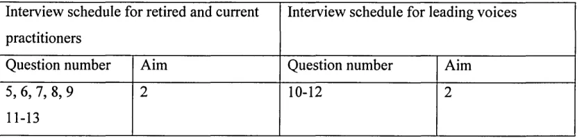
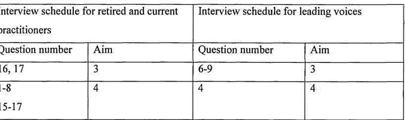
Chapter Six outlines the research design giving an academic account and justification for the research approach and methods employed. The study is based on data generated from interviews with 31 practitioners, 10 of whom are classed as leading voices in specialist clinical fields of diagnostic radiography.
about the quality of the generated data. Chapter Twelve concludes the thesis by itemising suggestions generated from the data and its analysis.
Summary
This research explores how an ideology, which appears to value science above groups of patients, has evolved in imaging. Despite an increase in the number of AHPs, there is scant literature on their emergence and development. The profession of radiography emerged in response to the increased need for the use of x-radiation in the treatment and diagnosis of patients. Simultaneously a medical specialism emerged, radiology. Radiography has subsequently been divided into therapeutic and diagnostic areas with the former being associated more with oncology and the latter with radiology. Over the years technology has influenced the culture, practice and language of radiography and currently patients can be examined using a variety of imaging modalities.
Throughout the history of radiography the roles of the diagnostic radiographer and the radiologist have changed in response to internal and external influences that have steered the identification of what constitutes specialist practice. Practice can be considered as special when it is of an exceptional or exclusive nature as specialism, and therefore its derivatives, speciality, specialist practice and specialisation are relative concepts. Areas of specialist practice that are easily identifiable are those categorised by the imaging modality used to acquire the images and those that have formed as a result of the delegation of radiological duties. Trauma and
CHAPTER TWO
Development of the radiography profession
Introduction
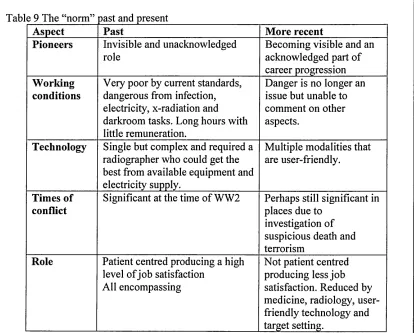
As the notion of specialism is relative, in order to explore how radiographic specialisation has changed over time, it is essential to examine what constitutes the concept of “norm” over time. Describing the key features of what might be considered usual is a primary objective of this research. This chapter discusses the contextual influences on radiographic practice, by
examining literature relating to professionalisation and occupational sociology, the medical and service contexts of radiographic practice and the professional contexts of radiographic education and eligibility to practice. The literature on professionalisation guides the interpretation of the history of diagnostic radiography whereas that on occupational sociology illustrates and gives a background to the predominantly functional approach to technical specialisation. The medical context demonstrates the power and controlling relationship of medicine through its interaction with radiographers (Reeves 2002). The service context explores the changing nature of service needs and modes of delivery and how this has impacted on radiographers. The professional context examines the educational dimension of radiography, and aspects of eligibility to practice. Where relevant, literature that helps to shed light on the formation of radiographic culture and practice is discussed. Finally, key influences on practice are suggested.
The professional responsibilities of the radiographer emphasise the control of work undertaken in an imaging room, including the selection of the procedure employed (Paterson and Price 1996). It is generally accepted that radiographers are not usually in total control of their work, however, radiographers working in ultrasound, reporting and barium sessions are beginning to exercise autonomy in key decisions regarding their practice, particularly in developing and defining practice. The formal blurring of work boundaries between radiographers and radiologists seemed to start with the proven ability of sonographers to work autonomously.
images may be required. This latter aspect of practice demonstrates how radiographers act as the gatekeepers of x-radiation, radiofrequency and magnetic field doses to patients and staff.
2.1 Professionalisation
Expertise can be constructed to fit as appropriate into particular occupational societies or closely related clusters of occupations such as radiography and radiology, which collectively provide an imaging service. Professionalisation is the main driver of institutionalised expertise in modem society (Larsonl977, Abbott 1988, Macdonald 1995). For the people involved, professionalism offers a continuous career, independence and an organised body of knowledge that is both continuously in demand and utilised through disciplined judgement (Abbott 1988). A profession is traditionally associated with a number of characteristics including:
• prolonged, specialised training • a body of abstract knowledge;
• controls and determines educational standards; • controls and determines fitness to practise; • broad recognition and approval of this authority; • a code of ethics;
• a professional culture supported by formal societies and associations.
(Freidson 1988)
Radiography has developed as a profession gradually incorporating such characteristics through opportunity and strategy. The process of professionalisation is a complex transformation that involves an increase in responsibility and autonomy which is embedded in the social effects of mass education, changes to class structure and social ideology (Larson 1977). Radiologists and physicists, mainly male and often from a higher social class, originally controlled the length and type of training for radiographers, the curriculum, syllabus content, assessments and the
radiographic professional body. Radiography has now evolved so that it is in a position to control all professional aspects through self-regulation employing the means which men used to rise to dominance - the professional project (requirement for qualifications and registration) and social closure (Macdonald 1995).
Professionalisation facilitates the formation of specialisms as evident in nursing. As nursing became more professionalised, a variety of specialisms begin to emerge in nursing, particularly in post-registration training. In parallel, health service developments increased, with a
Occupational segregation
In the health and caring services, occupational segregation was partially achieved by patriarchal discourse that promoted the subordination of female oriented professions through exclusion and control. Macdonald (1995) uses the caring professions as classic examples of patriarchal manipulation and the effects of the predominance of patriarchal value systems. Historically women were allowed to enter the health and caring workplace through any activity that was not seen to be of value to the scientific knowledge base of the male dominated professions. These activities became socially defined for women. Radiography emerged using activities rejected by medicine and science. Successful degradation of one profession by another leaves the lesser group open to intrusion from the outside world. At the same time the higher profession tends to be released from tedious routine tasks whilst increasing the status of their responsibilities (Abbott 1988). Radiographers took on the routine tasks requiring patient contact whilst radiologists captured those aspects of imaging that gave them a higher status with medical colleagues.
Gender
The traditional belief in male superiority has affected the mainly female world of radiography. The effects of gender discrimination emanate from cultural understandings that women should not be in a superior position to men of similar age or social class (Simpson & Simpson 1969). The social class or age of subordinates governs the level of acceptance of the occupational status of women. It is usual for women to experience a ceiling to their promotion in the
workplace that is dictated by the social group who would potentially form her subordinates. The disproportionate representation of women in senior positions is found throughout the NHS where women consitute the majority of the total workforce yet few attain a senior position. This is despite the fact that women tend to hold better post-basic qualifications than men (Finlayson & Nazroo 1998). Career advancement is also affected by discontinuous work histories brought about through childcare or care of elderly relatives. A workforce that is predominantly female can be viewed as unstable with low employer expectations. This philosophy can reinforce discrimination by lowering women’s career aspirations and performance thus maintaining a low status.
The work of women outside the home received little academic attention until the late 1950’s being systematically neglected as the scholarly gaze alighted on the wage earner rather than the household (Pahl 1984). In the late nineteenth and early twentieth centuries women were employed until they married when they gave up work outside the home to care for their
million, which was five times the number employed before WW2. Prior to the war married women in employment were referred to as ‘deviants’ as they were seen to be neglecting their household duties but after the war, those not in employment were made to feel guilty for not contributing in a time of need. The issue of gender is further explored in relation to the development of specialisation in radiography in para 3.3.
Functionality
The medical organisation of diagnostic tests dehumanises the patient producing a “diagnosed case” in order to translate signs and symptoms into a language that is taken into the medical knowledge system (Abbott 1988 p 46). In contrast, treatment is movement in the opposite direction reconstructing the patient. Diagnosis is therefore a reductionist process with the work of radiographers and radiologists often at its centre. As a radiographer’s career advances, there is the expectation that administrative tasks will replace the professional tasks (Katz 1969). Simpson & Simpson (1969) contrast this form of career development to the tasks of a professor who continues to research and the doctor who continues to treat patients where career
advancement does not include a greater emphasis on administrative duties.
Critical theory
Abbott (1988) alludes to the way in which a profession will defend its professional status against a profession occupying a higher status. There has been a tendency to give professions a low status where women are the bulk of the workforce, as in radiography (Etzioni 1969). Compared to medicine and similar to nursing, radiography has less training, autonomy, authority, academic worth and societal control producing a profession of lower status. Critical theory aims to produce knowledge of emancipatory interest and identify new possibilities through the exploration unexamined assumptions (Carr 2000). Critical theory provides a suitable lens through which the development of specialisation in diagnostic radiography can be viewed. This form of examination takes into account differentials of power through professional and social structures and guides the exploration of the lived experience of radiographers.
2.2 The medical context
Traditionally, the role of the radiographer is contextualised by the medical profession. Individual radiologists decide the boundaries of radiographic practice in their associated departments and, in doing so, increase the diversity of the range of skills and knowledge radiographers require. Originally the medical men performing x-ray work were called radiographers and their non-medical assistants were referred to as lay-radiographers (Denley
1967). It was soon decided that the medical men should be referred to as radiologists and the non-medical staff as radiographers. The traditional role of the radiographer was determined by the division of labour existing between medically qualified and non-medically qualified practitioners in x-ray departments, i.e. radiologists and radiographers, the radiographer
providing the image for the radiologist to interpret. The profession of radiology thus developed as a medical specialism and radiography developed with a more technical focus (Larkin 1983). In his presidential address in 1943, Major Duncan White, a radiologist who was President of the Society of Radiographers, clearly states the position of the radiographer in relation to the radiologist.
“You may have been admirably trained and be a first-class technician, producing consistently excellent radiographs, but the ultimate film is the
responsibility o f the radiologist. So you must conform to his wishes if there is to be that ideal departmental harmony and peace. Since the radiographic diagnosis o f disease rests upon alteration in density, outline and position o f the tissues examined, it is essential to adopt standard projections, constant as to centring, distance and kilovoltage applied to the tube together with the other factors
which influence density and distortion. ”
(White 1943 p73)
This quote is a fine example of patriarchal discourse employed by a radiologist when
particular gender stereotype existing at the end of the First World War (Larkin 1983). Prior to this, radiographers were mainly male. Some had formed their own private practices providing referring clinicians with clinical reports on radiographic images; many were employed in hospitals and functioned without medical support (Price & Paterson 1996). Radiologists,
perceiving radiographers as a threat, held financial and representational control of the Society of Radiographers. Through the British Medical Association (BMA) radiologists prohibited the reporting of radiographic findings to patients and referring clinicians by radiographers. Article 21 of the Articles of Association of the Society of Radiographers stipulated that radiographers could not issue written reports. At this time the use of x-radiation in health care was new and few doctors were able to maximise its benefits and needed guidance on x-ray procedure and subsequent findings. Medicine needed someone to be able to give them regular and timely guidance. Reacting to some discontent from referring clinicians about the issuing of reports, the decision was made that radiographers could describe the radiographic features of the image to the referring clinician to assist in making a diagnosis (Moodie 1970). The constituents of the radiographic image in this context have never been defined. Radiologists used radiographers to communicate with junior medical staff and to ensure the co-operation of medical colleagues in the control of the standard of requests for x-ray examinations. This quote neatly illustrates the use of, presumably female, radiographers.
“ ....let me suggest that you solicit the co-operation o f him who seeks your aid. In the majority o f institutions this will be a member o f the resident staff; as soon as possible after he has been appointed (radiographers may stay, but every hospital
has an ever-changing resident personnel) exercise all your tact and all your charm so that you promote that degree o f co-operation I have outlined. ”
(White 1943 p75)
Radiologists controlled the mental area of x-ray work, that which required a medical judgement, whilst radiographers performed the mechanical tasks. The mechanical tasks of positioning the patient required the radiographer to memorise standard patient postures and positions so that the radiologist could have sufficient anatomical and technical orientation to make an accurate diagnosis. Radiologists and radiographers placed emphasis on radiographers assessing the technical quality of the image rather than any diagnostic information it may contain (White 1943, Alexander 1949, Gould 1949, Forman 1957).
“ The interpretation is the province o f the radiologist and I would suggest that
you keep this little adage before you, ‘never try to appear what you are not’. ” (White 1943 p 74)
“In general, it is her foremost duty to carry out the instructions o f the
radiologist or radiotherapist to the best o f her ability. ”
(Forman 1957p95)
This suggests that some radiographers supported the status quo imposed by medicine. The ability of radiographers to make decisions about the clinical information presented on an image has been used strategically, and almost whimsically, by management and medicine. The first radiological acknowledgement of this was in 1971 when The Lancet published an article by Swinburne, a radiologist, who suggested that the radiographer's role might be expanded to include screening radiographs into "normal" and "abnormal" appearances (Swinburne 1971). Although the reporting of ultrasound examinations by radiographers began in the 1970s, this role expansion was not reflected elsewhere in radiography and demonstrates the use of radiographers at times of technological innovation. At the time of the introduction of x-ray imaging into the health services, the guidance provided by radiographers was essential to help referring clinicians make a diagnosis. Subsequently, the introduction of new imaging modalities has required radiographers, through their knowledge and understanding of normal and abnormal appearances, to support clinicians in the use of images with which the clinicians were not familiar.
Due to a shortage of radiologists, screening of radiographs into “normal” and “abnormal” appearances evolved into the “red dot system” for referrals from Accident and Emergency (A&E) departments (Jordan 1995). Radiographers place a red sticker onto radiographs
exhibiting abnormal radiographic appearances. This system can alert A&E staff to the fact that particular radiographs warrant closer inspection. In response to the expansion of the role of the radiographer, primarily in ultrasound, and with the support of the DHSS who were actively encouraging role development, Article 21 was changed at the Annual General Meeting of the Society in 1978. Radiographers could now issue written reports with the support of the
professional body (Jordan 1995). The Radiographers Board of the CPSM made similar changes to the statement of infamous conduct to permit reporting by suitably qualified radiographers.
The term “reporting” has been replaced by the term “film reading” in some radiological literature where radiographers are giving a professional opinion on a diagnosis. The reason for this is unclear. Perhaps it is to maintain the difference between the two professions and
radiographers are able to read screening mammograms at least as well as radiologists and do not take longer to do so (Wivell et al 2003).
A shortage of radiologists in the United Kingdom is currently a serious issue and radiologists are now looking more favourably at skill mix and delegation of tasks (RCR 1999). A delegated task formerly undertaken by radiologists is barium work. In line with release from tedious, routine tasks of a profession enjoying a higher status (Abbott 1988) the first to be delegated were barium enema studies (Bewell & Chapman 1996). A pilot study undertaken in 1991 demonstrated that radiographers could perform barium enema studies to a standard comparable with that of radiologists, and subsequently a training programme was set up and the progression of radiographers performing barium enemas was monitored. During this monitoring process it was discovered that one radiographer had had to stop performing barium enemas after a radiologist-performed barium enema resulted in the death of a patient. Although it may be too soon to arrive at concrete conclusions, Bewell & Chapman’s survey suggests that the
complication rate from radiographer-performed barium enemas is the same as that of
radiologists. Yet a radiographer was allegedly stopped from performing barium enema studies for reasons that were not related to his/her standard of performance. This clearly demonstrates how the local power of radiologists controls radiographic role expansion. Radiologists also control barium enema technique without the obvious use of a rigorous evidence base. The course providers advocate evidence-based practices such as screening the barium into the rectum in the lateral position to identify extravasations or intra-vaginal tube insertion. The monitoring survey showed that, on completion of the course, 20% of radiographers had adopted local technique set by the local radiologist that involved using the prone or supine position rather than the lateral. Similarly rectal balloons, not advocated by the course, were being used by 40% of radiographers but antispasmodic drugs, which were advocated, were not used by 15%. Paterson (1994) identifies two main barriers to radiographer role development,
radiological opposition and sufficient resources. In addition, the lack of evidence based practice in radiological practice may be yet another obstacle to radiographic role development.
practices employed in individual departments. Radiographers seem to identify themselves by the technology they use or their participation in medically delegated tasks. In order to move forward as a profession, radiographers need to identify more with a broader notion of health rather than be technologically focused (Castle 1988, DoH 2002). Radiographers may be required to change rapidly to accommodate social dimensions of health and become less reliant on technology and medicine for their identity.
2.3 The service context
The National Health Service (NHS) was formed in 1948 and its management structure remained unchanged until the early 1970s with the introduction of the “Grey Book” which removed the management of radiographic departments from radiographers and placed it in the hands of radiologists (Jordan 1995). Up until that time superintendent radiographers had managed x-ray departments. Although Jordan records the discontent this produced at the time, there are no studies to indicate the effect this may have had on the development of the profession. Similarly, there are no studies indicating the influence of the formation of the NHS on the development of the profession.
Although there is still medical influence, radiography is now being moulded to more suit service needs than the needs of medicine. Radiologists are formally delegating some of their traditional tasks to radiographers and, commonly, diagnostic radiographers now undertake barium sessions, intravenous injections and reporting of radiographic images, all of which were once solely within the practice of radiologists. Role expansion for radiographers enables improvements in service delivery to be achieved; for example, waiting lists for barium studies have been reduced by radiographers using equipment efficiently and effectively (Bewell & Chapman 1996). Boundaries of professional practice have an effect on the professional, the professions, the service and the patient (Williams 1996). Although presented as a linear influence, boundaries of practice are part of a complex network of interaction and reaction. For example service needs are affecting boundaries of practice where radiographers are required to play a more active role in critical care (DoH 2002). This in turn requires radiographers to examine their role and become more patient focused. Radiographic practice presents difficulties for analysis, given that the boundaries of practice vary, as the implementation of change is not uniform nationally. The role of a “consultant radiographer” is currently being developed. This role impacts on
pursue consultant status for radiographers will fail to provide the motivational influence required to keep those who are clinically focused. Radiology is now not the sole influence on radiography. Higher employer expectations may bring higher radiographer career aspirations and performance, upsetting the traditional balance of power between radiology and radiography.
2.4 The educational dimension
Prior to the formation of the NHS, each professional organisation had to set its own standards and, for radiographers this was, and still is, primarily achieved through the professional body for all radiographers, the Society of Radiographers. The Society was registered on August 6, 1920 (Moodie 1970). At this time there were about 40 radiographers in the UK (Kinloch 1980). During its inaugurate year, the Society planned for the conferment of certificates, diplomas and the collection of examination fees (Moodie 1970) beginning its quest for recognition as a profession. Originally trainee radiographers were treated as apprentices and training and educational was informal (Carter 1988). Totally under the control of the Society of
Radiographers, formal examinations were established in 1921 as a baseline for the quality of standards of practice. 45 candidates took the first examinations in January 1922; 17 were successful, 11 partly so and 17 failed (Kirby 1970). There appears to be some confusion over the numbers who passed as Kinloch (1980) reports that 20 candidates were successful.
Candidates who successfully completed the examinations were awarded the Membership of the Society of Radiographers (MSR). During its first decade, the Society formed an education subcommittee, which in 1931 discussed the possibility of issuing two diplomas, one for therapy and one for diagnosis and also discussed the possibility of a postgraduate course. Generally, candidates tended to be poorly prepared and, in response to this, the Society of Radiographers announced in 1937 that training centres must be formally recognised by the Society (Carter 1988). The education subcommittee placed a lot of emphasis on the fact that examinations and candidates needed credibility with radiologists; radiologists featured strongly in any
descriptions of teaching and assessment (Moodie 1970). There is resonance here with the control medicine had over nursing education where doctors wanted to ensure that nurses only had the training necessary to make them more efficient assistants for themselves (Parry 2001). Radiologists may have had similar concerns and through their control of radiographer education and training, maintained the status quo through their dominance of the radiographic curriculum. During WW2, refugee radiographers who had qualified and practised overseas were required to produce evidence “that they have been trained and employed under radiologists o f repute, ”
A series of clashes occurred between the Society of Radiographers and the Faculty of Radiology. The first was over the need to have a separate qualification for radiotherapy. The Faculty felt that the level of complexity of the physics required to achieve this award was too high, demonstrating radiological and scientific patriarchal discourse used to control knowledge (Macdonald 1995). Nevertheless, the radiotherapy certificate was implemented in 1945,
requiring candidates to achieve a high standard of knowledge of complex physical sciences and, from 1950, radiotherapy emerged as a distinct qualification from diagnostic radiography, (Radiography 1948). The Society fought for and won what was considered to be a higher level of physics for radiotherapy radiographers. This identifies the professional values deemed necessary at that time to progress the profession. The second clash was about the status of nursing qualifications. The Faculty wanted a nursing qualification as a prerequisite to
radiographic education, making the latter a post-qualification facet of nursing. Career paths in radiography and nursing were interchangeable at this point. The Society maintained that,
although nursing skills were advantageous, they were not necessary and radiography emerged as clearly separate to nursing from 1944 onwards (Moodie 1970). This clarity of separation was not universal as interchangeable career pathways for radiographers and nurses are reported as late as 1956 (Coombs 1981). When considered together, these clashes with the Faculty identify that the Society of Radiographers placed high professional value on scientific knowledge and by association, technology, with a lesser professional value on nursing or caring skills complying with male superiority.
Radiologists featured strongly in the formation of schools of radiography throughout the country and through this maintained a requirement for potential students to acquire nursing skills prior to starting the course. The reasons for this are given in an address by a radiologist at the North East Branch meeting of the Society of Radiographers.
“We find this invaluable as it teaches them how to handle sick and injured people in an efficient and kindly manner. They are familiarised with theatre
work and aseptic techniques: they become acquainted with the problems and difficulties o f ward people thus learning to establish a sympathetic relationship between the two departments and lastly, they almost unconsciously assimilate hospital etiquette which to many is so irksome and which is so important if there is to be any dignity in the hospital. ”
(Irwin 1951 p249)
The latter point in Irwin’s quote seems to emphasise the necessity to maintain a social order of work in which medicine and others can function. He refers to radiographers as being “important cogs in the great machine o f medicine”.
most had sat the required examinations, not all were members of the Society. The Diploma of the College of Radiographers (DCR) superseded this in 1977 on the formation of the College on
1st January 1977 (Jordan 1995). Radiographer training and education emerged firmly under the auspices of hospitals and, subsequently, the NHS. In 1970 the idea of transferring training out of the hospital environment and into the education sector was mooted and was met with much opposition from radiographers (Jordan 1995). At the same time discussions began on the formation of a degree programme for radiography. The first degree course was originally planned by Llandaff College in 1976 but because of severe opposition by the Welsh Office and the Department of Health, it had to be cancelled. Some 11 years later the first degree course ran in Dublin in 1987 and the first honours degree in Portsmouth started in 1989 with all
preregistration courses in radiography becoming of degree status by 1993 (Jordan 1995). Although radiographer education was slowly moving from the NHS into Higher Education, the move to degree status did not enjoy wholehearted support from the DoH and was severely opposed by its scientific officers whose attempts at bullying representatives from Portsmouth and the Society failed.
The first post qualification course was set up with the first “ordinary examination” held in 1937; there were ten candidates and only two were successful. Successful candidates were awarded the Fellowship of the Society of Radiographers. However this award could also be gained through a thesis or by special examination (Moodie 1970). In 1943, very few radiographers seemed interested in taking the Fellowship (White 1943). An announcement appeared in the May edition of Radiography in 1944 reporting that the first postgraduate course was held over an eleven-day period. This suggests that, although the first examinations were held in 1937, the candidates received little support or systematic education, which perhaps accounts for the fact that little interest was encouraged. Attendance at the first London-based course averaged 68 per day with delegates venturing from as far away as Inverness, Dublin, Pontefract and Newquay. The course content included neuroradiography, facio-maxillary radiography, skeletal
radiography, obstetric radiography, radiotherapy and clinical photography. Judging by the letters after each lecturer’s name, the course seems to have been delivered by four surgeons, five radiographers, three physicists and one physician (Radiography 1944). The Fellowship Diploma became endorsed after 1950 to indicate that the holder was eligible to teach. The Teacher’s Endorsement Examination was first held in 1956 (Denley 1967). Radiography was one of the few paramedic professions that had a higher qualification, the Higher Diploma of the Society of Radiographers (HDSR) (Miller 1975). The HDSR was first introduced in 1968 along with a more thorough examination for teachers (Denley 1967). Bentley (1973) criticises the HDSR fo