PHYTOCHEMICAL ANALYSIS AND ANTIBACTERIAL PROPERTIES
OF
TINOSPORA CORDIFOLIA
LEAVES EXTRACT
AGAINST
HUMAN BACTERIAL PATHOGENS
Mohammed Ali Hussein* and Bhutada S. A.
*Directorate General of Education of Anbar, Ministry of Education, Iraq.
Department of Microbiology, Sanjivani Arts, Commerce and Science College, Kopargaon,
Maharashtra, India.
ABSTRACT
Tinospora cardifolia is a large deciduous climbing shrub; the extract of
the plant is used as a remedy for many diseases. In the present study
the aqueous, methanol, ethanol and acetone extract of Tinospora
cordifolia, leaves extract were screened for the presence of
phytochemical components and tested for antibacterial activity against
Escherichia coli, Staphylococcus aureus, Klebsiella pneumonia,
Enterobacter aerogenes, Pseudomonas aeruginosa, Salmonella
typhimurium, Salmonella typhi, Staphylococcus epidermidis and
Proteus vulgaris. Results revealed the presence of anthraquinones,
alkaloids, saponins, tannins, glycosides and phenolics. The acetone
extracts had wide range of antibacterial activity against bacterial pathogens than the ethanol
and methanol extract, where as aqueous extract were slightly higher antibacterial activity as
ethanol extract. Antibacterial activity of various extract of leaves of Tinospora cordifolia
would lead to the establishment of some compounds that could be used to formulate new and
more potent antimicrobial drugs of natural origin.
KEYWORDS: Tinospora cordifolia, leaves extracts, Phytochemical Screening, antibacterial
activity.
INTRODUCTION
Plants are able to produce a large number of diverse bioactive compounds. High
concentrations of phytochemicals, which may protect against free radical damage,
accumulate in fruits and vegetables (Suffredini et al., 2004). Plants containing beneficial
Volume 7, Issue 11, 600-607. Research Article ISSN 2277– 7105
Article Received on 03 April 2018,
Revised on 24 April 2018, Accepted on 14 May 2018,
DOI: 10.20959/wjpr201811-12002
*Corresponding Author
Mohammed Ali Hussein
Directorate General of
Education of Anbar,
Ministry of Education,
phytochemicals may supplement the needs of the human body by acting as natural
antioxidants (Boots et al., 2008). Various studies have shown that many plants are rich source
of antioxidants. For instance, vitamins A, C, E, and phenolic compounds such as flavonoids,
tannins, and lignins, found in plants, all act as antioxidants(Suffredini et al., 2004).. The
consumption of fruits and vegetables has been linked with several health benefits, a result of
medicinal properties and high nutritional value (Valko et al., 2006).
Return to the nature is becoming importantidea in the last decades, because of increasing side
effects of drugs, drug tolerance in patinas and new recompenants in genetic materials of
bacteria which is responsible of drugsresistance. Thevariation in phytochemicals compounds
in different species of plants give us ability to use this material in different application, like
industrial, economic and medical application (Al-Saadi, 2012). As per the reports of World
Health Organization (WHO) nearly 80% of the world‟s population relies mainly on
plant-based-traditional-medicines to meet their primary healthcare needs (WHO, 2003). It‟s quite
interesting to note that a research paper entitled “A Neanderthal flower burial in northern
Iraq” published in the renowned journal named „Science‟ in the year 1975 revealed that fossil
studies have confirmed the use of plants „a means of therapy‟ in the Middle Paleolithic age
some 60,000 years ago (Solecki and Shanidar, 1975).
Emergence of multidrug resistant pathogens has been reported to be one of the leading causes
of death world (Reddy et al., 2009) wide with infectious diseases responsible for 68% of all
deaths globally in 2012 (WHO, 2000). Many infectious microorganisms' are resistant to
synthetic drugs and it has become the major concern for health institutions, pharmaceutical
companies and governments all over the world; thus there is need for an alternative therapy
(Tambekar and Dahikar, 2011).
Tinospora cardifolia is a large deciduous climbing shrub; the extract of the plant is used as a
remedy for many diseases including diabetes, hepatitis etc. The plant finds a special mention
for its use in tribal or folk medicine in different parts of the country. The drug has been
subjected to extensive phytochemical, pharmacological and clinical investigations and many
interesting findings have been reported (Nadkarni, 2005).
Many researchers had studied antimicrobial activity of other parts of plant like bark, leaves
activities of leaves of Tinospora cordifolia. To prove the validity of traditional medicine the
present work has been undertaken to evaluate the antimicrobial screening of leaves of
Tinospora cordifolia against the human bacterial pathogens.
MATERIALS AND METHODS
Sample Collection
Tinospora cordifolia leaves were collected from local region of Anbar City, Iraq in the month
of October and authenticated by Department of Horticulture, College of Agriculture,
University of Anbar, Ramadi, Anbar 31001, Iraq.
Preparation of plant material
Leaves were collected and dried at room temperature. The dried samples were powdered
separately. 100gm each of the sample was extracted separately with different solvents starting
with polar to non polar solvents in the order of aqueous, ethanol, methanol and acetone. The
crude residues were obtained by removing the solvents in rotary evaporator and each of the
extracts were resuspended in the respective solvents for further study.
Preparation of extracts
Solvent extraction method Thirty grams of dried powder of Tinospora cordifolia leaves were
extracted with aqueous, ethanol, methanol and acetone using soxhlet apparatus for 48 hrs.
The collected extracts were filtered with Whatman No.1 filter paper and used for estimation
of phytochemicals and antibacterial activity.
Phytochemical screening
Preliminary qualitative phytochemical screening was carried out with the following methods
(Khandelwal, 2001).
Test for Tannins
To 0.5 ml of extract solution, 1 ml of distilled water and 1 to 2 drops of ferric chloride
solution was added, observed for blue or green black coloration.
Test for Saponins
Two ml of distilled water was added to 2 ml of the test solution shaken well and observed for
Test for Flavonoids
A volume of 1.5 ml of 50% methanol was added to 4 ml of the extracts. The solution and
magnesium metal was added and warmed. Then, 5 to 6 drops of concentrated hydrochloric
acid was added to the solution and observed for red coloration.
Test for Steroids
(Salkwoski‟s test): Five drops of concentrated sulphuric acid (H2SO4) was added to 2 ml of
each extract and observed for red coloration.
Test for Glycosides
To 4 ml of extract solution and add few drops of glacial acetic acid, few drops of ferric
chloride and concentrated sulphuric acid and observed for a reddish brown coloration at the
junction of 2 layers and bluish green colour in upper layer.
Test for Alkaloids
To 4 ml of extract filtrate, a drop of Mayer‟s reagent was added along the sides of test tube.
Creamy yellow or white precipitate indicates that the test is positive.
Test for Anthraquinones
One gram of powdered plant material was taken and extracted with 10 ml of hot water for
five minutes and filtered. Filtrate was extracted with 10 ml of CCl4 then CCl4 layer was
taken off. Five ml water and 5 ml dilute ammonia solution was added. No free
anthraquinones were revealed as absence of appearance of pink to cherry red colour. One
gram of second sample of the same plant material was extracted with 10 ml of ferric chloride
solution and 5 ml of hydrochloric acid then it was heated on water bath for 10 minutes and
filtered. Filtrate was cooled and treated as mentioned above.
Test for phenolic compounds
Two ml of extract was diluted to 5 ml with distilled water. To this a few drops of neutral 5 %
ferric chloride solution was added. A dark green colour indicates the presence of phenolic
compounds
Bacterial cultures
The standard pathogenic bacterial cultures were procured from IMTECH, Chandigarh, India
in Mueller-Hinton Agar. The inoculum size of the bacterial culture was standardized
according to the National committee for Clinical Laboratory Standards (NCCLS, 2002)
guideline. The pathogenic bacterial culture was inoculated into sterile Nutrient broth and
incubated at 370C for 3h until the culture attained a turbidity of 0.5 McFarland units. The
final inoculum size was standardized to 105 CFU/mL with the help of SPC and
[image:5.595.190.407.229.377.2]Nephlo-turbidometer.
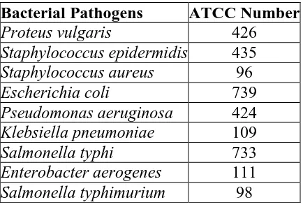
Table 1: Bacterial cultures used in study.
Bacterial Pathogens ATCC Number
Proteus vulgaris 426
Staphylococcus epidermidis 435
Staphylococcus aureus 96
Escherichia coli 739
Pseudomonas aeruginosa 424
Klebsiella pneumoniae 109
Salmonella typhi 733
Enterobacter aerogenes 111
Salmonella typhimurium 98
Preparation of disc for antibacterial activities
The aqueous, ethanol, methanol and acetone extracts were prepared in their respective
solvents and the sterile blotting paper disc (10 mm) were soaked in the diluted extract in such
concentration that the amount of solution absorbed by each disc was 1mg, 2mg, 3mg, 4mg,
5mg of each extracts of Tinospora cordifolia leaves. The prepared disc were dried in
controlled temperature to remove excess of solvent and used in study.
Antibacterial activity using disc diffusion method
The modified paper disc diffusion method was employed to determine the antibacterial
activity of aqueous, ethanol, methanol and acetone extracts. Turbidity of inoculums was
matched with McFarland turbidity standard (NCCLS, 2002). Inoculums were spread over the
Nutrient agar plate using a sterile cotton swab in order to get a uniform microbial growth.
Then the prepared antibacterial disc were placed over the lawn and pressed slightly along
with positive and negative controls. Ampicillin 10 mcg/disc (Hi-Media, Mumbai) were used
as positive control while disc soaked in various organic solvents and dried were placed on
lawns as negative control. The plates were incubated for 18h at 370C. The antibacterial
activity was evaluated and diameters of inhibition zones were measured. Experiment was
antibacterial activity was classified as strong (>20mm), moderate (16-19mm) and mild
(12-15mm) and less than 12mm was taken as inactive.
RESULTS AND DISCUSSION
Plants are important source of potentially useful structures for the development of new
chemotherapeutic agents. The first step towards this goal is the in vitro antibacterial activity
assay. Many reports are available on the antiviral, antibacterial, antifungal, anthelmintic,
antimolluscal and anti-inflammatory properties of plants. Some of these observations have
helped in identifying the active principle responsible for such activities and in the developing
drugs for the therapeutic use in human beings. However, not many reports are available on
the exploitation of antifungal or antibacterial property of plants for developing commercial
formulations for applications in crop protection. In the present study Phytochemical screening
of the leaves extract of Tinospora cordifolia in the present study also revealed presence of
terpenes and glycosides.
Table 2: Phytochemical analysis of leaves extract of Tinospora cordifolia.
Sr. No. Phytochemical
Constitutes Aqueous extract Ethanol extract Methanol extract Acetone Extract
1 Alkaloid + + + +
2 Flavonoids + ++ ++ +++
3 Glycosides + + + +
4 Saponins - ++ ++ +
5 Steroids - + + +
6 Tannins + ++ +++ +++
7 Anthroquinones - + + +
8 Phenolic compounds - +++ +++ +++
Table 3: Antibacterial activity of Tinospora cordifolia, extracts against bacterial
pathogens, (Zone of inhibition of growth in mm, average of 3 readings).
Medicinal Plants Solvent extract P. vulga ris S. epider midis S. aureus E. coli P. aeruginosa S. typhi E. aerogenes S. typhimurium Tinospora cordifolia
Aqueous 20 26 27 - 21 - - -
Ethanol 22 26 25 21 21 19 17 23
Methanol 22 32 27 22 23 17 19 18
Acetone 22 32 27 21 21 17 19 17
Negative control
Water - - - -
Ethanol - - - -
Methanol - - - -
Acetone - - - -
Positive control
Ampicillin
According to antibacterial profile (Table 3), maximum inhibitory effect of the aqueous
extract observed only on Staphylococcus epidermidis, Staphylococcus aureus, and moderate
antibacterial against Escherichia coli, Pseudomonas aeruginosa, Enterobacter aerogenes, but
mild inhibitory effect on Salmonella typhi, Salmonella typhimurium, Proteus vulgaris.
Methanol and ethanol extract showed strong antibacterial effect against Staphylococcus
epidermidis and Staphylococcus aureus and moderate antibacterial against Proteus vulgaris,
Escherichia coli, Enterobacter aerogenes, Salmonella typhi and Salmonella typhimurium but
mild effect on Pseudomonas aeruginosa. Acetone extract showed maximum inhibitory effect
on Staphylococcus aureus, Proteus vulgaris, Staphylococcus epidermidis, Pseudomonas
aeruginosa, Salmonella typhi, Salmonella typhimurium, but moderate inhibitory effect on
Escherichia coli, Enterobacter aerogenes. Several researchers have reported on the medicinal
properties of plants derived compounds. These classes of compounds are known to show
curative activity against several bacterial and it is not surprising that these plants extracts are
used traditionally by herbalist to cure bacteria related ill-health.
Tinospora cordifolia also exerted considerable antibacterial effect against tested pathogens.
However, it is ineffective against E. faecalis and S. aureus at lower concentrations with MIC
value of 500 μg. This plant has been subjected to chemical investigations extensively and a
number of chemical constituents belonging to different groups such as trepenoids, alkaloids,
lignans and flavonoids, tannins, cardiac glycosides and steroids have been reported
(Devprakash et al., 2011) which may account for the antimicrobial property of this agent.
In the present study the biological activity of the acetone extract of Tinospora cordifolia can
be attributed to the synergistic effect of the combination of flavonoids, steroids, terpinoids
and saponins.
CONCLUSION
The results obtained in this study thus suggests that the identified phytochemicals may be the
bioactive constituents responsible for the efficacy of leaves extract of Tinospora cordifolia
against fever, syphilitic, ulcer, inflammatory disease wounds, conjunctivitis etc. The results
of present investigation clearly indicate that the antibacterial and antifungal activity vary with
the species of the plants and plant material used. Thus, the study ascertains the value of
REFERENCES
1. Al-saadi. A.; Breesam. B. and al-turaihe. M. medical plants, AL radwan publisher., 2012.
2. Boots, A.W.; Haenen, G.R.; Bast, A. Health effects of quercetin: From antioxidant to
nutraceutical. Eur. J. Pharmacol, 2008; 585: 325–337.
3. Devprakash, Tinospora Cordifolia:- A Review On Its Ethnobotany, Phytochemical &
Pharmacological Profile. Asian Journal of Biochemical and Pharmaceutical Research,
2011; 4(1): 356-359.
4. Khan N. Comparative assessment of antibacterial activity of Tinospora cordifolia (Willd.)
Miers leaves and its callus. Der Pharmacia Sinica, 2013; 4(1): 56-59.
5. Khandelwal, KR. Preliminary photochemical screening, in: Practical Pharmacognosy
Techniques and Experiments. 8th edn. Nirali Publication, Pune, 2001; 149-156.
6. Nadkarni AK. Indian Materia Medica, Vol. 1 and II, 3 edn. M/s Popular Prakashan Pvt
Ltd, Bombay, 2005.
7. NCCLS (National Committee for Clinical Laboratory Standards), Performance Standards
for antimicrobial susceptibility testing. 8th Informational Supplement. M100 S12.
National Committee for Clinical Laboratory Standards, Villanova, Pa, 2002.
8. Prashant GM, Chandu GN, Murulikrishna KS, Shafiulla MD. The effect of mango and
neem, 2009.
9. Reddy P, Chadaga S, Noskin GA. Antibiotic considerations in the treatment of
multidrug-resistant (MDR) pathogens: a case-based review. JHospMed, 2009; 4(6): E8-15.
10.Solecki R. Shanidar IV A Neanderthal flower burial in Northern Iraq. Science, 1975;
190(4217): 880–881.
11.Suffredini, I.B.; Sader, H.S.; Gonçalves, A.G.; Reis, A.O.; Gales, A.C.; Varella, A.D.;
Younes, R.N. Screening of antibacterial extracts from plants native to the brazilian
amazon rain forest and atlantic forest. Braz. J. Med. Biol. Res., 2004; 37: 379–384.
12.Tambekar DH, Dahikar SB. Antibacterial activity of some Indian Ayurvedic preparations
against enteric bacterial pathogens, Journal of Advanced Pharmaceutical Technology &
Research, 2011; 2(1): 24-29.
13.Valko, M.; Rhodes, C.J.; Moncol, J.; Izakovic, M.; Mazur, M. Free radicals, metals and
antioxidants in oxidative stress-induced cancer. Chem. Biol. Interact, 2006; 160: 1–40.
14.WHO General guidelines for methodologies on research and evaluation of traditional