ISSN Online: 2151-1926 ISSN Print: 2151-1918
DOI: 10.4236/cm.2018.93008 Aug. 8, 2018 126 Chinese Medicine
Acetylshikonin Inhibits Colorectal Cancer
Growth via PI3K/Akt/mTOR Signaling Pathway
Yuzhen Zhu
1*, Yu Zhong
2*, Yu Zhou
3, Yanyan Liu
4, Qionglin Huang
2, Zhe Huang
5,
Yongcun Wang
6, Hua Ye
1, Xiaobing Zeng
1,7#, Xuebao Zheng
1,8#1Guangdong Key Laboratory for Research and Development of Natural Drugs, Guangdong Medical University, Zhanjiang, China
2Analysis Center of Guangdong Medical University, Zhanjiang, China
3Department of Gastroenterology, Affiliated Hospital of Guangdong Medical University, Zhanjiang, China 4Department of Clinical Laboratory, Affiliated Run Shaw Hospital of Zhejiang University School of Medicine, Hangzhou, China
5Department of Gastrointestinal Surgery, Affiliated Hospital of Guangdong Medical University, Zhanjiang, China
6Oncology Center, Affiliated Hospital of Guangdong Medical University, Zhanjiang, China
7Center Lab of Longhua Branch, Shenzhen People’s Hospital, 2nd Clinical Medical College of Jinan University, Shenzhen, China
8Mathematical Engineering Academy of Chinese Medicine, Guangzhou University of Chinese Medicine, Guangzhou, China
Abstract
Background: Acetylshikonin, a major constituent isolated from Arnebia euchroma, is a potential candidate for anti-colorectal cancer drugs. However, the potential activity and underlying mechanism of Acetylshikonin against colorectal cancer remain unclear. Methods: In this study, Acetylshikonin was isolated from the active CHCl3 extract of Arnebia euchroma using activity-guided screening, and elucidated by the extensive spectroscopic analysis and comparison with literature data. Human colorectal cancer cells HT29, DLD-1, HCT116 or Ca-co-2 were exposed to different concentrations of Acetylshikonin (6.25 - 100 μg/mL) for 24 or 48 h. Cell viability, cell apoptosis and cell cycle distribution were detected. The activity of Acetylshikonin and potential mechanism of the phosphoinositide 3-kinase (PI3K)/Akt/mammalian target of rapamycin (mTOR) pathway were evaluated in vitro and vivo. Results: We found that Acetylshikonin exhibited remarkable anti-proliferative activity in a dose-dependent manner against HT29 cells with the IC50 values of 60.82 μg/ml and 30.78 μg/ml at 24, 48 h, respectively. Moreover, Acetylshikonin induced cell cycle arrest at G0/G1 phase and early apoptosis through inhibition of PI3K/Akt/mTOR pathway. Furthermore, the assays of cell inhibition, early
*Yuzhen Zhu and Yu Zhong contributed equally to this work. How to cite this paper: Zhu, Y.Z., Zhong,
Y., Zhou, Y., Liu, Y.Y., Huang, Q.L., Huang, Z., Wang, Y.C., Ye, H., Zeng, X.B. and Zheng, X.B. (2018) Acetylshikonin Inhibits Colorectal Cancer Growth via PI3K/Akt/mTOR Signaling Pathway. Chi-nese Medicine, 9, 126-143.
https://doi.org/10.4236/cm.2018.93008
Received: June 19, 2018 Accepted: August 5, 2018 Published: August 8, 2018
Copyright © 2018 by authors and Scientific Research Publishing Inc. This work is licensed under the Creative Commons Attribution International License (CC BY 4.0).
http://creativecommons.org/licenses/by/4.0/
DOI: 10.4236/cm.2018.93008 127 Chinese Medicine
apoptosis and G0/G1 phase distribution showed that suppression of the PI3K/Akt pathway using LY294002 enhanced the anti-cancer effect of Acetylshikonin. Similarly, Acetylshikonin also decreased the growth of tumour in colorectal cancer xenografts in mice through PI3K/Akt/mTOR pathway. Conclusions: To sum up, these new findings provided a framework for further exploration of Acetylshikonin which possessed the potential antitumor activity by inhibiting PI3K/Akt/mTOR pathway.
Keywords
Arnebia euchroma, Acetylshikonin, Colorectal Cancer, Apoptosis, PI3K/Akt/mTOR Pathway
1. Background
Colorectal cancer (CRC) is one of the most common malignancies in the world, accounting for approximately 1.36 million new cases worldwide every year [1]. Although advances in detection and local control with surgery, radiation and chemotherapy, the overall survival rate of colorectal cancer patients has not improved significantly during the past several decades. Chemotherapy is a common therapeutic strategy after surgery, and the marketed chemotherapy drugs kill tumour cells through competitive inhibition of nucleotide synthesis or cytotoxicity. However, these drugs often cause vomiting, myelosuppression, drug resistance and other adverse effects [2]. Therefore, searching for novel anticancer components of traditional Chinese medicine has become a very interesting option for developing anti-colorectal cancer drugs.
Shikonin, a natural occurring naphthoquinone, has potent anti-tumour activity. Shikonin is able to inhibit cell proliferation and induces apoptosis in several human malignancies such as gastric cancer [3], non-small cell lung cancer [4], pancreatic cancer [5] and so on. However, certain limitations exist, including no clear target and liver and kidney toxicity [6]. Therefore, highly anti-cancer and low or even nontoxic shikonin-like compounds cause our concerns. Shikonin derivatives have exhibited antivirus, antioxidation, anti-inflammatory, anti-fertility, and other pharmacological effects in previous studies [7]. Additionally, shikonin derivatives have strong anti-tumour activities with few adverse effects, which make shikonin derivatives as promising anti-tumour agents [8].
DOI: 10.4236/cm.2018.93008 128 Chinese Medicine
framework for further exploration of Acetylshikonin which possessed the potential antitumor activity by inhibiting PI3K/Akt/mTOR pathway.
2. Methods
2.1. General
Optical rotations were measured using a JASCO P-1030 (Tokyo, Japan) auto-matic digital polarimeter. NMR spectra were recorded on a Bruker DPX-400 spectrometer (400 MHz for 1H NMR and 100 MHz for 13C NMR, Karlsruhe,
Germany) using standard Bruker pulse programs. Chemical shifts were showed as the δ-value with reference to tetramethylsilane (TMS) as an internal standard. Electrospray ionization mass spectrometry (ESI-MS) data were obtained on a Bruker Esquire LC 200-Ion trap mass spectrometer (Bruker, Karlsruhe, Germa-ny). And Sephadex LH-20 (Pharmacia, Sweden), silica gel (Qingdao Ocean Chemical Co., Ltd., Qingdao, China), and octadecylsilane (ODS) silica gel (Ma-cherey-Nagel, Duren, Germany) were used for column chromatography. HPLC was performed using an ODS silica gel column (Cosmosil 5C18-MS-II, 20 × 250 mm, Nacalai Tesque). All other chemicals were analytical or HPLC grade and obtained from Shanghai Chemical Reagents Co., Ltd. (Shanghai, China). Human colon cancer cell lines HT29, DLD-1, HCT116 and Caco-2 were purchased from the ATCC (PA, USA). These cells were grown in RPMI 1640 (Invitrogen, CA, USA) supplemented with 10% (v/v) fetal bovine serum (Invitrogen, CA, USA) and 1% penicillin-streptomycin (Invitrogen, CA, USA). MTT powder was ob-tained from Sigma (Aldrich, USA). The Cell Counting Kit-8 assay was purchased from Dojindo Laboratories (Kumamoto, Japan). A Cycle Test Plus DNA reagent kit and Annexin V-FITC Apoptosis Detection Kit were purchased from BD Bi-osciences Pharmingen (NJ, USA). Total RNA extraction kit was procured from Omega (R6834-02, GA, USA). The qRT-PCR was performed using EASY spin Plus Total RNA Extraction Kit (Aidlab, Beijing, China), Transcriptor First Strand cDNA Synthesis Kit (Roche, Basel, Switzerland) and SYBR Premix ExTaq kit (Takara, Dalian, China). Antibodies against PI3K, p-PI3K, Akt, p-Akt, mTOR, β-Actin were purchased from Cell Signalling Technology (MA, USA). BCA Protein Assay Kit, RIPA cell lysates, PMSF and Hematoxylin and Eosin Staining Kit were obtained from Beyotime (Shanghai, China). Irinotecan was obtained from Qilu Pharmaceutical Co. Ltd (Hainan, China).
2.2. Extraction and Isolation
ace-DOI: 10.4236/cm.2018.93008 129 Chinese Medicine
tate (3 L × 3) and water-saturated n-butanol (3 L × 3) gradually to afford 178.21 g, 62.01 g and 254.35 g of dried organic extracts, respectively.
Bioactivity-guided fractionation was used for the isolation work. On the basis of the bioactive results of the extracts, the chloroform fraction with the most po-tential activity was fractionated over a silica gel (200 - 300 mesh) column eluting with a gradually amount of MeOH in CHCl3 to give 5 fractions. The CHCl3-MeOH
(40: 1) elution (25.0 g) was further purified by a Sephadex LH-20 column (6 × 88 cm, 680 g; Solvent system: CHCl3-MeOH, 7: 3) to obtain three major fractions,
1a-1c. Fraction 1a (15.0 g) was applied to a preparative HPLC with 89% methanol (containing 0.1% CF3COOH, pH 3.0) to give fractions 1a1-1a3, and Acetylshikonin
(1122.15 mg) were purified by preparative HPLC (83% MeOH and 83% MeOH, containing 0.1% CF3COOH, pH 3.0) at 8 ml/min from Fraction 1a2 (1421.1 mg).
2.3. Acetylshikonin
Acetylshikonin was a red amorphous powder; ESI-MS m/z 331 [M - H]−;
[ ]
25
D
α
+ 151.2 (c 0.20, CHCl3); 1H NMR (CDCl3, 400 MHz) data: 6.99 (1H, s, H-3), 7.18
(2H, s, H-6, 7), 12.42 (1H, s, 6-OH), 12.58 (1H, s, 5-OH), 6.03 (1H, dd, J = 7.2, 4.4 Hz, H-1’), 2.46 (1H, m, H-2’), 2.62 (1H, m, H-2’), 5.12 (1H, t, J= 7.6 Hz, H-3’), 1.69 (3H, s, 5’-CH3), 1.58 (3H, s, 6’-CH3), 2.14 (3H, s, H-2’’); 13C NMR
(CDCl3, 100 MHz) data: 178.1 (C-1), 148.2 (C-2), 136.1 (C-3), 176.6 (C-4), 111.6
(C-5), 169.7 (C-6), 131.5 (C-7), 132.7 (C-8), 167.0 (C-9), 111.8 (C-10), 69.5 (C-1’), 32.8 (C-2’), 117.6 (C-3’), 132.9 (C-4’), 17.9 (C-5’), 25.7 (C-6’), 167.6 (C-1’’), 20.9 (C-2”).
2.4. Cell Culture
Four human colon carcinoma cell lines including HT29, DLD-1, HCT116, Ca-co-2 were cultured in the Dulbecco’s modified Eagle medium supplemented with 10% fetal bovine serum, 100 μg/ml penicillin, and 100 μg/mL streptomycin. The cells were incubated at 37˚C in a humidified atmosphere with 5% CO2.
2.5. Methyl Thiazolyl Tetrazolium (MTT) Assay
HT29 cells (5 × 10³ cells/well) were seeded in 96-well plates and cultured for 24
h. Cells were treated with various concentrations of Acetylshikonin (6.25, 12.5, 25, 50, 100 μg/mL) for 24 or 48 h. 20 μl MTT solution (0.5 mg/mL in PBS) was supplemented into each well and incubated for an additional 4 hours. The su-pernatant was then discarded, and 100 μL DMSO was added to dissolve the for-mazan crystals. Optical density was read at a wavelength of 490 nm on a micro-plate reader. Cytotoxicity of Acetylshikonin was assessed as described previous-ly, and calculated by the following formula:
( )
(
Control Sample)
(
Control Blank)
Inhibitory rate % = OD −OD OD −OD ×100%
2.6. Cell Proliferation Assay
DOI: 10.4236/cm.2018.93008 130 Chinese Medicine
according to the manufacturer’s protocol. Each cell line including Caco-2, HCT116, DLD-1, HT29 was seeded into 96-well plates (5 × 10³ cells/well). After
24 h, the medium was replaced with fresh medium and then adhered cells were treated with different concentrations of Acetylshikonin (6.25, 12.5, 25, 50, 100 μg/mL) for 48 h. The CCK-8 solution was added to each well and incubated for additional 2 h. The number of viable cells was quantified by assessing the absor-bance (450 nm) using Multiskan Spectrum Microplate Reader. The inhibitory rate of cell proliferation was calculated by the following formula:
( )
(
Sample Blank)
(
control Blank)
Inhibitory rate % = −1 OD −OD OD −OD ×100%
2.7. Apoptosis Detection Assay
HT29 (5 × 10⁵ cells/well) in 6-well culture plates were exposed to various
con-centrations of Acetylshikonin for 24 or 48 h. After that, we harvested the cells and supernatants, and washed them in PBS and resuspended 1 × 10⁶ cells/mL in
1 × Binding Buffer. 5 μL FITC-conjugated Annexin V and 5 μl Propidium lodide (PI) were added to the 100 μL cell suspension. After incubation for 15 minutes at room temperature in the dark, cells were then analyzed by flow cytometer. The amount of early apoptosis, late apoptosis, and necrosis was determined as the percentage of Annexin V+/PI−, Annexin V+/PI+, and Annexin V−/PI+ cells,
re-spectively. Each experiment was performed in triplicate.
2.8. Cell Cycle Analysis Assay
HT29 cells were trypsinized with 0.25% Trypsin EDTA at room temperature af-ter pretreatment with various concentration of Acetylshikonin for 24 h, and washed three times in buffer solution. Cell cycle distribution was detected by Cycletest Plus DNA Reagent kit. The percentages of cells in the different cell cycle phases (G0/G1, S, G2/M) were calculated using Flow cytometry.
2.9. Real-Time Polymerase Chain Reaction (RT-PCR)
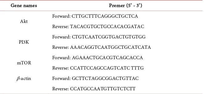
HT29 cells were treated with Acetylshikonin at different concentration of 0, 25, 50, 100 μg/ml for 48 h. Total RNA was extracted by Omega total RNA ex-traction kit and the total RNA was reverse transcribed using TaKaRa reverse transcription kit. Using reverse transcribed cDNA as the template and β-actin as the endogenous control. The amplification reactions were carried out ac-cording to the one-step SYBR1 prime Script1 RT-PCR II kit instructions. Am-plification was performed with an ABI 7500 real-time PCR thermocycler. Real-time PCR reaction system: 2 μl cDNA product, 0.4 μl each of forward and reverse primers, 10 μl Taq Master Mix, 6.8 μl RNase-free water, 0.4 μl ROX Reference Dye (50 ×), total volume 20 μl. The primer sequences that were used are shown in Table 1. Reaction conditions: pre-denaturation at 95˚C for 30 s, 95˚C for 5 s, 57˚C for 34 s and repeat for 35 cycles. Each sample repeated 3 times. The relative expression of each gene was calculated using the 2−∆∆Ct
DOI: 10.4236/cm.2018.93008 131 Chinese Medicine Table 1. Premer sequences of the genes.
Gene names Premer (5’ - 3’)
Akt Forward: CTTGCTTTCAGGGCTGCTCA Reverse: TACACGTGCTGCCACACGATAC
PI3K Forward: CTGTCAATCGGTGACTGTGTGG Reverse: AAACAGGTCAATGGCTGCATCATA
mTOR Forward: AGAAACTGCACGTCAGCACCA Reverse: CCATTCCAGCCAGTCATC TTTG β-actin Forward: GCTTCTAGGCGGACTGTTAC
Reverse: CCATGCCAATGTTGTCTCTT
2.10. Western Blot Analysis
HT29 (5 × 10⁵ cells/well) in 6-well culture plates were exposed to treat with
Acetylshikonin for 48 h. Both adherent and floating cells were collected. The tumour tissue protein was purified according to the reported method [11]. Western blotting used standard protocols. Proteins were separated by SDS-PAGE and transferred onto nitrocellulose membranes that were blocked with 5% non-fat milk in TBST, and in-cubated with primary antibodies: Akt, p-Akt, PI3K, p-PI3K, mTOR, β-actin (Cell Signaling Technology, MA, USA). Then we add HRP-conjugated polymer-tagged secondary antibodies (Abcam, MA, USA). Lastly, the protein bands were captured using Western blotting detection system (DNR Bio-Imaging Systems, Jerusalem, Israel). Signal intensity was quantified by densitometry with the Gel-pro Analyzer (Media Cybernetics, Rockville, MD, USA). All experiments were done in triplicate.
2.11. Inhibition of PI3K/Akt Pathway
HT29 cells were pretreated with LY294002 (10 mM, Selleck Chemicals, TX, USA), for 30 min, then co-treated with 50 μg/mL Acetylshikonin for further 48 h. Then cell viability, cell apoptosis, cell cycle arrest and expressions of PI3K/Akt proteins were measured in terms of the above experimental methods.
2.12. Tumour Xenograft in NOD/SCID Mice
A total of 16 male NOD/SCID mice with body weights of 10 - 12 g, were pur-chased from model animal research center of Nanjing University and main-tained in a specific pathogen-free environment. Animal experiments were ap-proved by the Guangdong Medical University Institutional Animal Care and Use Committee. After 2 weeks, mice were injected with DLD-1 cells (1 × 10⁷
cells in 0.2 mL volume) into the subcutaneous tissue of the back region. Tumour volume was measured by calliper every two day and calculated by the formula: (length × width × width)/2. Mice began to treat with drugs when tumours reached an average size of 62.5 mm3. The tumour-bearing mice were divided
Acetylshiko-DOI: 10.4236/cm.2018.93008 132 Chinese Medicine
nin (20 mg/kg in 1% DMSO, every two days), irinotecan (66.7 mg/kg, every four days). All mice were killed on Day 13, and the tumours were segregated, meas-ured, weighed, and stored in liquid nitrogen for later use.
2.13. Hematoxylin/Eosin (H & E) Staining
Tumours were collected, fixed in formalin and embedded in paraffin. Tissue sec-tions (5 μm in thickness) were prepared according to standard protocols for H&E staining. After staining, the tissue slices were viewed under microscopy with the Olympus DP controller software program (Tokyo, Japan).
2.14. Statistical Analysis
All experiments were performed at least three times, independently. The results were analysed using GraphPad Prism version 6.0 to perform one-way ANOVA. P value less than 0.05 was considered statistically significant.
3. Results and Discussion
3.1. Isolation and Identification of Acetylshikonin
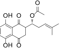
Acetylshikonin (Figure 1) was isolated from the extracts of A. euchroma by bio-assay-guided fractionation, and identified using spectral analysis by 1H and 13C
NMR and comparison with literature data [12].
3.2. Acetylshikonin Exhibited Anti-Proliferative Activity against
Human Colorectal Cancer Cells
To explore the effects of Acetylshikonin on colorectal cancer cells proliferation, HT29 cells were exposed to different concentration of Acetylshikonin for 24 or 48 h, and cell viability was measured using the MTT assay. As shown in Figure 2(a) and Figure 2(b), Acetylshikonin showed significant inhibition against the HT29 cell with IC50 values of 60.82 and 30.78 μg/ml at 24 and 48 h, respectively.
[image:7.595.316.435.606.706.2]Also, human colorectal cancer cells including Caco-2, HCT116, DLD-1 and HT29 were treated with Acetylshikonin for 48 h, and cell proliferation analysis was conducted by Cell Counting Kit-8. It was found that Acetylshikonin led to a dose-dependent inhibition of cell proliferation in all four cancer cell lines. Taken together, these results suggest that Acetylshikonin inhibits the growth of colorectal cancer cells in vitro.
Figure 1. Chemical structure of Acetylshikonin.
OH
OH O
O
O
DOI: 10.4236/cm.2018.93008 133 Chinese Medicine
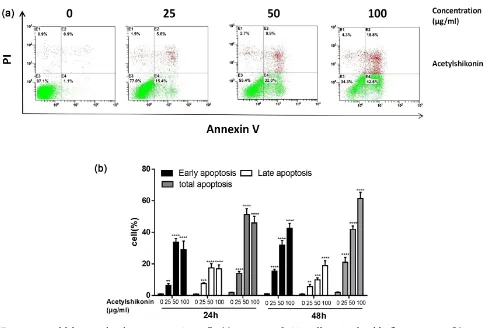
3.3. Acetylshikonin Induced Apoptosis in HT29 Cells
[image:8.595.73.527.177.304.2]Flow cytometry was performed to detect the apoptosis manner with Annexin V and PI staining. Figure 3(a) and Figure 3(b) showed that the early apoptosis ratio increased from 15.4% to 42.6% with the treatment of Acetylshikonin (25, 50, 100 μg/ml) in HT29 cells in a dose-dependent manner. Thus, Acetylshikonin could induce apoptosis, especially early apoptosis in HT29 cells.
Figure 2. Effects of Acetylshikonin on colon carcinoma cells proliferation. (a): Cell inhibitory was measured using the MTT assay after treating cells with different concentrations (6.25, 12.5, 25, 50, 100μg/ml) of Acetylshikonin for 24 and 48 h. (b): HT29, HCT116, DLD-1 and Caco-2 cells were treated with various concentrations of Acetylshikonin for 48 h. Cell inhibitory was ana-lyzed by CCK-8 assay. Results were obtained from three independent experiments, and the dots represent mean ± SD.
[image:8.595.56.540.362.690.2]DOI: 10.4236/cm.2018.93008 134 Chinese Medicine
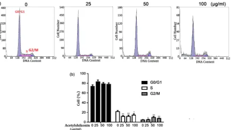
3.4. Acetylshikonin Induced Cell Cycle Arrested
at G0/G1 in HT29 Cells
The effect of Acetylshikonin on regulation of the cell cycle in HT29 cells was studied using Cycletest Plus DNA Reagent kit, and the results were evaluated by flow cytometry. As shown in Figure 4(a) and Figure 4(b), an increasing pro-portion of HT29 cells in G0/G1 phase was observed after treated with an in-creasing concentration of Acetylshikonin, while the percentage of HT29 cells in S or G2/M phase decreased.
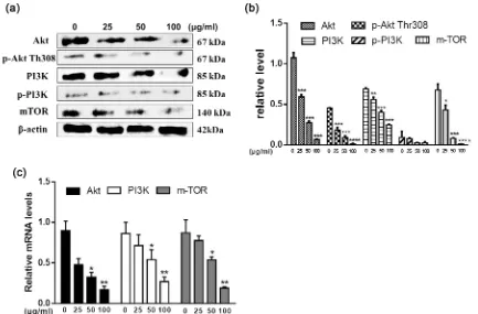
3.5. Activation of PI3K/Akt/mTOR Signaling Pathway Was
Essen-tial for Acetylshikonin-Induced Apoptosis
To further elucidate underlying mechanism of Acetylshikonin proapoptotic effect on HT29 cells, we evaluated whether PI3K/Akt/mTOR signaling pathway is involved in the apoptosis. Total protein from 0, 25, 50, 100 μg/ml Acetylshikonin treated HT29 cells for 48 h were collected, and apoptotic proteins such as Akt, p-Akt, PI3K, p-PI3K, mTOR were determined by Western blot. With the increasing concentration of Acetylshikonin, the expression of these proteins including PI3K, p-PI3K, Akt, p-Akt, mTOR downregulated (Figure 5(a) and Figure 5(b)). Also, the results of RT-PCR showed that Acetylshikonin down-regulated the expression of these mRNAs including PI3K, Akt, mTOR in HT29 cells in a dose-dependent manner (Figure 5(c)).
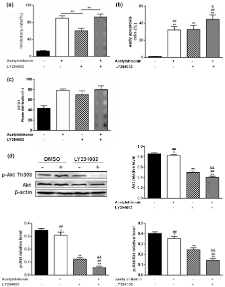
[image:9.595.66.542.409.677.2]To further determine the relationship between cell apoptosis and activation of
DOI: 10.4236/cm.2018.93008 135 Chinese Medicine Figure 5. Effects of Acetylshikonin on Akt/PI3K/mTOR signalling pathway in HT29 cells. (a): Western blot ana-lysed the protein expression of Akt, p-Akt, PI3K, p-PI3K, mTOR in HT29 cells when treated with or without Ace-tylshikonin. (b): Histograms demonstrate the relative expression level of proteins. (c): Akt, PI3K and mTOR gene expression were measured by real-time PCR treated with different concentrations of acetylshikonin for 48 h. **P < 0.001, *P < 0.05 versus control (no Acetylshikonin) group.
PI3K/Akt/mTOR pathway, HT29 cells was exposed to Acetylshikonin and LY294002 (a PI3K/Akt inhibitor) for 48 h. HT29 cells treated with LY294002 plus 50 μg/ml Acetylshikonin had higher cell inhibitory rates and total apoptotic cells than those only treated with Acetylshikonin. But there was little change observed in the G0/G1 phase distribution (Figure 6(a) and Figure 6(b)). Similarly, Acetylshikonin plus LY294002 treatment caused a significant decrease in the ratio of p-Akt308/Akt, compared with that treated with Acetylshikonin alone (Figure 6(c) and Figure 6(d)).
These results demonstrated that apoptosis is induced by Acetylshikonin in HT29 cells via PI3K/Akt/mTOR signaling pathway.
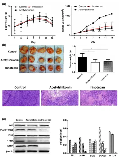
3.6. Acetylshikonin Inhibited the Growth of Xenografted Tumours
in Nude Mice by PI3K/Akt/mTOR Signaling Pathway
Based on the above results, we could thus conclude that Acetylshikonin is a promising anti-colorectal cancer agent. To further investigate the anti-tumour effect of Acetylshikonin, we established a subcutaneously transplanted colon carcinoma model using DLD-1 cells. After solid tumors were palpable (about 62.5 m3), mice were divided randomly into three groups and treated with
DOI: 10.4236/cm.2018.93008 137 Chinese Medicine
days), Acetylshikonin (20 mg/kg in 1% DMSO, every two days), irinotecan (66.7 mg/kg, every four days). No severe side effects were observed during the drug injected. The tumors were excised and weighed on day 13 when mice sacrificed. As shown in Figure 7(a) and Figure 7(b), both tumor weight and volume were significantly decreased in Acetylshikonin-treated group comparing with the control group. Furthermore, hematoxylin/eosin (H&E) staining indicated that Acetylshikonin promoted necrosis and induced the disappearance of tumor cells. Compared to the control group, the expression of these proteins including PI3K, p-PI3K, Akt, p-Akt and mTOR in the tumour tissue were dramatically decreased in Acetylshikonin-treated group (Figure 7(c)). Thus, Acetylshikonin suppressed the growth of colorectal tumours by PI3K/Akt/mTOR signaling pathway in vivo.
Traditional Chinese medicine, envisioned as safer alternatives for their chem-ical counterparts, has been used for preventing cancer for centuries. Currently, effective components isolated from traditional Chinese medicines have become an important approach to discover anti-cancer drugs [13].
In our study, Acetylshikonin was extracted and isolated from the dried roots of A. euchroma. Acetylshikonin dramatically suppressed the proliferation of HT29 cells in the time and dose-dependent manner. The results of CCK-8 assay also revealed that Acetylshikonin at 25 - 100 µg/ml exhibited significant inhibi-tion of cell proliferainhibi-tion in human colorectal cancer cells including HT29, DLD-1, HCT116 and Caco-2. These findings indicated that Acetylshikonin has the potential to develop into a novel anti-tumor drug for colorectal cancer.
Apoptosis is a genetically programmed process leading to cell death, which primarily functions to eliminate senescent or altered cells that are useless or harmful to health. In contrast, abnormal changes in apoptotic mechanisms that promote deficient programmed cell death are closely related to the occurrence and development of tumours [14]. Recent evidence has indicated that selective induction of apoptosis in tumour cells is the most direct and efficacy method to treat tumours [15]. In our study, to further clarify the inhibitory effects of Acetylshikonin on colorectal cancer, Annexin V/PI staining was applied to detect the effect of Acetylshikonin on HT29 cells apoptosis. The proportion of total apoptotic cells (particularly early apoptosis) induced by Acetylshikonin was significantly increased dose-dependently.
Cell cycle progression plays an important role in tumour growth, and its reg-ulation is an effective strategy to control tumour growth. It has been reported that the active ingredients of traditional Chinese medicine can arrest tumour cells at different cell cycle, which in turn inhibit tumour cell growth, proliferation, and induce apoptosis [16] [17]. In present study, Acetylshikonin blocked cell cycle progression at G0/G1 phase, which demonstrated that Acetylshikonin interfered with the normal tumour cell cycle and inhibited tumour growth.
DOI: 10.4236/cm.2018.93008 139 Chinese Medicine
and angiogenesis [22]. Previous studies have indicated that abnormal activation of PI3K/Akt/mTOR signaling pathway is commonly involved in the develop-ment and progression of various tumours [23] [24] [25], especially colorectal cancer [26]. Akt is one of core target proteins to downstream of PI3K, which relies on the phosphorylation at the Thr308 site of the catalytic domain and the Ser473 site of the regulatory domain of the C-terminus. In this study, we found Acetylshikonin down-regulated Akt phosphorylation at Thr308. However, the precise molecular mechanism by which Acetylshikonin deactivates Akt should be further investigated. Combining Acetylshikonin with LY294002 largely increased the rate of cell inhibitory, early apoptosis and G0/G1 phase distribution of HT29. As a targeted anti-cancer drug LY294002, has also been reported in several cancers, Acetylshikonin may also be useful for improving anti-cancer activity of PI3K/Akt/mTOR-targeting drugs.
Further experiments were conducted in vivo based on the preliminary in vitro research. Thus, we established the transplanted colon carcinoma model of human DLD-1 cells in NOD/SCID mice to observe the anti-tumor effect of Acetylshikonin in vivo. Acetylshikonin obviously inhibited the growth of colon tumours with no body weight changes. Additionally, there was little difference in weight and pathology of tumours between the Acetylshikonin-treated group and irinotecan positive group. Furthermore, Acetylshikonin could inhibit PI3K/Akt/mTOR pathway in vivo by Western blot analysis, which is in ageement with the results in vitro.
4. Conclusions
In conclusion, our study demonstrated that Acetylshikonin possessed the anti-cancer activity in human colorectal cancer. Acetylshikonin effectively inhibited cell pro-liferation, induced cell cycle arrested at G0/G1 and promoted colorectal cancer cells apoptosis in vitro. Colorectal cancer growth was also suppressed by Acetylshikonin in vivo. Meanwhile, activation of PI3K/Akt/mTOR signaling pathway played a crucial role in the treatment with Acetylshikonin. Collectively, these findings reveal that Acetylshikonin may serve as an anti-tumour candidate for colorectal cancer treatment.
Authors’ Contributions
XBZ and YZZ developed the concept of this study. YZZ, XBZ, YZ, YZ, YYL, QLH, ZH, YCW and HY designed and performed experiments and analysed da-ta. YZZ, XBZ and XBZ prepared the draft and final version of the manuscript. All authors read and approved the final manuscript.
Acknowledgements
DOI: 10.4236/cm.2018.93008 140 Chinese Medicine
Province (2016A030310356, 2014A030307001), the Science and Technology Fund of Zhanjiang (2014A01014), Research Foundation of Guangdong Medical University (M2016021, M2013003) and Traditional Chinese Medicine Research Foundation of Guangdong Provincial Bureau (20151260).
Conflicts of Interest
The authors declare that they have no conflict of interest.
Availability of Data and Materials
The datasets used and/or analyzed during the current study are available from the corresponding author on reasonable request.
Consent for Publication
All of authors consent to publication of this study in Journal of Chinese Medi-cine.
Ethics Approval and Consent to Participate
Animal protocol was conducted according to the animal procedure approved by Animal Ethics Committee, Guangdong Key Laboratory for Research and Development of Natural Drugs, Guangdong Medical University (SYXK (GUANGDONG) 2016-0013).
References
[1] Benard, F., Barkun, A.N., Martel, M. and von Renteln, D. (2018) Systematic Review of Colorectal Cancer Screening Guidelines for Average-Risk Adults: Summarizing the Current Global Recommendations. World Journal of Gastroenterology, 24, 124-138. https://doi.org/10.3748/wjg.v24.i1.124
[2] Miguel, A., Azevedo, L.F., Araujo, M. and Pereira, A.C. (2012) Frequency of Ad-verse Drug Reactions in Hospitalized Patients: A Systematic Review and Me-ta-Analysis. Pharmacoepidemiology and Drug Safety, 21, 1139-1154.
https://doi.org/10.1002/pds.3309
[3] Liang, W., Cai, A., Chen, G., Xi, H., Wu, X., Cui, J., Zhang, K., Zhao, X., Yu, J., Wei, B. and Chen, L. (2016) Shikonin Induces Mitochondria-Mediated Apoptosis and Enhances Chemotherapeutic Sensitivity of Gastric Cancer through Reactive Oxygen Species. Scientific Reports,6, Article ID: 38267. https://doi.org/10.1038/srep38267 [4] Li, X., Fan, X.X., Jiang, Z.B., Loo, W.T., Yao, X.J., Leung, E.L., Chow, L.W. and Liu,
L. (2017) Shikonin Inhibits Gefitinib-Resistant Non-Small Cell Lung Cancer by In-hibiting TrxR and Activating the EGFR Proteasomal Degradation Pathway. Phar-macological Research, 115, 45-55. https://doi.org/10.1016/j.phrs.2016.11.011 [5] Wang, Y., Zhou, Y., Jia, G., Han, B., Liu, J., Teng, Y., Lv, J., Song, Z., Li, Y., Ji, L.,
Pan, S., Jiang, H. and Sun, B. (2014) Shikonin Suppresses Tumour Growth and Synergizes with Gemcitabine in a Pancreatic Cancer Xenograft Model: Involvement of NF-κB Signalling Pathway. Biochemical Pharmacology, 88, 322-333.
https://doi.org/10.1016/j.bcp.2014.01.041
DOI: 10.4236/cm.2018.93008 141 Chinese Medicine the Inhibition Risk of Shikonin on Cytochrome P450 via Cocktail Inhibition Assay.
Toxicology Letters, 281, 74-83. https://doi.org/10.1016/j.toxlet.2017.09.014 [7] Huang, G., Zhao, H.R., Meng, Q.Q., Zhang, Q.J., Dong, J.Y., Zhu, B.Q. and Li, S.S.
(2018) Synthesis and Biological Evaluation of Sulfur-Containing Shikonin Oxime Derivatives as Potential Antineoplastic Agents. European Journal of Medicinal Chemistry, 143, 166-181. https://doi.org/10.1016/j.ejmech.2017.11.031
[8] Ning, X., Qi, H., Li, R., Li, Y., Jin, Y., McNutt, M.A., Liu, J. and Yin, Y. (2017) Dis-covery of Novel Naphthoquinone Derivatives as Inhibitors of the Tumour Cell Spe-cific M2 Isoform of Pyruvate Kinase. European Journal of Medicinal Chemistry, 138, 343-352. https://doi.org/10.1016/j.ejmech.2017.06.064
[9] Park, S.H., Phuc, N.M., Lee, J., Wu, Z., Kim, J., Kim, H., Kim, N.D., Lee, T., Song, K.S. and Liu, K.H. (2017) Identification of Acetylshikonin as the Novel CYP2J2 In-hibitor with Anti-Cancer Activity in HepG2 Cells. Phytomedicine, 24, 134-140.
https://doi.org/10.1016/j.phymed.2016.12.001
[10] Cho, S.C. and Choi, B.Y. (2015) Acetylshikonin Inhibits Human Pancreatic PANC-1 Cancer Cell Proliferation by Suppressing the NF-κB Activity. Biomolecules & Therapeutics, 23, 428-433. https://doi.org/10.4062/biomolther.2015.102
[11] Zou, Y., Lin, J.T., Li, W.Y., Wu, Z.G., He, Z.W., Huang, G.L., Wang, J., Ye, C.G., Cheng, X.Y., Ding, C.C., Zheng, X.B. and Chi, H.G. (2016) Huangqin-Tang Ameli-orates Dextran Sodium Sulphate-Induced Colitis by Regulating Intestinal Epithelial Cell Homeostasis, Inflammation and Immune Response. Scientific Reports, 6, Ar-ticle ID: 39299. https://doi.org/10.1038/srep39299
[12] Ko, F.N., Lee, Y.S., Kuo, S.C., Chang, Y.S. and Teng, C.M. (1995) Inhibition on Platelet Activation by Shikonin Derivatives Isolated from Arnebia euchroma. Bio-chimica et Biophysica Acta (BBA), Molecular Cell Research, 1268, 329-334.
https://doi.org/10.1016/0167-4889(95)00078-7
[13] Qi, F., Zhao, L., Zhou, A., Zhang, B., Li, A., Wang, Z. and Han, J. (2015) The Ad-vantages of Using Traditional Chinese Medicine as an Adjunctive Therapy in the Whole Course of Cancer Treatment instead of Only Terminal Stage of Cancer. Bi-oScience Trends, 9, 16-34. https://doi.org/10.5582/bst.2015.01019
[14] Marquez-Jurado, S., Diaz-Colunga, J., Das Neves, R.P., Martinez-Lorente, A., Al-mazan, F., Guantes, R. and Iborra, F.J. (2018) Mitochondrial Levels Determine Va-riability in Cell Death by Modulating Apoptotic Gene Expression. Nature Commu-nications, 9, 389. https://doi.org/10.1038/s41467-017-02787-4
[15] Fulda, S. (2018) Cell Death-Based Treatment of Glioblastoma. Cell Death & Disease, 9, 121. https://doi.org/10.1038/s41419-017-0021-8
[16] Zheng, T., Que, Z., Jiao, L., Kang, Y., Gong, Y., Yao, J., Ma, C., Bi, L., Dong, Q., Zhao, X. and Xu, L. (2017) Herbal Formula YYJD Inhibits Tumour Growth by In-ducing Cell Cycle Arrest and Senescence in Lung Cancer. Scientific Reports, 7, Ar-ticle No. 4984.https://doi.org/10.1038/s41598-017-05146-x
[17] Chen, X., Wu, Q.S., Meng, F.C., Tang, Z.H., Chen, X., Lin, L.G., Chen, P., Qiang, W.A., Wang, Y.T., Zhang, Q.W. and Lu, J.J. (2016) Chikusetsusaponin IVa Methyl Ester Induces G1 Cell Cycle Arrest, Triggers Apoptosis and Inhibits Migration and Invasion in Ovarian Cancer Cells. Phytomedicine, 23, 1555-1565.
https://doi.org/10.1016/j.phymed.2016.09.002
DOI: 10.4236/cm.2018.93008 142 Chinese Medicine
https://doi.org/10.1038/s41419-017-0132-2
[19] Pan, Y., Wang, N., Xia, P., Wang, E., Guo, Q. and Ye, Z. (2018) Inhibition of Rac1 Ameliorates Neuronal Oxidative Stress Damage via Reducing Bcl-2/Rac1 Complex Formation in Mitochondria through PI3K/Akt/mTOR Pathway. Experimental Neurology, 300, 149-166.https://doi.org/10.1016/j.expneurol.2017.10.030
[20] Wang, F., Wang, Q., Zhou, Z.W., Yu, S.N., Pan, S.T., He, Z.X., Zhang, X., Wang, D., Yang, Y.X., Yang, T., Sun, T., Li, M., Qiu, J.X. and Zhou, S.F. (2015) Plumbagin In-duces Cell Cycle Arrest and Autophagy and Suppresses Epithelial to Mesenchymal Transition Involving PI3K/Akt/mTOR-Mediated Pathway in Human Pancreatic Cancer Cells. Drug Design Development and Therapy, 9, 537-560.
[21] Ackermann, M.A., King, B., Lieberman, N.A.P., Bobbili, P.J., Rudloff, M., Berndsen, C.E., Wright, N.T. and Hecker, P.A. (2017) Kontrogianni-Konstantopoulos A. Nov-el Obscurins Mediate Cardiomyocyte Adhesion and Size via the PI3K/AKT/mTOR Signalling Pathway. Journal of Molecular and Cellular Cardiology, 111, 27-39.
https://doi.org/10.1016/j.yjmcc.2017.08.004
[22] Xie, Y., Naizabekov, S., Chen, Z. and Tokay, T. (2016) Power of PTEN/AKT: Mole-cular Switch between Tumour Suppressors and Oncogenes. Oncology Letters, 12, 375-378. https://doi.org/10.3892/ol.2016.4636
[23] Courtney, K.D., Corcoran, R.B. and Engelman, J.A. (2010) The PI3K Pathway as Drug Target in Human Cancer. Journal of Clinical Oncology, 28, 1075-1083.
https://doi.org/10.1200/JCO.2009.25.3641
[24] Tonlaar, N., Galoforo, S., Thibodeau, B.J., Ahmed, S., Wilson, T.G., Yumpo Carde-nas, P., Marples, B. and Wilson, G.D. (2017) Antitumour Activity of the Dual PI3K/MTOR Inhibitor, PF-04691502, in Combination with Radiation in Head and Neck Cancer. Radiotherapy and Oncology, 124, 504-512.
https://doi.org/10.1016/j.radonc.2017.08.001
[25] Pandurangan, A.K. (1970) Potential Targets for Prevention of Colorectal Cancer: A Focus on PI3K/Akt/mTOR and Wnt Pathways. Asian Pacific Journal of Cancer Prevention, 14, 2201-2205.https://doi.org/10.7314/APJCP.2013.14.4.2201
[26] Liu, Y., Bi, T., Wang, Z., Wu, G., Qian, L., Gao, Q. and Shen, G. (2016) Oxymatrine Synergistically Enhances Antitumour Activity of Oxaliplatin in Colon Carcinoma through PI3K/AKT/mTOR Pathway. Apoptosis, 21, 1398-1407.
DOI: 10.4236/cm.2018.93008 143 Chinese Medicine
Abbreviations
CHCl3, Chloroform
MTT, Methyl Thiazolyl Tetrazolium CCK-8, Cell Counting Kit-8
RT-PCR, Reverse Transcription Polymerase Chain Reaction CRC, Colorectal Cancer
NMR, Nuclear Magnetic Resonance TMS, Tetramethylsilane
ODS, Octadecylsilane
ATCC, American Type Culture Collection
qRT-PCR, Quantitative Reverse Transcription Polymerase Chain Reaction DMSO, Dimethyl Sulphoxide
PI, Propidium lodide H&E, Hematoxylin/eosin
PI3K, Phosphatidyl Inositol (-3) Kinase
p-PI3K, Phosphorylated Phosphatidyl Inositol (-3) Kinase Akt, Protein Kinase B