ISSN Online: 2162-4461 ISSN Print: 2162-4453
Noninvasive Prenatal Testing for Fetal
Chromosomal Abnormalities Using Massively
Parallel Sequencing: Clinical Experience from
7910 Korean Pregnancies
Seon Young Yun, Hyuk Jung Kwon, Amit Goyal, Katiyar P. Shashank, Heesu Im, Joungsu Joo,
Jin-Sik Bae, Min Seob Lee, Sunghoon Lee
*EONE-DIAGNOMICS Genome Center (EDGC), Incheon, South Korea
Abstract
Objective: The purpose of this study is to review the clinical experience and performance of noninvasive prenatal testing (NIPT) method, using cell-free DNAto detect chromosomes 21, 18, 13, X, and Y abnormalities in over 7910 clinical samples from South Korean population. Method: Pregnant women between 1st of November 2015 to 18th of February 2018, with obstetric clinical
findings participated in the study. NIPT was performed based on masivelly parallel sequencing with 0.3× low coverage paired-end sequencing using cell-free DNA in maternal plasma. Further invasive prenatal testing was recommended for pregnant women with positive NIPT results. Results: Of the total 7910 participants, 7890 (99.75%) were tested for NIPT and the re-maining 20 (0.25%) were below the Quality Control (QC) standards. T13, T18, XXX, XXY and XYY had 100% of sensitivity, specificity, positive predic-tive values (PPV) and accuracy. The overall sensitivity was 100% and specific-ity, PPV and accuracy of all chromosomal abnormalities with further valida-tion were 99.92%, 94.25%, and, 99.92% respectively. Conclusion: Our NIPT results showed high positive predictive value for the detection of autosomal trisomies and sex chromosome aneuploidies in our sample cohort.
Keywords
Cell-Free DNA, Trisomy, Clinical Performance, Mosaicism, CPM, Fetal Abnormality, Noninvasive Prenatal Testing, NIPT
1. Introduction
In 2017, there were about 360,000 newborn babies in South Korea. Although the
How to cite this paper: Yun, S.Y., Kwon, H.J., Goyal, A., Shashank, K.P., Im, H., Joo, J., Bae, J.-S., Lee, M.S. and Lee, S. (2018) Noninvasive Prenatal Testing for Fetal Chromosomal Abnormalities Using Mas-sively Parallel Sequencing: Clinical Expe-rience from 7910 Korean Pregnancies. Open Journal of Genetics, 8, 42-53. https://doi.org/10.4236/ojgen.2018.83005
Received: June 24, 2018 Accepted: August 21, 2018 Published: August 24, 2018
Copyright © 2018 by authors and Scientific Research Publishing Inc. This work is licensed under the Creative Commons Attribution International License (CC BY 4.0).
http://creativecommons.org/licenses/by/4.0/ Open Access
number of newborns is decreasing annually, the demand for NIPT test increases due to maternal aging, which is considered as common cause of chromosomal aneuploidies. Trisomy 21, also known as Down syndrome, is the most common chromosomal aneuploidy with around 0.001% occurrence rate in general popu-lation, however the risk increases up to 0.02% for women with high maternal age (above 45 years old) [1] [2] [3] [4]. The trisomy 18 (Edwards syndrome) and trisomy 13 (Patau syndrome) with estimated prevalence of 1/6000, 1/16,000, re-spectively, are reported to be most commonly occurring chromosomal aneup-loidies. Traditional invasive methods of fetus prenatal screening, e.g. amniocen-tesis and chorionic villus sampling, have a high miscarriage risk as well as low detection rate of 50% - 95% at a 5% false-positive rate. Recent technology ad-vancement in Next Generation Sequencing (NGS) and Bioinformatics led to a novel Non-Invasive Prenatal Test (NIPT) method to analyze fetus aneuploidy using cell-free DNA (cfDNA) in the plasma of pregnant women. Although the results may vary depending on the analytical methods, many studies have re-ported that NIPT introduces a higher sensitivity and specificity than the conven-tional first trimester screening (FTS) [4] [5] [6]. The analysis uses massive paral-lel sequencing data and applies statistical normalization to each chromosome read count. Sequence reads are mapped to the human reference genome and used to calculate z-score after normalizing [7]. Most of the published NIPT stu-dies rely on the z-score calculation, which represents the quantitative variations of the chromosome of interest with the normal reference samples in dataset and the results are shown as positive or negative by checking if the z-score exceeds the predefined threshold. However, NIPT testing using the single z-score for target chromosomes such as chr21, chr18, and chr13 might be affected by un-usual changes in reads that map to non-targeted chromosomes. Additionally, maternal mosaicism or copy number alteration and low fetal fraction can also cause the false NIPT results. Therefore, a proper data normalization, fetal frac-tion detecfrac-tion and better z-score calculafrac-tion algorithm is required to reduce such false results.
To overcome the above limitation in conventional methods, we developed two algorithms: Double-Z score algorithm and Multi-Z algorithm [8]. Double-Z al-gorithm calculates a z-score within a target sample, by comparing the number of GC corrected reads across all chromosomes. Double-Z algorithm generates more distinguishable z-score by calculating again excluding chromosome 13, 18 and 21. Multi-Z algorithm in contrast shows better performance in low coverage samples. Multi-Z algorithm generates multiple z-scores by making the 2-demensional z-score matrix of each of 22 autosomal chromosomes (in 22 rows) as a reference, which are then normalized between all autosomal chromo-somes. We also applied size-based cfDNA and maternal DNA fragment separa-tion algorithm to increase the accuracy by reducing the effect of maternal mo-saicism and copy number alternation [9]. Introduction of size-based fetus cfDNA enrichmentin above two algorithms showed better detection perfor-mance (close to 100%) for autosomal as well as sex chromosome aneuploidy.
This study is based on clinical data of 7910 NIPT test samples of South Korean population. The main objective of this study is to show the clinical performance metrics in detecting chromosomal aneuploidies as a primary screening method.
2. Method
2.1. Patients and Sample Collection
This is a prospective multicenter clinical study of NIPT cases from 107 clinical centers in Korea from November 1, 2015 to February 18, 2018. NIPT was offered to the women with high risk of aneuploidy or abnormal ultrasound findings above the age of 17 years and a minimum gestation age of 10 weeks. All pregnant women went through prenatal genetic counseling and an informed consent was conducted before blood sampling.
10 mL of maternal peripheral blood was collected into a Cell-Free DNA tube (VanGenes, Xiamen, China) in clinical centers. The blood samples were deli-vered under room temperature to the sequencing facility. Plasma was prepared within 4 days of blood collection using a two-step centrifugation protocol. The whole-blood sample was first centrifuged at 1900 g for 15 min at 22˚C. The su-pernatant was transferred to sterile 1.5-mL tubes, which were centrifuged again at 16,000 g for 15 min at 22˚C. The final supernatant was transferred to a new 5.0-mL polypropylene tube, which was stored at −80˚C if DNA extraction was not performed immediately. cfDNA was isolated from 2 mL plasma by using the QIAsymphony DSP Virus/Pathogen Kit (Qiagen, Hilden, Germany) according to the manufacturer’s instructions.
2.2. Library Preparation and DNA Sequencing
NGS libraries were prepared from 50 μL of cfDNA solution using the NEBNext® UltraTM II DNA Library Prep Kit and Multiplex Oligos for Illumina® according
to the manufacturer’s instructions (New England Biolabs, Ipswich, USA, Cat# E7645L and E6609S). After quantification on the QIAxcel Advanced System (Qiagen, Hilden, Germany), the libraries from 70 different samples were pooled and with 75-cycle paired-end multiplex sequencing on Illumina NextSeq500 platforms (Illumina, San Diego, CA, USA).
2.3. Quality Control
Guidelines for quality control are provided for each process from the sample collection to the report generation. Quality-control failed blood samples(due to hemolysis) were excluded and re-sampling was done if feasible. Samples with lower amount of plasma (<2.5 mL) were also re-sampled. The quality parameters of the testable samples are as follows: 1) the yield should be more than 30 ng, 2) more than 90% reads must be over the sequencing quality threshold 30 (Q30) 3) sequenced reads must have over 41.5% ± 1.5% GC content, 4) the amount of sequencing reads mapped to the human reference genome (hg19, GRCh37) must be more than 95%, and 5) the minimal amount of properly paired reads must be
at least 10 million. Subsequent analysis was performed only on the quality con-trol criteria passed reads.
2.4. Data Analysis and Statistical Analysis
We used paired-end sequencing reads to identify the size distribution of fetal and maternal DNA in cell-free maternal plasma. First, paired-end reads from short cfDNA fragments of less than 150 base pairs (bp), “fetal reads”, were se-lected to enrich cfDNAs derived from fetus cells while those from larger frag-ments of over 185 bp, “maternal reads” were used to enrich cfDNAs from ma-ternal cells. Second, we calculated z-scores using GC corrected fetal and mater-nal reads across all chromosomes and amater-nalyzed the presence of aneuploidies of both fetal and maternal. For sex chromosomal analysis, the gender was classified using the male-specific regions of chromosome Y [10] [11] followed by the aneuploidy detection using the sex-chromosome specific Z scores. The fetal frac-tions were obtained by regression method through the fetus-specific region and cfDNA size distribution. The precision of the results was improved through the male-specific region of the Y chromosome, especially for the male fetus.
2.5. Report Delivery and Clinical Outcome Follow-up
The NIPT clinical report is delivered within 5 - 10 calendar days from the date of sample collection. The results of the quality control, fetal fraction and the chro-mosome aneuploidy are provided in the clinical report. For the positive report, further confirmatory tests are recommended such as amniocentesis (AC), cho-rionic villus sampling (CVS) or quantitative fluorescent polymerase chain reac-tion (QF-PCR). These follow-up tests are used as the gold standard to calculate sensitivity (detection rate), specificity (1-False Positive Rate), positive predictive value (PPV) and 95% CIs on the assumption of a standard normal distribution. Follow-up counseling is provided by the clinician based on the outcome of the screening and/or confirmative tests. In addition to the fetus, NIPT test can also detect chromosomal abnormalities in the mother, which are reported in the NIPT report as additional findings. The entire procedure, including all the steps described so far, is illustrated in Figure 1.
2.6. NIPT Performance Calculation
An indicator of the accuracy of NIPT is the sensitivity and specificity. However, an important indicator to consider when interpreting NIPT results is PPV, which is an indicator that is affected by the prevalence of the disease. We used the results of follow-up examinations to calculate the sensitivity, specificity, PPV, and 95% confidence interval (CI) for NIPT positive results. For clinical outcome data, the sensitivity was calculated based on true positives (TP), true negatives (TN), false positives (FP), and false negatives (FN) count as [TP/(TP + FN)], the specificity was calculated as [TN/(FP + TN)], the PPV was calculated as [(TP)/(TP + FP)], and the Accuracy was calculated as [(TP + TN)/All cases].
Figure 1. The entire process from sampling to delivery of NIPT result. All the positive cases were recommended for further confirmative invasive test while negative cases are monitored to ensure a complete follow-up result.
3. Results
3.1. Participants
Between November 1 of 2016 and February 18 of 2018, total 7910 pregnant women samples were collected for NIPT from 107 clinical centers in South Ko-rea. The demographic characteristics of the pregnant women in this study are depicted in Table 1. NIPT was performed as a screening service for a total of 7910 pregnant women, including 7792 singleton and 118 twin pregnancies. The median maternal age and gestational age at the time of test was 35 (range, 16 - 47) years and 15 (range, 10 - 31) weeks, respectively. Of the total sample, 62.19% are samples over 35 years old and 67.5% samples were with a BMI less than 25. The majority (99.91%) of samples were collected during the first and second trimester. Also, 68 pregnant women in this study had a family history of chro-mosomal abnormalities.
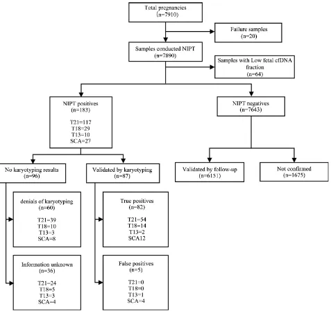
Figure 2 shows the NIPT results and clinical outcomes. In this dataset, 20
samples’ analysis was canceled due to improper cfDNA concentration, hemolysis,
quality control failure, and premature gestational age. Further, a total of 64 sam-ples failed due to low fetal fraction while the NIPT service could not provide any accurate and conclusive analysis for the 84 samples and classified as failures.
Table 1. Demographic characteristics of the study population.
Characteristic (n = 7910) Total NIPT positives (n = 183)
Maternal age (yrs) 35 (16 - 47) 36 (25 - 46)
16 - 24 yrs (n, %) 144 (1.82%) 0 (0.00%)
25 - 29 yrs (n, %) 724 (9.15%) 8 (4.37%)
30 - 34 yrs (n, %) 2123 (26.84%) 45 (24.59%)
35 - 40 yrs (n, %) 4346 (54.94%) 97 (53.01%)
>40 yrs (n, %) 573 (7.24%) 33 (18.03%)
Gestational age (wks) 15 (10 - 31) 13 (10 - 20)
First trimester (9 - 13 wks) 3469 (43.86%) 99 (54.10%)
Second trimester (14 - 27 wks) 4434 (56.06%) 84 (45.90%)
Third trimester (≥28 wks) 7 (0.09%) 0 (0.00%)
BMI 22 (15.1 - 45.8) 21 (15.6 - 34.5)
Underweight (<18.5 bmi) 363 (4.59%) 10 (5.46%)
Normal (18.5 - 22.9 bmi) 3661 (46.28%) 99 (54.10%)
Overweight (23 - 24.9 bmi) 1315 (16.62%) 26 (14.21%)
Obese Class I (25 - 29.9 bmi) 1429 (18.07%) 22 (12.02%)
Obese Class II (30 - 35 bmi) 338 (4.27%) 5 (2.73%)
Obese Class III (>35 bmi) 53 (0.67%) 0 (0.00%)
Unknown 751 (9.49%) 21 (11.48%)
Singleton pregnancy 7792 (98.51%) 179 (97.81%)
Twin pregnancy 118 (1.49%) 4 (2.19%)
Other high-risk factors
1 risk factor 7142 (90.29%) 154 (84.15%)
Serum screening test High risk group 2029 (28.41%) 47 (30.52%)
Ultrasound abnormal findings 238 (3.33%) 26 (16.88%)
Advanced maternal age 2928 (41.00%) 43 (27.92%)
Family history of aneuploidy 68 (0.95%) 1 (0.65%)
Other 1879 (26.31%) 37 (24.03%)
More than 2 risk factors 734 (9.28%) 29 (15.85%)
Unknown 34 (0.43%) 0 (0.00%)
Figure 2. Summarizing study participants and NIPT results and clinical outcome. A total of 7910 pregnant women underwent NIPT in several obstetrical center. Among 7890 pregnancies, 64 pregnancies with low fetal fraction were excluded and 183 pregnancies were positive and 7643 pregnancies were negative. Only 87 of the positive samples were further analyzed and 82 of them were true positive samples.
3.2. Follow-Up Investigation of NIPT Positive Cases
Among the 7826 samples obtained from the NIPT results, 185 (2.36%) were re-ported to have chromosomal aneuploidy and 87 of them (47.54%) were validated by karyotyping. Of the 87 validated samples, 82 are TP and 5 are FP (Figure 2). Further analysis of the five FPs, as listed in Table 2, depicts that the false posi-tives are caused due to the partial duplication of maternal chr21 (in case of T21) and mosaic aneuploidy of maternal chromosome or probably Confined placental mosaicism (in case of XO). However, all these 5 false-positive cases were classi-fied as normal after performing size-based fetus and maternal cfDNA analysis.
Several rare cases have been found in our TP cases, e.g. one T21 case (Table 2) was detected as Robertsonian translocation even though the karyotyping test had diploid, one T18 case was proven as pseudoisodicentric (psuidic) [7]. In one sample of mosaic XO, 46, X, del (Xp) was observed in 24 out of 30 cells and 46, X, i (Xq) karyotype was observed in the remaining 6 cells (Table 2).
Clinical information for NIPT positive samples is shown in Table 1. The NIPT positive sets were higher in the mean maternal age and more than two risk factors as compared to the total set including NIPT negative results.
3.3. Follow-Up Investigation of NIPT Negative Cases
Assuming that 6151 pregnancies over 10 weeks of gestation gave a birth, no false negatives were reported at the time of writing this paper, no abnormal com-ments have been reported from the monthly diagnosis for the remaining uncon-firmed samples.
3.4. Clinical Performance of NIPT Results
[image:8.595.210.536.461.712.2]A total of 6238 cases including 87 NIPT positive results validated by karyotyping and 6151 NIPT negative results confirmed by insurance policy, were analyzed for sensitivity, specificity, PPV, accuracy, and incidence analysis in Table 3. There were 54 T21, 14 T18, 3 T13, and 7643 euploid cases. T18, T13, XXX, XXY and XYY had 100% sensitivity, specificity, PPV and accuracy while T21 had 99.98% specificity and XO had 99.94% specificity. The overall sensitivity, speci-ficity, PPV, and accuracy of all chromosomal abnormalities were 100.00%,
Table 2. Summary of clinical tracing results of false-positive and cytogenetic variant
NIPT results.
False-positive NIPT results (n = 3)
Results MA (yrs) (wks) GA (bmi) BMI (%) FF confirmation Cytogenetic Remark
T13 37 11 Overweight (23.28) 20.7 46, XY Vanishing Twin
XO 36 11 + 3 Normal (20.59) 5.6 46, XX Family history of aneuploidy
XO 35 17 + 1 Normal (19.7) 9.9 46, XX
Cytogenetic variant NIPT results (n = 3)
T18 34 11 + 5 Normal (21.9) 12.4 46, XY, psu idic (18) (p11.31) Ultrasound result (NT 3.9 mm)
T21 41 13 Unknown 10.2 46, XY, +21, der (21; 21) (q10; q10) Ultrasound result (NT 3.9 mm)
XO 37 16 + 5 Overweight (23.7) 7.2 [24]/46, X, i (X) (q10) [6] 46, XX, del (X) (p11.1)
Notes: MA, maternal age; yrs, years; GA, gestational age; wks, weeks; BMI, body mass index; FF, fetal fraction.
Table 3. Overall performance of NIPT results in Korean pregnant women.
Positive NIPT cases with confirmatory prenatal karyotype (n = 6238)
Trisomy TP (n) (n) FP FN (n) Sensitivity % (95% CI) Specificity % (95% CI) (95% CI) PPV % Accuracy % (95% CI) Incidence % (95% CI)
T21 54 1 0 (93.40 - 100) 100 (99.91 - 100) 99.98 (88.38 - 99.74) 98.18 (99.91 - 100) 99.98 (0.66 - 1.14) 0.87
T18 14 0 0 (76.84 - 100) 100 (99.94 - 100) 100 100 (99.94 - 100) 100 (0.12 - 0.38) 0.22
T13 2 0 0 (15.81 - 100) 100 (99.94 - 100) 100 100 (99.94 - 100) 100 (0.00 - 0.12) 0.03
XO 6 4 0 (54.07 - 100) 100 (99.84 - 99.98) 99.94 (36.03 - 79.98) 60 (99.84 - 99.98) 99.94 (0.04 - 0.21) 0.10
XXX 1 0 0 (2.5 - 100) 100 (99.94 - 100) 100 100 (99.94 - 100) 100 (0.00 - 0.09) 0.02
XXY 4 0 0 (39.76 - 100) 100 (99.94 - 100) 100 (39.58 - 100) 100 (99.94 - 100) 100 (0.02 - 0.16) 0.06
XYY 1 0 0 (2.5 - 100) 100 (99.94 - 100) 100 100 (99.94 - 100) 100 (0.00 - 0.09) 0.02
Total 82 5 0 (95.6 - 100) 100 (99.81 - 99.97) 99.92 (87.23 - 97.52) 94.25 (99.81 - 99.97) 99.92 (1.05 - 1.63) 1.31
All positive NIPT cases (n = 6238)
T21 117 1 0 (96.90 - 100) 100 (99.93 - 100) 99.99 (94.28 - 99.88) 99.15 (99.93 - 100) 99.99 (1.24 - 1.79) 1.50
T18 29 0 0 (88.06 - 100) 100 (99.95 - 100) 100 100 (99.95 - 100) 100 (0.25 - 0.54) 0.37
T13 9 0 0 (66.37 - 100) 100 (99.95 - 100) 100 100 (99.95 - 100) 100 (0.05 - 0.22) 0.12
XO 12 4 0 (73.54 - 100) 100 (99.87 - 99.99) 99.95 (52.97 - 88.88) 75 (99.87 - 99.99) 99.95 (0.08 - 0.27) 0.15
XXX 5 0 0 (47.82 - 100) 100 (99.95 - 100) 100 100 (99.95 - 100) 100 (0.02 - 0.15) 0.06
XXY 6 0 0 (54.07 - 100) 100 (99.95 - 100) 100 100 (99.95 - 100) 100 (0.03 - 0.17) 0.08
XYY 2 0 0 (15.81 - 100) 100 (99.95 - 100) 100 100 (99.95 - 100) 100 (0.00 - 0.09) 0.03
Total 180 5 0 (97.97 - 100) 100 (99.85 - 99.98) 99.93 (93.75 - 98.86) 97.30 (99.85 - 99.98) 99.94 (1.98 - 2.66) 2.30
Theoretical PPV
Boundaries condition Trisomy 21 (% (n)) Trisomy 18 (% (n)) Trisomy 13 (% (n)) (% (n)) XO (% (n)) XXX (% (n)) XXY (% (n)) XYY (% (n)) Total Lower boundary
All unconfirmed cases
considered as FP 46.15 (54/117) 48.28 (14/29) (2/9) 22 (6/12) 50 (1/5) 20 66.67 (4/6) (1/2) 50 45.56 (82/180) Upper boundary
All unconfirmed cases
considered as TP 99.15 (116/117) 100 (29/29) 90 (9/9) 66.7 (8/12) (5/5) 100 (6/6) 100 (2/2) 100 97.22 (175/180)
Notes: CI, confidence interval; NIPT, noninvasive prenatal testing; TP, true positive; FP, false positive; FN, false negative; PPV, positive predictive value.
99.92%, 94.25%, and 99.92% respectively. The three most frequent incidents were T21, T18, and XO in that order.
Assuming that all positive results are TP, the overall specificity, sensitivity, PPV, and accuracy of all chromosomal abnormalities were 99.93%, 100.00%, 97.30%, and 99.94%, respectively.
A theoretical PPV was also calculated under the two boundary conditions that all invalidated NIPT positive cases were either assumed to be a TPs or an FPs (Table 3).
This provided the range of PPV for T21, T18, T13, XO, XXX, XXY, and XYY as 46% - 100%, 48% - 100%, 30% - 90%, 50% - 85%, 20% - 100%, 66% - 100% and 50% - 100%, respectively.
4. Discussion
4.1. Summary
This study summarizes the demography result of the clinical use of NIPT during Nov. 2015 to Feb. 2018 in South Korea. Among 7910 patient samples, 98.9% (7826 of 7910) were analyzed successfully while 0.3% (20 of 7910) failed by QC thresholds and 0.8% (64 of 7910) failed due to low fetal fraction. At analysis lev-el, inconclusive or failed reports were generated mostly due to the low fetal frac-tion and the higher sequencing bias.
In our patient cohort, we saw a positive correlation between maternal age and the risk probability. The risk for aneuploidy starts to increase at the early 40s and the highest risk samples are in the oldest pregnancy age group.
The performance of NIPT in detecting autosomal trisomy and sex chromo-some aneuploidy was compared in Table 3. Of the 7643 negative cases, 6151 samples are considered to be true negative since they have already passed their gestation period till the time of manuscript preparation and no adverse report has been found in follow-up. The lack of karyotyping confirmation of all positive results limits the generation of sufficient performance statistics.
The adequate proportion of fetus originated fragments in maternal plasma is the key factor to make a reliable analysis result, and an approximately minimum 4% fetus fraction is required for accurate analysis [12]. In our study, fetal frac-tion decreased with increasing maternal weight, and more than 56.25% of sam-ples with lower fetal fraction had a Body Mass Index (BMI) of more than 23.0. Accurate determination of fetal fraction is very important for NIPT screening test as it helps the clinician to make an informed decision. Identification of the FF for the male fetus is easier and more accurate as the algorithm can extract the male-specific chromosome Y cfDNA from the sample and calculate the norma-lized value to predict the FF. In this study, a correlation study between the FF values obtained both from the chromosome Y-based FF and fragment size based linear regression algorithm was performed, which revealed a high (0.85) r2
coef-ficient of correlation. A high coefcoef-ficient of correlation is significant as we can determine the FF of female fetus using the fragment size based linear regression
algorithm. However, it is necessary to improve the accuracy of the FF detection to make it accurate, which will be the main area of focus in our future studies.
In the case of Turner syndrome, the false-positive rate can be high due to the maternal XO mosaicism or loss of maternal X chromosome by aging. In our study, we could observe some samples have the loss of X chromosome in mater-nal DNA. We applied cfDNA size separation method to reduce the false decision rate by distinguishing fetal X monosomy from maternal mosaic aneuploidy.
4.2. Limitations of Study
Among 183 patients, we did not receive karyotyping results for 96 samples, of which 60 participants refused for karyotyping while no follow-up information can be obtained for the remaining 36 cases. Many NIPT algorithms have false negative results for samples with low fetal fraction, so if we have a high z-score in the negative case with a strong fetus trisomy signal, we report it as “suspected” to confirm using the karyotyping. Since the maternal mosaic aneuploidy as well as the low fetal fraction may affect the overall result, karyotyping confirmation is essential for all the positive and suspected cases.
5. Conclusion
This study reports a high statistical confidence with the improved method of NIPT testing. The relatively high cost of NIPT and the absence of reimburse-ment in South Korea are main drawbacks for pregnant women. It is important that NIPT should be used effectively and ethically to be an ultimate first tier screening service. In clinics, patients should be given precise information about the limitations of NIPT testing, and the appropriate genetic counseling is needed. At the same time, NIPT service providers must continually improve ex-perimental methods and algorithms to reduce the probability of false positives and false negatives. In addition, it is crucial that all positives cases must be con-firmed by fetal karyotyping before the termination of pregnancy.
Conflicts of Interest
The authors declare no conflicts of interest regarding the publication of this pa-per.
References
[1] Savva, G.M., Morris, J.K., Mutton, D.E. and Alberman, E. (2006) Maternal Age-Specific Fetal Loss Rates in Down Syndrome Pregnancies. Prenatal Diagnosis, 26, 499-504. https://doi.org/10.1002/pd.1443
[2] Morris, J.K., Mutton, D.E. and Alberman, E. (2002) Revised Estimates of the Ma-ternal Age Specific Live Birth Prevalence of Down’s Syndrome. Journal of Medical Screening, 9, 2-6. https://doi.org/10.1136/jms.9.1.2
[3] Haddow, J.E. (1990) Prenatal Screening for Open Neural Tube Defects, Down’s Syndrome, and Other Major Fetal Disorders. Seminars in Perinatology, 14, 488-503. [4] Fairbrother, G., Johnson, S., Musci, T.J. and Song, K. (2013) Clinical Experience of
Noninvasive Prenatal Testing with Cell-Free DNA for Fetal Trisomies 21, 18, and 13, in a General Screening Population. Prenatal Diagnosis, 33, 580-583.
https://doi.org/10.1002/pd.4092
[5] Manotaya, S., Xu, H., et al. (016) Clinical Experience from Thailand: Noninvasive Prenatal Testing as Screening Tests for Trisomies 21, 18 and 13 in 4736 Pregnan-cies. Prenatal Diagnosis, 36, 224-231. https://doi.org/10.1002/pd.4775
[6] Taneja, P.A., Snyder, H.L., et al. (2015) Noninvasive Prenatal Testing in the General Obstetric Population: Clinical Performance and Counseling Considerations in over 35000 Cases. Prenatal Diagnosis, 36, 237-243. https://doi.org/10.1002/pd.4766 [7] Fan, H.C., Blumenfeld, Y.J., Chitkara, U., Hudgins, L. and Quake, S.R. (2008)
Non-invasive Diagnosis of Fetal Aneuploidy by Shotgun Sequencing DNA from Maternal Blood. Proceedings of the National Academy of Sciences of the United States of America, 105, 16266-16271. https://doi.org/10.1073/pnas.0808319105
[8] Kwon, H.J., Goyal, A., et al. (2017) Multiple z-Score Based Method for Noninvasive Prenatal Test Using Cell-Free DNA in Maternal Plasma. Open Journal of Genetics, 7, 1-8. https://doi.org/10.4236/ojgen.2017.71001
[9] Chan, K.C., Zhang, J., Hui, A.B., Wong, N., Lau, T.K., Leung, T.N., Lo, K.W., Huang, D.W. and Lo, Y.M. (2004) Size Distributions of Maternal and Fetal DNA in Maternalplasma. Clinical Chemistry, 50, 88-92.
https://doi.org/10.1373/clinchem.2003.024893
[10] Mangs, A.H. and Morris, B.J. (2007) The Human Pseudoautosomal Region (PAR): Origin, Function and Future. Current Genomics, 8, 129-136.
https://doi.org/10.2174/138920207780368141
[11] Skaletsky, H., Kuroda-Kawaguchi, T., Minx, P.J., Cordum, H.S., Hillier, L., Brown, L.G., et al. (2003) The Male-Specific Region of the Human Y Chromosome Is a Mosaic of Discrete Sequence Classes. Nature, 423, 825-837.
https://doi.org/10.1038/nature01722
[12] Wang, E., Batey, A., Struble, C., Musci, T., Song, K. and Oliphant, A. (2013) Gesta-tional Age and Maternal Weight Effects on Fetal Cell-Free DNA in Maternal Plas-ma. Prenatal Diagnosis, 33, 662-666.