ORIGINAL RESEARCH
Diffusion Anisotropy Changes in the Brains of
Professional Boxers
L. Zhang L.A. Heier R.D. Zimmerman B. Jordan A.M. Ulug˘
BACKGROUND AND PURPOSE: Professional boxing may result in brain injury. We hypothesize that quantitative MR diffusion imaging may be useful in determining early white matter changes.
METHODS: Forty-nine professional boxers (age 30⫾4.5 years) and 19 healthy control subjects (age 32⫾9.5 years) were imaged on a clinical 1.5T scanner. None of the subjects had neurologic disorder or deficit. The average diffusion constant (Dav) and diffusion anisotropy (FA) were determined pixel by pixel. Regional diffusion measurements were done in the corpus callosum (CC) and internal capsule (IC). The whole brain diffusion constant (BDav) was also determined. Studentttest was used to analyze the diffusion difference between boxers and the healthy control subjects.P⬍.05 was considered statistically significant.
RESULTS: Of the 49 professional boxers, 42 had normal conventional MRIs. The remaining 7 boxers had abnormal MR imaging findings dominated by nonspecific white matter disease. There was a significant difference in diffusion and anisotropy measurements in all the boxers compared with the healthy control subjects. In the boxer group, BDavincreased and FA decreased significantly in the CC and posterior limb of IC. The measured FA and Davinversely correlated in regions of CC and IC in boxers but not in healthy control subjects. BDavalso robustly correlated with both FA and Davin the splenium of CC in boxers.
CONCLUSION:Increased BDavand the decreased FA in the CC and IC may represent preclinical signs of subtle brain injury in professional boxers.
P
rofessional boxing is a controversial sport, and research has raised ethical concerns1based on biomedical findings indicating traumatic brain injury (TBI).2-12Long-term TBIdue to boxing (usually termed “dementia pugilistica,”9
“punch drunk,” and formally, chronic traumatic encephalop-athy) represents a cumulative process of repetitive head blows in contact sports.2,8-10MR imaging has been widely used to
reveal brain damage including atrophy, focal or diffuse non-specific white matter lesions, hemorrhage, infarcts, and demy-elination.13,14These microstructural changes may affect the diffusivity of water molecules within the extracellular space.15
Routine MR imaging methods may be negative or nonspecific in symptomatic patients. Diffusion tensor imaging (DTI) is very sensitive to the microscopic motion of water molecules within the extracellular space, so it can be used to characterize abnormalities of the white matter21-23in many diseases,
in-cluding early changes of TBI as seen in humans and in animal research studies.24-30Diffusion measurements were found to be reflective of the clinical severity and prognosis of TBI,18
suggesting that diffusion parameters can be used as markers in TBI evaluation. DTI also allows quantitative analysis of the directional diffusion properties of water molecules, thus indi-cating the integrity of organized tissue microstructure,26,27
which is used in many situations for the measurement of the directional diffusion of white matter tracts relating to tissue orientation and integrity in white matter.28,29-31
White matter is of concern in professional boxing because of its anatomic predominance and physical function. Early
detection of white matter damage may help in improving the efficacy of neuroprotective treatment of professional boxers. The purpose of this study is to find a convenient marker for the white matter changes induced by professional boxing using DTI, which may be useful in monitoring the neurologic integ-rity of boxers in the long run. Such a marker may also useful in evaluating patients with TBIs not caused by boxing, such as those sustained in car crashes.
Methods
Subjects
Forty-nine professional boxers (age, 30⫾4.5 years) and 19 healthy control subjects (age, 32⫾9.5 years) were included in this study. All the subjects were free of neurologic disease. The MR imaging was performed on a 1.5T clinical MR scanner with a quadrature head coil. Clinical MR images included: axial T1-weighted (repetition time [TR]/echo time [TE] 500 ms/minimum), axial T2-weighted (TR/TE, 4000 ms/102 ms); fluid-attenuated inversion recovery (FLAIR) (TR/ TE/inversion time [TI], 10,000/162/2200 ms; matrix, 256⫻192), and diffusion-weighted imaging (TR/TE, 10,500 ms/minimum; matrix, 128⫻128; section thickness, 5 mm). There were no gaps between the
Received July 7, 2005; accepted after revision December 22.
From the Department of Radiology (L.Z., L.A.H., R.D.Z., A.M.U.), Weill Medical College of Cornell University, New York, NY; Burke Rehabilitation Hospital (B.J.), White Plains, NY; and New York State Athletic Commission (B.J.), New York, NY.
Address correspondence to: Aziz M. Ulug˘, PhD, Department of Radiology, Box 141, Weill Medical College of Cornell University, 1300 York Ave, New York, NY 10021; e-mail: [email protected]
[image:1.585.301.537.325.441.2]sections. The conventional MR images were evaluated by the staff neuroradiologists. The study was approved by the institutional review board.
The subjects were also imaged using a single shot, echo-planar DTI sequence. Diffusion-weighted images from 26 gradient direc-tions were acquired. An additional 6 images without diffusion weighting were also collected. The diffusion tensor was deter-mined on a pixel-by-pixel basis. In the DTI protocol, the maxi-mumb-value per axis was 820s/mm2. Using TR/TE, 12,000 s/100 ms, 30 contiguous sections with 5-mm thickness covering the en-tire brain were acquired. The FOV was 22 cm and imaging acqui-sition matrix was 128⫻128. The detailed methodology was re-ported elsewhere.26
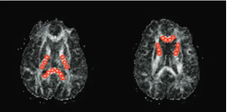
In all subjects, a computer program was used to calculate the global BDavfrom the diffusion distribution histograms as reported previously.32This program distributed the pixels into 250 bins with a bin width of 0.02 (10⫺5cm2/s). This histogram was fitted to a triple Gaussian curve using commercial software (KaleidaGraph; Abelbeck/ Synergy Software, Reading, Pa). The average diffusion constant (Dav) and diffusion anisotropy maps (FA) were then calculated pixel by pixel. Region of interest (ROI) measurements were done on these diffusion maps. The ROIs were drawn manually inside the structures studied. The regional Davand FA of the genu and splenium of the corpus callosum (CC) and the anterior and posterior limbs of the internal capsule (IC) were measured (Fig 1).
Studentttest was used to determine the diffusion difference be-tween boxers and the healthy control subjects.P⬍.05 was considered statistically significant.
Using diffusion tensor tractography, we also visualized overall trackable white matter fibers in all subjects. We visually compared the tractography results of the boxers with those of the control subjects.
Results
Conventional MR Imaging Findings
Forty-two (86%) boxers had a normal clinical MR imaging. Four (8%) had focal or diffuse nonspecific subcortical white
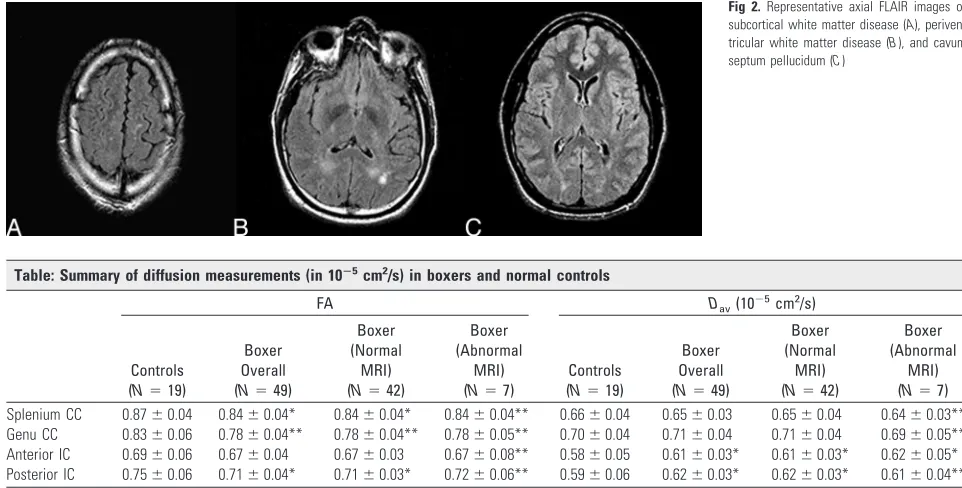
matter disease or periventricular white matter disease, mostly in the frontal and parietal lobes. One (2%) had chronic hem-orrhage, 1 (2%) had all the above, and 1 (2%) had a cavum septum pellucidum (Fig 2).
Diffusion Coefficient and Anisotropy Measurements
Quantitative diffusion measurements are summarized in the Table and illustrated in Fig 3. The BDavwas increased by 2.6%
(P⬍.05) in all the boxers and in 2.4% (P⬍.05) of boxers with normal results on MR imaging (P⬍.05), in agreement with a previous study done on a different boxer population.16
The splenium of the CC had the highest anisotropy mea-sured by FA, followed by genu in both boxers and healthy control subjects. The regional diffusion anisotropy mea-surements of all the boxers showed decreased anisotropy in the genu of the CC (6.2%,P⬍.005), splenium of CC (3.2%,
P ⬍ .05), anterior limb of the IC (3.8%, P ⬎ .05), and posterior limb of the IC (5.4%, P⬍.05) compared with healthy control subjects (Fig 2). In boxers with normal MR imaging results, the regional anisotropy measurements also showed the same trend of decrease with the same signifi-cance level.
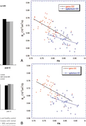
Pearson correlation analysis revealed that the regional diffusion changes of Davsignificantly correlate to the
cor-responding FA (Fig 4A) in the genu (P⬍.00001), the sple-nium (P⬍.01) of the CC, and in the anterior IC (P⬍.001). The same analysis revealed similar robust correlations be-tween the Dav and FA in the genu (P ⬍ .0005) and the
splenium (P ⬍ .05) of CC in boxers with a normal MR imaging (Fig 4B). The correlation between FA and Davin
the anterior IC was significant (P⬍.05). The same analysis found no correlation between the measurements of FA and Davin healthy control subjects. BDavwas significantly
corre-lated to the FA (negatively) and the Dav(positively) of the
sple-nium in boxers, whereas no such correlation was found in the healthy control subjects.
Fig 2.Representative axial FLAIR images of subcortical white matter disease (A), periven-tricular white matter disease (B), and cavum septum pellucidum (C)
Table: Summary of diffusion measurements (in 10ⴚ5cm2/s) in boxers and normal controls
FA Dav(10⫺
5cm2/s)
Controls (N⫽19)
Boxer Overall (N⫽49)
Boxer (Normal
MRI) (N⫽42)
Boxer (Abnormal
MRI) (N⫽7)
Controls (N⫽19)
Boxer Overall (N⫽49)
Boxer (Normal
MRI) (N⫽42)
Boxer (Abnormal
MRI) (N⫽7) Splenium CC 0.87⫾0.04 0.84⫾0.04* 0.84⫾0.04* 0.84⫾0.04** 0.66⫾0.04 0.65⫾0.03 0.65⫾0.04 0.64⫾0.03** Genu CC 0.83⫾0.06 0.78⫾0.04** 0.78⫾0.04** 0.78⫾0.05** 0.70⫾0.04 0.71⫾0.04 0.71⫾0.04 0.69⫾0.05** Anterior IC 0.69⫾0.06 0.67⫾0.04 0.67⫾0.03 0.67⫾0.08** 0.58⫾0.05 0.61⫾0.03* 0.61⫾0.03* 0.62⫾0.05* Posterior IC 0.75⫾0.06 0.71⫾0.04* 0.71⫾0.03* 0.72⫾0.06** 0.59⫾0.06 0.62⫾0.03* 0.62⫾0.03* 0.61⫾0.04**
Note:—FA indicates diffusion anisotropy;Dav, average diffusion constant; CC, corpus callosum; IC, internal capsule. Values are means (SD). *P⬍.05; **P⬍.005, compared with the normal controls.
BRAIN
ORIGINAL
[image:2.585.50.531.42.286.2]DTI-Based Tractography
We also visualized the white matter fiber tracts over the entire brain using diffusion tensor-based tractography in all boxers. DTI-based tractography showed less trackable white matter fibers in the CC as well as the whole brain in boxers compared with healthy control subjects (Fig 5). Two boxers had repeated studies more than 6 months apart. Both repeated studies showed decreased trackable fibers in the whole brain and CC compared with their initial scans (Fig 6).
Discussion
It is well established that active boxing exposes professional boxers to intracranial injury33with a wide variety of pathol-ogy, including that seen in Alzheimer disease,34though most
of the imaging findings are normal or borderline.11,13,16,35,36
Professional boxers included in this study showed a variety of brain findings ranging from normal (86%) to nonspecific white matter disease mainly affecting the periventricular fron-tal-parietal white matter.7-10,16
In this study, we focused on the CC and IC for 2 reasons:
(1) both CC and IC are oriented and tight white matter bun-dles, and (2) they are also the largest white matter tracts in the brain. For the diffusion analysis, in all boxers, BDavand Davin
both limbs of the IC increased significantly compared with those of the healthy control subjects. The measured increase in BDavof this study is in agreement with a previous study done
on a different boxer population.16Considering that most of
the conventional MR images were normal and the abnormal conventional MR images only had nonspecific findings, this increase in measured diffusion values suggests that DTI is more sensitive than routine MR imaging in detecting the early response to brain injury induced by boxing; hence, the quan-titative diffusion measurements may be used to monitor the brain.
The splenium of CC has the highest anisotropy as mea-sured by FA followed by the genu of CC in both boxers and
Fig 3.Comparison of FA (A) and Dav(B) measurements between boxers and healthy control subjects. Compared with healthy control subjects, boxers overall and boxers with normal MR imaging have decreased FA in splenium CC (P⬍.05), genu CC (P⬍.005), and posterior IC (P⬍.05) and increased BDav(P⬍.05) and Davin both the anterior and posterior limbs
of IC (P⬍.05). Fig 4.Robust correlations of Davand FA in CC of all boxers (A) and boxers with normal MR imaging (B). For the genu CC, Dav⫽1.23⫺0.66⫻FA,r⫽0.59,P⬍.001; for the splenium CC, Dav⫽0.95⫺0.35⫻FA,r⫽0.37,P⬍.01.
B, The group of boxers with normal MR imaging. For the genu CC (dots), Dav⫽1.19⫺ 0.61⫻FA,r⫽0.55,P⬍.001; for the splenium CC (circles): Dav⫽0.94⫺0.34⫻FA,
[image:3.585.231.527.42.474.2] [image:3.585.57.302.46.431.2]healthy control subjects, which agrees with a previous re-port.37Well myelinated fibers and more oriented bundles may contribute to its high anisotropy.38The robust correlation
be-tween the regional diffusion characteristics of Davand the FA
of the splenium to the BDavindicates its remarkable
involve-ment in the brain injury, which contributes to the increased global diffusion in the boxers.
Reduced anisotropy in DTI reflects the impaired neurofila-ment alignneurofila-ment, the integrity of the myelin sheath, and axonal loss in highly oriented tissue.26,39,40The decreased FA in the CC and posterior limb of IC in boxers overall and in boxers with normal MR imaging suggests early impairment of the axon fibers going through the CC and IC. Water diffusion is less curbed by a damaged myelin sheath. The discrepancy that diffusion did not increase significantly compared with healthy control subjects, whereas the anisotropy decreased in the CC, suggests that FA is more sensitive to the pathologic involve-ment of the CC in chronic traumatic encephalopathy. This discrepancy was not found in the IC in this study, though anisotropy in the IC has been reported to be sensitive in de-tecting other pathologic processes.41This suggests that (1) the
severity of the diffusion anisotropy changes may vary in dif-ferent pathologic situations; (2) the sensitivity of anisotropy changes measured by FA is partially dependent on the intrinsic microstructural characteristics of the local anatomy; (3) the CC is more susceptible to head blows in boxing, and the dif-fusion changes occur earlier in this area; and (4) the difdif-fusion anisotropy changes may provide additional markers to evalu-ate the boxers’ neurologic function.
In all the boxers, FA showed robust negative correlation with the corresponding Davin the entire CC and in both limbs
of IC. Unlike the boxers, no correlation was found between FA and Dav in healthy control subjects. Where fiber tracts are
damaged, water diffusion is less restricted, which may result in increased Davand decreased anisotropy.42For the boxers with
normal MR imaging results, the FA-Davcorrelations are only
significant in the CC. This suggests that the CC is more sus-ceptible to the damage caused by boxing.
All boxers had decreased trackable fibers globally com-pared with healthy control subjects. This suggests global white matter damage. The marked decrease in the number of
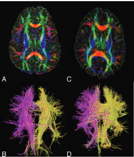
track-Fig 5.Diffusion anisotropy maps and diffusion tensor imaging-based white matter trac-tography of a representative boxer (AandB; 27 years old) and a control (CandD; 29 years old). Intensity is proportional to anisotropy and color shows the direction. The boxer has decreased anisotropy in CC and anterior and posterior limb of IC compared with the healthy control subjects. Fiber tracking showed overall fewer trackable white matter fibers in this boxer’s brain (B) compared with that of a control subject (D). The difference in fibers through the corpus callosum is particularly striking.
[image:4.585.54.286.41.312.2] [image:4.585.300.534.43.542.2]able fibers in CC suggests that CC is more susceptible to the blows to the head that characterize this sport.
Two boxers had repeat studies within 1 and 2 years, respec-tively. The second study of both boxers had decreased track-able fibers within the whole brain compared with their initial scans, with more marked deficit in the CC. Because we have not scanned control subjects serially during the same time period, we cannot exclude the possibility that the effects are due to experimental instability.
Conclusions
Quantitative diffusion analysis can detect diffusion anisotropy changes in the brains of professional boxers even when the clinical MR images are normal. Increased BDavmay represent
overall microstructural impairment of the central nervous sys-tem as a result of chronic traumatic brain injury. The anisot-ropy measured by FA is more sensitive than diffusion mea-sured by Dav in reflecting earlier impairment of the white
matter. The regional decreased anisotropy measurements in the boxers point to specific white matter injury. The paucity of white matter fibers on the fiber tracking points to subtle but widespread white matter damage that is not detectable on clin-ical MR images. Quantitative DTI shows promise as a clinclin-ical marker for early TBI in boxers. It is also expected to be a useful tool in the study of TBI in general.
References
1. Spriggs M.Compulsory brain scans and genetic tests for boxers– or should boxing be banned?J Med Ethics2004;30:515–16
2. Ross RJ, Casson JR, Siegel O, et al.Boxing injuries: neurologic, radiologic and neuropsychologic evaluation.Clin Sports Med1987;6:41–51
3. Jordan BD, Relkin NR, Ravdin LD, et al.Apolipoprotein E epsilon4 associated with chronic traumatic brain injury in boxing.JAMA1997;278:136 – 40 4. Moseley IF.The neuroimaging evidence for chronic brain damage due to
box-ing.Neuroradiology2000;42:1– 8
5. Rodriguez G, Vitali P, Nobili F.Long-term effects of boxing and judochoking techniques on brain function.Ital J Neurol Sci1998;19:367–72
6. Nicholl J, Coleman P, William B.The epidemiology of sports and exercise related injury in the United Kingdom.Br J Sports Med1995;29:232–38. 7. Holzgraefe M, Lemme W, Funke W, et al.The significance of diagnostic
imag-ing in acute and chronic brain damage in boximag-ing: a prospective study in am-ateur boxing using magnetic resonance imaging (MRI).Int J Sports Med1992; 13:616 –20
8. Jordan BD.Chronic traumatic brain injury associated with boxing.Semin Neurology2000;20:179 – 85
9. Bodensteiner J, Schaefer G.Dementia pugilistica and cavum septi pellucidi: born to box.Sports Med1997;24:361– 65
10. Jordan BD.Chronic traumatic injury associated with boxing.Semin Neurol
2000;20:179 –18
11. Jordan BD, Zimmerman RD.Computerized tomography and magnetic reso-nance imaging comparison in boxers.JAMA1990;263:1670 –74
12. Bigler ED.Quantitive magnetic resonance imaging in traumatic brain injury.
J Head Trauma Rehabil2001;16:1–21
13. Casson IR. Siegel O, Sham R, et al.Brain damage in modern boxers. JAMA
1984;251:2663– 67
14. Corsellis JA, Bruton CJ, Freeman-Browne D.The aftermath of boxing.Psychol Med1973;3:270 –303
15. Le Bihan D.Molecular diffusion, tissue microdynamics and microstructure.
NMR Biomed1995;8:375– 86
16. Zhang L, Ravdin LD, Relkin N, et al.Increased diffusion in the brain of profes-sional boxers: a preclinical sign of traumatic brain injury?AJNR Am J Neuro-radiol2003;24:52–57
17. Jones D, Dardis R, Ervine M, et al.Cluster analysis of diffusion tensor magnetic resonance images in human head injury.Neurosurgery2000;47:306 –14 18. Schaefer PW, Huisman TAGM, Sorensen AG, et al.Diffusion-weighted MR
imaging in closed head injury: high correlation with initial Glasgow Coma scale score and score on modified Rankin scale at discharge.Radiology2004; 233:58 – 66
19. Le Bihan D, Turner R, Douek P, et al.Diffusion MR imaging: clinical applica-tions.AJR Am J Roentgenol1992;159:591–99
20. Assaf Y, Holokovsky A, Berman E, et al.Diffusion and perfusion magnetic resonance imaging following closed head injury in rats.J Neurotrauma1999; 16:1165–76
21. Mori S, van Zijl PC.Fiber tracking: principles and strategies—a technical review.NMR Biomed2002;15:468 – 80
22. Yamada K, Kizu O, Mori S, et al.Brain fiber tracking with clinically feasible diffusion-tensor MR imaging: initial experience.Radiology2003;227:295–301 23. Wakana S, Jiang H, Nagae-Poetscher LM, et al.Fiber tract-based atlas of human
white matter anatomy.Radiology2004;230:77– 87
24. Mamata H, Jolesz FA, Maier SE.Characterization of central nervous system structures by magnetic resonance diffusion anisotropy.Neurochem Int2004; 45:553– 60
25. Sotak CH.Nuclear magnetic resonance (NMR) measurement of the apparent diffusion coefficient (ADC) of tissue water and its relationship to cell volume changes in pathological states.Neurochem Int2004;45:569 – 82
26. Ulug AM, Moore DF, Bojko AS, et al.Clinical use of diffusion tensor imaging for diseases causing neuronal and axonal damage.AJNR Am J Neuroradiol
1999;20:1044 – 48
27. Lee SK, Kim DI, Mori S, et al.Diffusion tensor MRI visualizes decreased sub-cortical fiber connectivity in focal sub-cortical dysplasia.Neuroimage2004;22: 1826 –29
28. Yamada K, Mori S, Nakamura H, et al.Fiber-tracking method reveals sensori-motor pathway involvement in stroke patients.Stroke2003;34:E159 – 62 29. Wakana S, Jiang H, Nagae-Poetscher M, et al.Fiber tract-based atlas of human
white matter anatomy.Radiology2004;230
30. Chepuri NB, Yen YF, Burdette JH, et al.Diffusion anisotropy in the corpus callosum.AJNR Am J Neuroradiol2002;23:803– 08
31. Ulug AM, van Zijl PCM.Orientation-independent diffusion imaging without tensor diagonalization: anisotropy definitions based on physical attributes of the diffusion ellipsoid.J Magn Reson Imaging1999;9:804 –13
32. Chun T, Filippi CG, Zimmerman RD, et al. Diffusion changes in the aging human brain.AJNR Am J Neuroradiol2000;21:1078 – 83
33. Roberts AH.Brain damage in boxers. A study of the prevalence of traumatic encephalopathy among ex-professional boxers.London: Pitman Medical; 1969.
34. Corsellis JAN.Boxing and the brain.BMJ1989;105–109
35. Jordan BD, Jahre C, Hauser WH, et al.CT of 388 active professional boxers.
Radiology1992;185:509 –12
36. Fernando MS, O’Brien JT, Perry RH, et al.Neuropathology Group of MRC CFAS. Comparison of the pathology of cerebral white matter with post-mor-tem magnetic resonance imaging (MRI) in the elderly brain.Neuropathol Appl Neurobiol2004;30:3853–95
37. Chepuri NB, Yen YF, Burdette JH, et al.Diffusion anisotropy in the corpus callosum.AJNR Am J Neuroradiol2002;23:803– 08
38. Moody DM, Bell MA, Challa VR.The corpus callosum, a unique white matter tract: anatomic features that may explain sparing in Binswanger disease and resistance to flow of fluid masses.AJNR Am J Neuroradiol1988;9:1051–59 39. Werring DJ, Clark CA, Barker GJ, et al.Diffusion tensor imaging of lesions and
normal-appearing white matter in multiple sclerosis.Neurology1999;52: 1626 –32
40. Pierpaoli C, Jezzard P, Basser PJ, et al. Diffusion tensor MR imaging of the human brain.Radiology1996;201:637– 48
41. Higano S, Zhong J, Shrier DA, et al.Diffusion anisotropy of the internal cap-sule and the corona radiata in association with stroke and tumors as measured by diffusion-weighted MR imaging.AJNR Am J Neuroradiol2001;22:456 – 63 42. Ulug AM, Grunewald T, Lin MT, et al.Diffusion tensor imaging in the