Original Article
Gastrointestinal stromal tumors with exon 8 c-
kit
gene
mutation might occur at extragastric sites and have
metastasis-prone nature
Takashi Ito1,2, Masahiro Yamamura3, Toshihiro Hirai4, Takashi Ishikawa5, Tatsuo Kanda6, Takuya Nakai7,
Mizuka Ohkouchi1, Yuka Hashikura1, Koji Isozaki1, Seiichi Hirota1
1Department of Surgical Pathology, Hyogo College of Medicine, Hyogo, Japan; 2Department of Pathology, Sanda
Municipal Hospital, Hyogo, Japan; 3Department of Clinical Oncology, Kawasaki Medical School, Okayama, Japan; 4Department of Surgery, Division of Gastroenterology, Kawasaki Medical School, Okayama, Japan; 5Division of
Digestive and General Surgery, Niigata University Graduate School of Medical and Dental Sciences, Niigata, Japan; 6Department of Surgery, Sanjo General Hospital, Niigata, Japan; 7Department of Surgery, Faculty of
Medicine, Kinki University, Osaka, Japan
Received September 22, 2014; Accepted November 8, 2014; Epub October 15, 2014; Published November 1, 2014
Abstract: Gastrointestinal stromal tumors (GISTs) are the most common mesenchymal tumors of the human gut. Most sporadic GISTs have somatic gain-of-function mutations of the c-kit gene. The mutations are frequently found at exon 11, sometimes at exon 9 and rarely at exon 13 or 17. Recently, exon 8 c-kit gene mutations were reported in very minor proportion of sporadic GISTs. We also found 3 GISTs with exon 8 c-kit gene mutations in approximately 1,000 sporadic GISTs examined. In the present report, we showed the clinicopathological data of those GISTs. One case had a deletion of codon 419 of aspartate, and 2 cases had a substitution of 3 amino acids of codon 417 to codon 419 to tyrosine. The former was the same mutation recently reported in 2 GIST cases, but the latter has not been reported in any GISTs. All three cases occurred at extragastric sites and two of three showed distant metasta-sis. Since the remaining case was regarded as high risk for recurrence, imatinib adjuvant treatment has been done
without evidence of metastasis. Our results confirmed the idea that exon 8 mutations are minor but actually existing
abnormalities in sporadic GISTs, and suggested that such GISTs have a feature of extragastric development and a metastasis-prone nature. Since the exon 8 mutations appeared to be really sensitive to imatinib as shown in the present case study, accurate genotyping including exon 8 of the c-kit gene is necessary in GISTs to predict response to imatinib in both the unresectable/metastatic and adjuvant settings.
Keywords: c-kit gene, exon 8, gain-of-function mutation, GIST, imatinib, sunitinib
Introduction
Gastrointestinal stromal tumors (GISTs) are the most common mesenchymal tumors of the human gut. Most GISTs express a receptor tyro-sine kinase (TK), KIT [1, 2], encoded by proto-oncogene c-kit [3-6]. Interstitial cells of Cajal (ICCs), which are present in the gastrointestinal (GI) wall and regulate the GI motility through their spontaneous impulse generation [7], are also positive for KIT. Since ICCs are the only proper cells in GI tract that express KIT, GISTs are now considered to originate from ICCs or precursor of ICCs.
KIT consists of an extracellular (EC) domain
and acute leukemias [1, 11-15]. In sporadic GISTs, most of them have somatic gain-of-func-tion mutagain-of-func-tions of the c-kit gene [1]. The muta-tions are frequently found at exon 11 encoding JM domain (70-80%), at exon 9 encoding EC domain (approximately 10%) and rarely at exon 13 encoding TK I domain and exon 17 encoding TK II domain (less than 2% each) [16, 17]. In approximately a half of c-kit gene mutation-negative GISTs, the mutations of platelet-derived growth factor receptor alpha (PDGFRA) gene which shows a quite similar structure to KIT are observed at exon 18 encoding TK II domain (approximately 10%), rarely at exon 12 encoding JM domain (less than 2%), and more rarely at exon 14 encoding TK I domain (less than 1%) [18, 19]. On the other hand, several types of germline gain-of-function mutations of the c-kit gene have been detected in approxi-mately 20 families with multiple GISTs [20-23]. Although the mutations in the familial GISTs are also most frequently detected at exon 11, one family with the mutation at exon 8 encoding EC domain, 3 families with the mutation at exon 13 and 4 families with the mutation at exon 17 have been reported [20-23]. Development of multiple GISTs with ICC hyperplasia is common-ly observed in patients with the familial GISTs, but some families have mast cell neoplasms and/or hyperpigmentation of the digital, peri-oral and perineal regions.
Imatinib is a selective TK inhibitor for KIT and PDGFRA [24], which is clinically used for treat-ment of metastatic or unresectable GISTs [25]. It generally shows a remarkable effect on most GIST cases, but its effectiveness is well-known to be dependent on the types of the c-kit and PDGFRA gene mutations [26, 27]. Most GISTs with exon 11 c-kit gene mutations show best response to imatinib treatment whereas the particular type of the PDGFRA gene mutation at exon 18, a substitution of aspartate to valine at codon 842 (Asp842Val), is resistant to imatinib [19]. Imatinib is now being used for GISTs after complete resection when they are regarded as high risk tumors for recurrence [28]. On the other hand, sunitinib and regorafenib are multi-targeted TK inhibitors. After the failure of ima-tinib treatment, suniima-tinib is administered as a second-line drug [29] and regorafenib as a third-line one in GISTs [30].
In contrast with sporadic GISTs, most of the c-kit gene mutations are present at exon 17 in sporadic mast cell neoplasms. However, some sporadic mast cell neoplasms have mutations at exon 9, at exon 11 and even at exon 8 [31]. Recently, moreover, exon 8 c-kit gene muta-tions were also reported in very minor propor-tion of sporadic GIST cases [32]. Types of exon 8 c-kit gene mutations reported in mast cell neoplasms are various [31], but deletion of aspartate at codon 419 (Del-Asp419) is the only type of exon 8 c-kit gene mutation report-ed in GISTs [32]. In the present study, we exam-ined whether GISTs have exon 8 c-kit gene mutations in approximately 1,000 sporadic cases, and found 3 such cases. One case had Del-Asp419 and two had substitution of 3 amino acids of codon 417 to codon 419 to tyro-sine, hereafter designated as ThrTyrAsp (417-419) Tyr. Here, we showed clinicopathological features of these GISTs.
Materials and methods
Analyses of c-kit and PDGFRA gene mutations
[image:2.612.89.289.73.260.2]We have analyzed c-kit and PDGFRA gene mutations in approximately 1,000 authentic GIST cases. When fresh-frozen samples were available, total RNA was extracted using RNeasy Mini Kit (QIAGEN, Valencia, CA). Almost all coding regions of the c-kit and PDGFRA genes were amplified by polymerase chain
Table 1. Clinicopathological characteristics of GISTs with exon 8 c-kit gene mutations
Case No. Age Sex Size (cm) Site KIT CD34 Mitosis/50HPFs Metastasis Survival period (months)a
1 53 M 8.0 SI + + 51 + (B) 159 (dead) 2 56 M 3.5 D + _ 13 + (L, P) 102 (dead) 3 41 M 8.5 D + + 32 _b 29 (alive)
HPF, high power field; M, male; SI, small intestine; D, duodenum; B, bone; L, liver; P, peritoneum. aSurvival period after the first GIST resection; bThis case is during adjuvant imatinib therapy.
reaction (PCR) after reverse transcription of the extracted RNA as described previously [1, 19]. When fresh-frozen samples were not available, genomic DNA was extracted from paraffin sec-tions using QIAamp DNA Mini Kit (QIAGEN). Exons 8, 9, 11, 13 and 17 of the c-kit gene and exons 12, 14 and 18 of the PDGFRA gene were amplified by PCR. Primers used for PCR were as described previously [17, 19], and a forward primer of 5’-CCATTTCTGTTTTCCTGTAG-3’ and a reverse primer of 5’-CTCTGCATTATAAGCAGT- GC-3’ were used for genomic DNA analysis at exon 8 of the c-kit gene. Direct sequencing of the amplified products was carried out with ABI BigDye terminator ver.3.1 (Applied Biosystems, Foster City, CA) and ABI Prism 3100-Avant Genetic Analyzer (Applied Biosystems). The informed consent for the present study was obtained, and the study was approved by the institutional review boards.
Histology and immunohistochemistry of GISTs
Resected GIST tissues were fixed with 10% for-malin and embedded in paraffin. Sections (3 micrometer thick) were cut and used for hema-toxylin and eosin staining and immunohisto-chemistry. Immunohistochemistry was per-formed using ENVISION+ KIT HRP (DAB) system (DAKO, Glostrup, Denmark). Rabbit polyclonal antibody against human KIT (A4502; DAKO) and mouse monoclonal antibody against human CD34 (Novocastra Laboratories, New- castle upon Tyne, UK) were used as the primary antibodies.
Results
Detection of exon 8 c-kit gene mutations
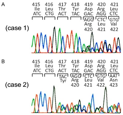
We examined whether exon 8 c-kit gene muta-tions were detected in approximately 1,000 sporadic GISTs. Among them, one GIST had Del-Asp419 and 2 GISTs had ThrTyrAsp (417-419) Tyr (Figure 1A and 1B).
Clinicopathologicl features of GISTs with exon 8 c-kit gene mutations
Brief clinicopathological features of 3 GIST cases with exon 8 c-kit gene mutations were shown in Table 1. Characteristically, all GISTs with the mutation occurred outside the stom-ach (2 at the duodenum and 1 at the small intestine). All three tumors consisted of spindle shaped cells (Figure 2A, 2D and 2G). Immu- nohistochemistry revealed that the tumor cells of all cases were diffusely and strongly positive for KIT (Figure 2B, 2E and 2H). CD34 was posi-tive in cases 1 and 3 but negaposi-tive in case 2 (Figure 2C, 2F and 2I). All three GISTs had high mitotic figures (Table 1), suggesting that they were highly aggressive. In fact, two of three GISTs showed metastasis. The patient of case 3 has been receiving imatinib adjuvant therapy until now, and no recurrence is evident.
Clinical history of patients with exon 8 c-kit gene mutations
rein-troduction of imatinib or sunitinib, irradiation, and metastatectomy, the drug-resistant rem- aining lesion could not be controlled. He died 13 years and 3 months after the first operation for small intestinal GIST. Sequencing of c-kit
cDNA derived from the resected samples revealed that the sunitinib-resistant lesion had second Asp822Lys at exon 17 in addition to Del-Asp419. Imatinib- and sunitinib-resistant characteristics are considered to be resulted from this second mutation.
Case 2: A 56-year-old Japanese man underwent partial resection of the duodenum for duodenal GIST (3.5 cm in diameter) which showed ThrTyrAsp (417-419) Tyr at exon 8 of the c-kit
gene. Two years after the complete resection of duodenal GIST, multiple liver metastases were
[image:4.612.92.523.71.399.2]found by abdominal CT. Imatinib administration (400 mg/day) was started, and the metastatic foci had been controlled for 18 months. Because of regrowth of one of the metastatic foci in the liver, partial hepatectomy was per-formed. In spite of continuation of imatinib administration after the metastatectomy, peri-toneal masses developed. Although the perito-neal lesions were resected, multiple peritoperito-neal masses developed again. He received sunitinib therapy with 4 month control period. After the failure of sunitinib, regorafenib was adminis-tered on clinical trial, but it showed no apparent effect. He died 8 years and 4 months after the first operation for duodenal GIST. Sequencing of c-kit cDNA derived from the imatinib-resis-tant hepatic tumor and peritoneal tumor revealed that both lesions had Asp910Tyr at
exon 18 in addition to ThrTyrAsp (417-419) Tyr at exon 8. This second mutation is considered to be a cause of imatinib-resistant character.
Case 3: A 41-year-old Japanese man received pancreaticoduodenectomy for duodenal GIST (8.5 cm in diameter) which had ThrTyrAsp (417-419) Tyr at exon 8 of the c-kit gene. Since it was judged as a high risk tumor for recurrence, adjuvant imatinib treatment (400 mg/day) has been done for 29 months after the operation. At present, he is under imatinib treatment and there is no evidence of recurrence.
Discussion
We reported here 3 cases of sporadic GISTs with exon 8 c-kit gene mutations. Frequency of the mutations is low (approximately 0.3%), but at least 3 GISTs really had two types of the mutations such as Del-Asp419 and ThrTyrAsp (417-419) Tyr.
Exon 8 c-kit gene mutations was first reported in acute myeloid leukemia in 1999 [33]. In acute myeloid leukemia [33, 34], various types of exon 8 c-kit gene mutations are known including ThrTyr (417&418) His, ThrTyrAsp (417-419) Asn, ThrTyrAsp (417-(417-419) Ile, ThrTyrAsp (417-419) Phe, ThrTyrAsp (417-419) Tyr, ThrTyrAsp (417-419) Val, ThrTyrAsp (417-419) ArgAla, ThrTyrAsp (417-419) ArgGly, TyrAsp (418&419) Gly, TyrAsp (418&419) Ser, Asp- 419Phe, Del-Asp419 and AspArg (419&420) PhePheAspGly. In pediatric mastocytosis, on the other hand, exon 8 c-kit gene mutations were reported in 2010 [31]. The types of the exon 8 c-kit gene mutations reported in pediat-ric mastocytosis are Del-Asp419, Ins-PhePhe between codon418 and codon419, ThrTyrAsp (417-419) Tyr and Cys443Tyr [31]. Many types of exon 8 c-kit gene mutations are common in acute myeloid leukemia and pediatric mastocy-tosis. Although acute myeloid leukemia does not appear to have any highly frequent muta-tion types, Del-Asp419 appears to be the most frequent mutation type in pediatric masto- cytosis.
Exon 8 c-kit gene mutations in GISTs were first reported in familial GIST cases with germline c-kit gene mutation in 2005 [23], and the type of the mutation was Del-Asp419. The patients in the family members had not only multiple GISTs but also mastocytosis. Recently, 2 spo-radic GIST cases with Del-Asp419 were found
[32]. Therefore, Del-Asp419 was the only muta-tion type reported in GISTs with exon 8 c-kit
gene mutation. In the present study, we report-ed one GIST case with Del-Asp419 and 2 GISTs cases with ThrTyrAsp (417-419) Tyr. Described above, ThrTyrAsp (417-419) Tyr has been report-ed in acute myeloid leukemia and sporadic pediatric mast cell neoplasms [31, 33, 34]. Thus, these are the first reported GIST cases with ThrTyrAsp (417-419) Tyr among sporadic and familial GIST cases.
In the previous report, 2 sporadic GISTs with exon 8 c-kit gene mutations occurred at the small bowel [32]. In the present study, 3 GISTs with exon 8 c-kit gene mutations were present at the duodenum in 2 cases and at the small intestine in 1 case. Since all of the reported GIST cases with exon 8 c-kit gene mutations developed at the small intestine or duodenum, those GISTs appear to arise from extragastric sites. However, the number of GIST cases with exon 8 c-kit gene mutations is too small to draw conclusions, and further effort to collect those GIST cases is needed.
In the previous report of sporadic GISTs with exon 8 c-kit gene mutations, one GIST devel-oped multiple metastatic foci in the peritoneum [32]. The other case did not show metastasis. However, the patient had been receiving adju-vant imatinib therapy because the tumor was regarded as intermediate to high risk for recur-rence [32]. In the present study, 2 cases of GISTs with exon 8 c-kit gene mutations showed distant metastasis; one was in bone and the other was in liver and peritoneum. The other case in this study does not show metastasis, but the patient also has been receiving adju-vant imatinib therapy because the tumor was classified as high risk for recurrence. These results suggested that GISTs with exon 8 c-kit
were demonstrated to be sensitive to imatinib in vitro [25, 34]. As described above, imatinib was reported to be administered to one spo-radic GIST case with Del-Asp419 because of being regarded as intermediate to high risk for recurrence [32]. The patient did not show recur-rence for 24 months under adjuvant imatinib treatment, but the case cannot clearly demon-strate whether the mutation is sensitive to ima-tinib in vivo. On the other hand, our 2 cases with distant metastasis showed apparent clini-cal effect of imatinib. Recently, imatinib tends to be selectively used only for GISTs with ima-tinib-sensitive mutations. In many GIST cases, the mutations are usually examined only at exons 9, 11, 13 and 17 of the c-kit gene and exon 12, 14, 18 of the PDGFRA gene. Therefore, GISTs with exon 8 c-kit gene mutation could be erroneously regarded as so-called wild-type GISTs which are usually resistant to imatinib, and those cases might not be considered to be candidates for imatinib treatment. We have to examine whether the exon 8 c-kit gene muta-tions are present in so-called wild-type GISTs to prevent loss of opportunity for imatinib treat-ment in those patients.
Asp816Val c-kit gene mutation at exon 17 is often observed in sporadic mast cell neo-plasms [11, 12]. However, any GISTs have not been reported to possess this mutation. Namely, there is tumor-type specificity in Asp816Val. However, this is an exceptional cor-relation between tumor-types and particular mutations. Many types of the c-kit gene muta-tions are common in mast cell neoplasms and GISTs. Indeed, both of mast cell neoplasms and GISTs have the same exon 8 c-kit gene muta-tions, at least Del-Asp419 and ThrTyrAsp (417-419) Tyr. The cause of tumor-type specificity in Asp816Val remains to be clarified.
Finally, we showed that GISTs with exon 8 c-kit
gene mutations might have features of extra-gastric development and metastasis-prone nature. Since the exon 8 c-kit gene mutations appeared to be sensitive to imatinib, accurate genotyping including not only exons 9, 11, 13 and 17 but also exon 8 of the c-kit gene is nec-essary to predict response to imatinib in both unresectable/metastatic and adjuvant set- tings.
Acknowledgements
This work was partly supported by Grant-in-Aid for Scientific Research (B) from the Japan
Society for the Promotion of Science (Grant No. 23390094).
Disclosure of conflict of interest
Seiichi Hirota received research fund from Novartis Pharma K.K. Japan.The other authors have no conflict of interest.
Address correspondence to: Dr. Seiichi Hirota, Department of Surgical Pathology, Hyogo College of Medicine, 1-1 Mukogawa-Cho, Nishinomiya, Hyogo 663-8501, Japan. Tel: 798-45-6667; Fax: +81-798-45-6671; E-mail: hiros@hyo-med.ac.jp
References
[1] Hirota S, Isozaki K, Moriyama Y, Hashimoto K, Nishida T, Ishiguro S, Kawano K, Hanada M, Kurata A, Takeda M, Tunio GM, Matsuzawa Y, Kanakura Y, Shinomura Y, Kitamura Y. Gain-of-function mutations of c-kit in human gastroin-testinal stromal tumors. Science 1998; 279: 577-580.
[2] Kindblom LG, Remotti HE, Aldenborg F, Meis-Kindblom JM. Gastrointestinal pacemaker cell tumor (GIPACT): gastrointestinal stromal tu-mors show phenotypic characteristics of the interstitial cells of Cajal. Am J Pathol 1998; 152: 1259-1269.
[3] Yarden Y, Kuang WJ, Yang-Feng T, Coussens L, Munemitsu S, Dull TJ, Chen E, Schlessinger J, Francke U, Ullrich A. Human proto-oncogene
c-kit: a new cell surface receptor tyrosine kinase
for an unidentified ligand. EMBO J 1987; 6:
3341-3351.
[4] Besmer P, Murphy JE, George PC, Qiu F, Ber-gold PJ, Lederman L, Snyder Jr HW, Broudeur D, Zuckerman EE, Hardy WD. A new acute transforming feline retrovirus and relationship of its oncogene v-kit with the protein kinase gene family. Nature 1986; 320: 415-421. [5] Qiu FH, Ray P, Brown K, Barker PE, Jhanwar S,
Ruddle FH, Besmer P. Primary structure of
c-kit: relationship with the CSF-1/PDGF receptor kinase family--oncogenic activation of v-kit in-volves deletion of extracellular domain and C terminus. EMBO J 1988; 7: 1003-1011. [6] Geissler EN, Ryan MA, Housman DE. The
dom-inant-white spotting (W) locus of the mouse encodes the c-kit proto-oncogene. Cell 1988; 55: 185-192.
[7] Thomsen L, Robinson TL, Lee JC, Farraway LA, Hughes MJ, Andrews DW, Huizinga JD. Interstitial cells of Cajal generate a rhythmic pacemaker current. Nat Med 1998; 4: 848-851.
Cosman D, Lyman SD. Identification of a ligand
for the c-kit proto-oncogene. Cell 1990; 63: 167-174.
[9] Flanagan JG, Leder P. The kit ligand: a cell
sur-face molecule altered in steel mutant fibro -blasts. Cell 1990; 63: 185-194.
[10] Zsebo KM, Williams DA, Geissler EN, Broudy YC, Martin FH, Atkins HL, Hsu RY, Birkitt NC, Okino KH, Murdock DC, Jacobson FW, Langley KE, Smith KA, Takeishi T, Cattanach BM, Galli SJ, Suggs SV. Stem cell factor is encoded at the Sl locus of the mouse and is the ligand for the c-kit tyrosine kinase receptor. Cell 1990; 63: 213-224.
[11] Nagata H, Worobec AS, Oh CK, Chowdhury BA, Tannenbaum S, Suzuki Y, Metcalfe DD.
Identification of a point mutation in the cata -lytic domain of the protooncogene c-kit in pe-ripheral blood mononuclear cells of patients who have mastocytosis with an associated he-matologic disorder. Proc Natl Acad Sci U S A 1995; 92: 10560-10564.
[12] Longley BJ, Tyrrell L, Lu SZ, Ma YS, Langley K, Ding TG, Duffy T, Jacobs P, Tang LH, Modlin I. Somatic c-kit activating mutation in urticaria pigmentosa and aggressive mastocytosis: es-tablishment of clonality in a human mast cell neoplasm. Nat Genet 1996; 12: 312-314. [13] Tian Q, Frierson HF Jr, Krystal GW, Moskaluk
CA. Activating c-kit gene mutations in human germ cell tumors. Am J Pathol 1999; 154: 1643-1647.
[14] Went PT, Dirnhofer S, Bundi M, Mirlacher M, Schraml P, Mangialaio S, Dimitrijevic S, Kononen J, Lugli A, Simon R, Sauter G. Prevalence of KIT expression in human tu-mors. J Clin Oncol 2004; 22: 4514-4520. [15] Curtin JA, Busam K, Pinkel D, Bastian BC.
Somatic activation of KIT in distinct subtypes of melanoma. J Cin Oncol 2006; 24: 4340-4346.
[16] Rubin BP, Singer S, Tsao C, Duensing A, Lux ML, Ruiz R, Hibbard MK, Chen CJ, Xiao S, Tuveson DA, Demetri GD, Fletcher CD, Fletcher JA. KIT activation is a ubiquitous feature of gas-trointestinal stromal tumors. Cancer Res 2001; 61: 8118-8121.
[17] Kinoshita K, Isozaki K, Hirota S, Nishida T, Chen H, Nakahara M, Nagasawa Y, Ohashi A, Shinomura Y, Kitamura Y, Matsuzawa Y. C-kit
gene mutation at exon 17 or 13 is very rare in sporadic gastrointestinal stromal tumors. J Gastroenterol Hepatol 2003; 18: 147-151. [18] Heinrich MC, Corless CL, Duensing A,
McGreevey L, Chen CJ, Joseph N, Singer S,
Griffith DJ, Haley A, Town A, Demetri GD,
Fletcher CD, Fletcher JA. PDGFRA Activating Mutations in Gastrointestinal Stromal Tumors. Science 2003; 299: 708-710.
[19] Hirota S, Ohashi A, Nishida T, Isozaki K, Kinoshita K, Shinomura Y, Kitamura Y. Gain-of-function mutations of platelet-derived growth factor receptor alpha gene in gastrointestinal stromal tumors. Gastroenterology 2003; 125: 660-667.
[20] Nishida T, Hirota S, Taniguchi M, Hashimoto K, Isozaki K, Nakamura H, Kanakura Y, Tanaka T, Takabayashi A, Matsuda H, Kitamura Y. Familial gastrointestinal stromal tumours with germline mutation of the KIT gene. Nat Genet 1998; 19: 323-324.
[21] Isozaki K, Terris B, Belghiti J, Schiffmann S, Hirota S, Vanderwinden JM. Germline-activating mutation in the kinase domain of KIT gene in familial gastrointestinal stromal tumors. Am J Pathol 2000; 157: 1581-1585. [22] Hirota S, Nishida T, Isozaki K, Taniguchi M,
Nishikawa K, Ohashi A, Takabayashi A, Obayashi T, Okuno T, Kinoshita K, Chen H, Shinomura Y, Kitamura Y. Familial gastrointes-tinal stromal tumors associated with dyspha-gia and novel type germline mutation of KIT gene. Gastroenterology 2002; 122: 1493-1499.
[23] Hartmann K, Wardelmann E, Ma Y, Merkelbach-Bruse S, Preussner LM, Woolery C, Baldus SE, Heinicke T, Thiele J, Buettner R, Longley BJ. Novel germline mutation of KIT associated with familial gastrointestinal stromal tumors and mastocytosis. Gastroenterology 2005; 129: 1042-1046.
[24] Buchdunger E, Cioffi CL, Law N, Stover D, Ohno-Jones S, Druker BJ, Lydon NB. Abl pro-tein-tyrosine kinase inhibitor STI571 inhibits in vitro signal transduction mediated by c-kit and platelet-derived growth factor receptors. J Pharmacol Exp Ther 2000; 295: 139-145. [25] Demetri GD, von Mehren M, Blanke CD, Van
den Abbeele AD, Eisenberg B, Roberts PJ, Heinrich MC, Tuveson DA, Singer S, Janicek M, Fletcher JA, Silverman SG, Silberman SL, Capdeville R, Kiese B, Peng B, Dimitrijevic S, Druker BJ, Corless C, Fletcher CD, Joensuu H.
Efficacy and safety of imatinib mesylate in ad -vanced gastrointestinal stromal tumors. N Engl J Med 2002; 347: 472-480.
[26] Chen H, Isozaki K, Kinoshita K, Ohashi A, Shinomura Y, Matsuzawa Y, Kitamura Y, Hirota S. Imatinib inhibits various types of activating mutant kit found in gastrointestinal stromal tu-mors. Int J Cancer 2003; 105: 130-135. [27] Heinrich MC, Corless CL, Demetri GD, Blanke
[28] Joensuu H, Eriksson M, Hall KS, Hartmann JT, Pink D, Schütte J, Ramadori G, Hohenberger P, Duyster J, Al-Batran SE, Schlemmer M, Bauer S, Wardelmann E, Sarlomo-Rikala M, Nilsson B, Sihto H, Monge OR, Bono P, Kallio R, Vehtari A, Leinonen M, Alvegård T, Reichardt P. One vs three years of adjuvant imatinib for operable gastrointestinal stromal tumor: randamized trial. JAMA 2012; 307: 1265-1272.
[29] Demetri GD, van Oosterom AT, Garrett CR, Blackstein ME, Shah MH, Verweij J, McArthur G, Judson IR, Heinrich MC, Morgan JA, Desai J, Fletcher CD, George S, Bello CL, Huang X,
Baum CM, Casali PG. Efficacy and safety of
sunitinib in patients with advanced gastroin-testinal stromal tumour after failure of ima-tinib: a randamised controlled trial. Lancet 2006; 368: 1329-1338.
[30] Demetri GD, Reicherdt P, Kang YK, Blay JY, Rutkowski P, Gelderblom H, Hohenberger P, Leahy M, von Mehren M, Joensuu H, Badalamenti G, Blackstein M, Le Cesne A, Schöffski P, Maki RG, Bauer S, Nguyen BB, Xu J, Nishida T, Chung J, Kappeler C, Kuss I, Laurent D, Casali PG; GRID study investigators.
Efficacy and safety of regorafenib for advanced
gastrointestinal stromal tumours after failure of imatinib and sunitinib (GRID): an interna-tional multicentre randamised placebo-con-trolled phase 3 trial. Lancet 2013; 381: 295-302.
[31] Bodemer C, Hermine O, Palmérini F, Yang Y, Grandpeix-Guyodo C, Leventhal PS, Hadj-Rabia S, Nasca L, Georgin-Lavialle S, Cohen-Akenine A, Launay JM, Barete S, Feger F, Arock M, Catteau B, Sans B, Stalder JF, Skowron F, Thomas L, Lorette G, Plantin P, Bordigoni P, Lortholary O, de Prost Y, Moussy A, Sobol H, Dubreuil P. Pediatric mastocytosis is clonal dis-ease associated with D816V and other activat-ing c-KIT mutations. J Invest Dermatol 2010; 130: 804-805.
[32] Huss S, Kunstlinger H, Wardelmann E, Kleine MA, Binot E, Merkelbach-Bruse S, Rüdiger T, Mittler J, Hartmann W, Büttner R, Schildhaus HU. A subset of gastrointestinal stromal tu-mors previously regarded as wild-type tutu-mors carries somatic activating mutations in KIT exon 8 (p.D419del). Mod Pathol 2013; 26: 1004-1012.
[33] Gari M, Goodeve A, Wilson G, Winship P, Langabeer S, Linch D, Vandenberghe E, Peake I, Reilly J. c-kit proto-oncogene exon 8 in-frame deletion plus insertion mutations in acute my-eloid leukaemia. Br J Haematol 1999; 105: 894-900.