Original Article
Association of natriuretic peptide polymorphisms with
left ventricular dysfunction in southern Han Chinese
coronary artery disease patients
Zhijun Wu1*, Min Xu1*,Haihui Sheng2, Yuqing Lou3, Xiuxiu Su1, Yanjia Chen1, Lin Lu1, Yan Liu1, Wei Jin1
1Department of Cardiology, Ruijin Hospital, Shanghai Jiao Tong University School of Medicine, Shanghai, China; 2National Engineering Center for Biochip at Shanghai, Shanghai, China; 3Department of Pulmonary, Shanghai Chest Hospital, Shanghai Jiao Tong University, Shanghai, China. *Equal contributors.
Received June 3, 2014; Accepted July 16, 2014; Epub September 15, 2014; Published October 1, 2014
Abstract: Background: Left ventricular dysfunction (LVD) occurs with myocardial ischemia and coronary artery dis-ease (CAD). The natriuretic peptide system has compensatory vasodilatory, natriuretic and paracrine effects on LVD and subsequent heart failure. The aim of this study was to investigate the relationship between natriuretic peptide polymorphisms and risk of LVD in CAD patients. Methods: We recruited 747 consecutive Southern Han Chinese patients with angiographically confirmed CAD, 201 had a reduced left ventricle ejection fraction (LVEF ≤45%, LVD group) and 546 had a preserved left ventricle ejection fraction (LVEF >45%). The reduced and preserved LVEF groups were matched by gender and age. Taqman assays were performed to identify five polymorphisms in the
NPPA-NPPB locus (rs5065, rs5063, rs632793, rs198388 and rs198389). Results: Single-locus analyses found no significant difference in the allele and genotype frequencies of the reduced and preserved LVEF group, even after adjusting for confounding factors. Subgroup analyses performed by hyperlipidemia (HLP) demonstrated 3 polymorphisms, rs632793 (OR = 0.31, 95% CI 0.1-0.93, P = 0.04), rs198388 (OR = 0.26, 95% CI 0.09-0.79, P = 0.02) and rs198389 (OR = 0.26, 95% CI 0.09-0.80, P = 0.02) were associated with the reduced risk of LVD. No CAD-susceptible haplotypes were identified. Multifactor dimensionality reduction analysis did not detect any gene-to-gene interactions among the five loci. Three loci (rs5063, rs632793 and rs198388) formed the best model with the maximum testing accuracy (39.89%) and cross-validation consistency (10/10). Conclusion: Three NPPA-NPPB polymorphisms (rs632793, rs198388 and rs198389) were associated with reduced risk of LVD in CAD patients with HLP.
Keywords: Left ventricular dysfunction, coronary artery disease, natriuretic peptide, polymorphism, heart failure
Introduction
Left ventricular dysfunction (LVD) is an early
stage of heart failure (HF) characterized by a
reduced ejection fraction and a depressed level of left ventricular wall motility [1]. Coronary artery disease (CAD) and myocardial infarction (MI), the cardiac polygenic disorders [2-4], are contributing factors to the development of LVD. Other diseases, such as cardiomyopathy, hypertension (HTN) and valvular disease, may
also lead to LVD. LVD with subsequent HF is characterized by a continuous interaction
between the underlying myocardial dysfunction and a series of compensatory mechanisms. A host of hemodynamic and neurohormonal fac-tors, such as the adrenergic and
renin-angio-tensin-aldosterone systems (RAAS) are trig-gered to modulate left ventricle remodeling of the vascular tree once LVD occurs. One of the key neuroendocrine axes is the natriuretic
pep-tide system. This system consists of five pep -tides, atrial peptide (ANP), urodilatin (an isoform of ANP), B-type natriuretic peptide (BNP), C-type natriuretic peptide (CNP), and dendroaspis natriuretic peptide (DNP) [5]. These peptides share a similar molecular structure and biologi-cal functions (natriuresis, diuresis and
vasodila-tion) [6]. ANP and BNP are mainly synthesized
line with the degree of cardiac dysfunction and left ventricular failure, making them useful markers in evaluating the severity of these phe-nomena [8]. Circulating natriuretic peptide lev-els are helpful to assess the prognosis in patients with CAD and MI, with or without LVD [9, 10]. The persistence of elevated levels of natriuretic peptides for several months after MI suggests a continued risk of pathologic
remod-eling and the development of LVD and HF [11].
The NPPA ( natriuretic peptide precursor A) and NPPB ( natriuretic peptide precursor B) genes lie in tandem 9.7kb apart on chromosome 1.
Mice with homozygous mutations of the NPPA
gene have right and left ventricular hypertro-phy. This hypertrophy is increased dispropor-tionately (relative to controls) in response to transverse aortic constriction, suggesting that ANP negatively regulates matrix remodeling in the myocardium [12]. In contrast, NPPB gene
knockout mice exhibit multifocal fibrotic lesions in the cardiac ventricles. These increase in size
and number in response to ventricular
over-load, indicating that BNP acts as an anti-fibrotic
factor [13]. Several polymorphisms within the NPPA-NPPB locus have been reported to be correlated with inter-individual variation in cir-culating natriuretic peptide concentrations [14, 15], contributing to ambulatory cardiovascular
disease states such as stroke [16], HF [17],
CAD [18] and HTN [15, 19]. The
nonsynony-mous coding polymorphism rs5065 is signifi -cantly associated with increased circulating BNP and amino-terminal BNP (NT-proBNP)
lev-els in severe HF patients [20]. The rs5065 and
rs5063 polymorphisms correlate with incr- eased left ventricular mass index and left ven-tricular septal thickness in HTN patients [21]. The minor NPPA-NPPB allele, rs632793, rs198388 and rs198389 are associated with increased circulating ANP and BNP concentra-tions and a reduced rate of cardiovascular readmission in CAD patients [15, 22]. Surprisingly, these polymorphisms have not been evaluated as markers for LVD. Therefore, we investigated the relationship between com-mon NPPA-NPPB polymorphisms and the pres-ence of LVD in CAD patients.
Materials and methods
Ethics statement
The ethics committee of Ruijin Hospital, Shanghai Jiao Tong University School of
Medicine approved this study. All the authors followed the guidelines of the World’s Association Declaration of Helsinki. Written informed consent for the study was obtained from each patient.
Study cohort
A total of 747 consecutive patients undergoing coronary angiography for the diagnosis and intervention of CAD were admitted to Ruijin Hospital from January 2008 to December 2012. Coronary angiography was performed via the femoral or radial artery approach, according to the clinical standards of the American College of Cardiology/American Heart Association guidelines for coronary angiogra-phy [23]. Standard Judkins techniques were used for angiography [24]. Physicians perform-ing the studies were blinded to the study
proto-col. CAD was defined as ≥50% luminal obstruc -tion at least one or more major coronary epicardial coronary vessel. The patients were
grouped according to the number of significant -ly stenotic vessels, single-, double- or triple-vessel disease. Any patient with cardiomyopa-thy, congenital heart disease, pulmonary heart disease, valvular heart disease, stroke, infec-tion, tumor or immune system disorders was excluded. Left ventricular (LV) function was cal-culated by the echocardiography staff just before angiography. Imaging (e.g., M-mode, 2D)
and color-flow Doppler echocardiography were performed. LV ejection fraction (EF), volumes
and internal dimensions were measured according to the American Society of Echocardiography recommendations [25]. LVD
was defined as (modified Simpson’s rule) LVEF≤ 45% [26]. All patients were interviewed to
ascertain their sociodemographic, economic and health status characteristics as well as life-style (e.g., habits, tobacco usage). Ultrasound staff was blinded to polymorphism analysis results. Subjects were residents of Shanghai, a population that is primarily of Southern Han Chinese ethnicity.
Data collection
Complete clinical history and information on conventional cardiovascular risk factors were obtained by reviewing the patients’ medical
records. HTN was defined as a systolic blood pressure >140 mmHg, a diastolic blood pres
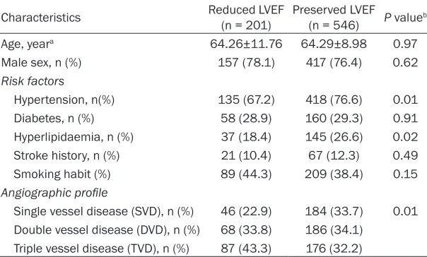
Table 1. The baseline characteristics of study population
Characteristics Reduced LVEF(n = 201) Preserved LVEF(n = 546) P valueb
Age, yeara 64.26±11.76 64.29±8.98 0.97
Male sex, n (%) 157 (78.1) 417 (76.4) 0.62
Risk factors
Hypertension, n(%) 135 (67.2) 418 (76.6) 0.01
Diabetes, n (%) 58 (28.9) 160 (29.3) 0.91
Hyperlipidaemia, n (%) 37 (18.4) 145 (26.6) 0.02
Stroke history, n (%) 21 (10.4) 67 (12.3) 0.49
Smoking habit (%) 89 (44.3) 209 (38.4) 0.15
Angiographic profile
Single vessel disease (SVD), n (%) 46 (22.9) 184 (33.7) 0.01
Double vessel disease (DVD), n (%) 68 (33.8) 186 (34.1) Triple vessel disease (TVD), n (%) 87 (43.3) 176 (32.2)
aData are expressed as mean ± SD. bThe unpaired t-test is used for age and the x2 test is
used for other categorical characteristics.
drugs. Smoking habit was classified as smok -ers (ex-smoker and current smok-ers) or
non-smokers. Diabetes mellitus (DM) was defined
as a fasting serum glucose level above 7.0 mmol/L, a two-hour postprandial glucose greater than 11.1 mmol/L, or the use of dia-betic medications. The diagnosis and manage-ment of Hyperlipidemia (HLP) was performed according to the National Cholesterol Education Program Adult Treatment Panel III Guidelines
(NCEP ATIII) [27]. Stroke was defined and clas
-sified based according toTrial of Org 10172 in Acute Stroke Treatment (TOAST) criteria [28]. DNA isolation and genotyping
Blood was drawn using EDTA-collection tubes. Genomic DNA was extracted from peripheral blood leukocytes according to standard phe-nol-chloroform methods, and stored at -20°C until batch genotyping. Taqman assay was used to genotype SNPs (Single nucleotide poly-morphisms). SNP Taqman probes and primers were designed using the Applied Biosystems Assay-by-Design Service for SNP genotyping.
The sample DNA was amplified by PCR follow
-ing the recommendations of the manufacturer. Thermal cycling was done on a Gene Amp PCR System 9700 thermal cycler (Applied
Biosystems, Foster City, CA, USA). Allele detec -tion and genotype determina-tion were
per-formed using the fluorescence mode of an ABI
PRISM 7900HT Sequence Detector (Applied
Biosystems, Foster City, CA, USA). Genotyping
was performed without knowledge of the
case-control status of the sub-jects. Ten percent of samples from patients and controls were se- quenced to estimate the quality of genotyping. No discrepancy was found.
Five SNPs located in the
NPPA-NPPB locus (rs- 5065, rs5063, rs63- 2793, rs198388 and rs198389) were inve- stigated.
Statistical analysis Continuous variables we- re reported as the mean ± standard deviation (SD) or median with 5th and 95th percentiles. Categorical measures were reported as
per-centages. The chi-square test or Fisher’s exact test were used to examine the goodness of fit
between the observed allelic frequencies and the allelic frequencies expected by Hardy Weinberg Equilibrium (HWE). HWE was deter-mined using the web program- http://ihg2.
helmholtz-muenchen.de/cgi-bin/hw/hwa1.pl.
Differences in the allelic and genotypic fre-quencies of cases and controls were evaluated using the chi-square test. An unpaired t-test was used to test group differences of continu-ous variables. Genotype frequency of the
sub-jects specified by different genetic models
(allelic, additive, dominant, recessive and
homozygote comparison) was analyzed by mul -tivariate logistic regression adjusted for con-founding factors. A two-tailed P<0.05 was
regarded as statistically significant. Data man -agement and statistical analyses were per-formed using SPSS software version 20.0 (SPSS, Chicago, IL, USA).
Linkage disequilibrium (LD) and haplotype blocks within 5 SNPs of the NPPA-NPPB locus
were identified using the online computer plat -form SHEsis- http://analysis.bio-x.cn/myAnaly-sis.php [29]. LD coefficients were calculated
using the formula D’=D/Dmax or D/Dmin.
Haplotypes with a frequency higher than 1% were examined and significance was estimated
Table 2. Genotype distributions and allele frequencies of the 5 examined polymorphisms between
the reduced and preserved LVEF groups in CAD patients and the risk prediction under various genetic
models
SNP ID
(rs number) Genotypeand allele Cas-es Con-trol PΧ2a Genetic models
Unadjusted Adjustedb
OR 95% CI P value OR 95% CI P value
rs5065 GG 0 0 0.75 Allelic comparison 0.81 0.22-2.97 0.75 0.84 0.23-3.11 0.80 AG 3 10 Dominant model 0.81 0.22-2.98 0.75 0.84 0.23-3.12 0.80 AA 198 535 Recessive model –c – – – – –
G (%) 0.7 0.9 0.75 Homozygote comparison – – – – – –
A (%) 99.3 99.1 Additive model 0.81 0.22-2.98 0.75 0.84 0.23-3.12 0.80
rs5063 TT 4 7 0.78 Allelic comparison 1.09 0.74-1.61 0.65 1.06 0.72-1.58 0.76 CT 32 86 Dominant model 1.06 0.69-1.62 0.79 1.04 0.67-1.60 0.87 CC 165 452 Recessive model 1.56 0.45-5.39 0.48 1.43 0.40-5.13 0.58
T (%) 10.0 9.2 0.65 Homozygote comparison 1.57 0.45-5.42 0.48 1.46 0.41-5.20 0.56
C (%) 90.0 90.8 Additive model 1.09 0.75-1.58 0.66 1.06 0.72-1.55 0.77
rs632793 GG 6 9 0.50 Allelic comparison 1.07 0.76-1.50 0.69 1.06 0.75-1.50 0.75 AG 42 120 Dominant model 1.01 0.69-1.48 0.95 1.02 0.69-1.50 0.93 AA 153 416 Recessive model 1.83 0.64-5.22 0.26 1.56 0.53-4.56 0.42
G (%) 13.4 12.7 0.69 Homozygote comparison 1.81 0.64-5.18 0.27 1.54 0.52-4.52 0.43
A (%) 86.6 87.3 Additive model 1.07 0.77-1.49 0.70 1.06 0.75-1.48 0.75
rs198388 TT 5 14 0.50 Allelic comparison 0.84 0.61-1.17 0.30 0.81 0.58-1.13 0.22 CT 45 145 Dominant model 0.8 0.56-1.16 0.25 0.78 0.53-1.14 0.20 CC 151 386 Recessive model 0.97 0.34-2.72 0.95 0.85 0.30-2.44 0.76
T (%) 13.7 15.9 0.30 Homozygote comparison 0.91 0.32-2.58 0.86 0.82 0.28-2.35 0.71
C (%) 86.3 84.1 Additive model 0.84 0.61-1.17 0.30 0.81 0.59-1.13 0.22
rs198389 GG 6 10 0.43 Allelic comparison 0.96 0.68-1.33 0.79 0.95 0.68-1.34 0.77 AG 42 132 Dominant model 0.89 0.61-1.29 0.54 0.89 0.61-1.31 0.56 AA 153 402 Recessive model 1.64 0.59-4.58 0.34 1.49 0.52-4.27 0.45
G (%) 13.4 14.0 0.79 Homozygote comparison 1.58 0.56-4.41 0.39 1.44 0.51-4.13 0.49
A (%) 86.6 86.0 Additive model 0.96 0.69-1.33 0.79 0.95 0.68-1.33 0.77
aP values were calculated by x2 test for differences in genotypes and alleles between the two groups. bORs adjusted for age, gender, HTN, DM, HLP, Stroke history, smoking habit and angiographic profile. cdata not available.
main functions in Haplo.stats were implement-ed: Haplo.em was used to calculate maximum likelihood estimates of haplotype probability using a “progressive insertion” algorithm that progressively inserted batches of loci into hap-lotypes of growing lengths. Haplo.cc and Haplo. glm were used to calculate crude and adjusted
odds ratios (ORs) and 95% confidence intervals
(CIs) for each haplotype, respectively. These two approaches computed the regression of a trait on haplotypes and other covariates based
on a generalized linear model [30, 31]. Haplo.
score was used to calculate score statistics and test the difference in haplotype frequen-cies between cases and controls on the basis of simulated P (Psim) values that were obtained from 1000 replicates [32].
Gene-to-gene interactions in the occurrence of LVD were evaluated using the open-source
mul-tifactor dimensionality reduction (MDR) soft-ware version 3.0 (www.epistasis.org) [33, 34]. MDR collapsed high-dimensional multilocus-genotype variables into a single dimensional multilocus-genotype variable by sorting the genotypes into two levels, high- or low- risk. All possible combinations of these 5 polymor-phisms were tested. A probabilistic naïve Bayes
classifier with 10-fold cross-validation was
used to estimate the prediction accuracy and the empirical P-value. The data are divided into 10 divisions equally. 9/10 of the data is regard-ed as training set and then the remaining 1/10 is tested. A single best model with maximal testing accuracy and cross-validation consis-tency was determined by measuring the num-ber of times of 10 divisions of the data. The P-value <0.05 was considered statistically
Table 3. Stratified analyses of the 5 NPPA-NPPB polymorphisms with the risk of LVD in CAD patients with HLP under the additive genetic model
Study ID
(rs number) Group Unadjusted Adjusted
a
OR 95% CI P value OR 95% CI P value
rs5065 HLP –b – – – – –
Non-HLP 0.91 0.24-3.49 0.89 0.97 0.25-3.77 0.96 rs5063 HLP 0.69 0.28-1.71 0.42 0.69 0.26-1.81 0.45 Non-HLP 1.24 0.82-1.88 0.31 1.19 0.78-1.82 0.41 rs632793 HLP 0.35 0.12-1.00 0.05 0.31 0.10-0.93 0.04 Non-HLP 1.31 0.91-1.89 0.15 1.30 0.90-1.89 0.16 rs198388 HLP 0.30 0.10-0.86 0.03 0.26 0.09-0.79 0.02 Non-HLP 0.99 0.69-1.40 0.93 0.97 0.68-1.39 0.86 rs198389 HLP 0.31 0.11-0.88 0.03 0.26 0.09-0.80 0.02 Non-HLP 1.17 0.82-1.68 0.39 1.17 0.81-1.69 0.40 aORs adjusted for age, gender, HTN, DM, Stroke history, smoking habit and
angio-graphic profile. bdata not available.
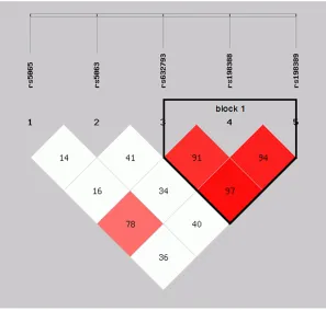
Figure 1. Block structure of linkage disequilibrium (LD) for five genotyped single nucleotide polymorphisms (SNPs) in the NPPA-NPPB locus. Stronger correla-tions between these SNPs are noted by red color in the intersecting squares linking each pair of SNPs.
Results
Patient characteristics
Clinical characteristics of CAD patients are shown in Table 1. Of the total 747 CAD patients,
201 had reduced (≤45%) LVEF and 546 had pre
-served LVEF (>45%). The reduced LVEF group was
matched with the preserved
LVEF group for age (P = 0.97) and gender (P = 0.62).
There was no significant dif -ference in the distribution of other confounding fac-tors including DM (P = 0.91), smoking habit (P = 0.15) and stroke history (P
= 0.49). The reduced LVEF group had a significantly
lower proportion of HTN (P = 0.01) and HLP (P = 0.02)
than the preserved LVEF
group. The angiographic
profile of the reduced LVEF group was significantly dif -ferent from that of the
pre-served LVEF group (P = 0.01) (Table 1).
Single-locus analysis
Ninety-nine percent of the samples were successfully
identified. The genotype
and allele frequencies of these 5 polymorphisms, as well as their risk prediction under different genetic models, are listed in Table 2. No deviation from HWE was observed in both the
reduced LVEF group and the preserved LVEF group
(P>0.05). There was no dif -ference in the allele or gen-otype frequencies of the two groups. The risk esti-mates for LVD with these 5 polymorphisms did not change after adjusting for age, gender, HTN, DM, HLP, stroke history, smoking habit and angiographic
profile.
Stratification analyses were performed to
investigate the interactive effect of NPPA-NPPB polymorphisms and confounding factors on
[image:5.612.90.387.310.594.2]Table 4. Haplotype frequencies of the 5 NPPA-NPPB polymorphisms and their risk prediction of LVD in CAD patients
Haplotype Cases (%) Controls (%) Psim Unadjusted Adjusteda
OR 95% CI P value OR 95% CI P value
Totalb
A-C-G-T-G 7.09 7.76 0.95 1.02 0.66-1.58 0.96 1.05 0.67-1.63 0.84
A-T-A-C-A 5.44 4.44 0.47 1.19 0.70-2.02 0.47 1.24 0.72-2.12 0.44
A-T-G-T-G 4.03 3.93 0.90 1.01 0.54-1.87 0.90 0.92 0.49-1.74 0.80
A-C-A-C-A 79.13 78.86 0.86 Reference Reference
Block1c
A-T-A 1.26 2.40 0.17 0.51 0.19-1.34 0.16 0.41 0.15-1.11 0.08
G-T-G 11.93 12.03 0.99 0.97 0.68-1.38 0.96 0.97 0.68-1.38 0.86
A-C-A 84.81 83.44 0.51 Reference Reference
Psim: simulated P value. aORs adjusted for age, gender, HTN, DM, HLP, Stroke history, smoking habit and angiographic profile. bAlleles in total haplotype were arrayed in order of rs5065, rs5063, rs632793, rs198388 and rs198389. cAlleles in block 1
haplotype were arrayed in order of rs632793, rs198388 and rs198389.
Table 5. Summary of the multifactor dimensionality reduction analysis
Best combination of each model Testing accuracy Cross-validation consistency P valuea
rs198388 0.3963 9 0.8834
rs632793, rs198388 0.3788 5 0.9281
rs5063, rs632793, rs198388 0.3989 10 0.7708
aP value based on 1000 permutations.
were significantly associated with LVD
(rs632793: P = 0.04, OR=0.31, 95% CI
0.10-0.93; rs198388: P = 0.02, OR = 0.26, 95% CI
0.09-0.79; rs198389: P = 0.02, OR=0.26, 95%
CI 0.09-0.80). No such difference was found in the non-HLP population.
Haplotype analysis
Considering these 5 polymorphisms were locat-ed on the same chromosome, we performlocat-ed a linkage analysis (Figure 1). Strong linkage pat-terns were observed between rs632793,
rs198388 and rs198389 (D’≥0.91), suggest -ing that these 3 loci in the NPPA-NPPB locus were in the same block (block 1, Figure 1). Haplotype analyses were performed to investi-gate combinational effects of these 5 polymor-phisms on LVD risk (Table 4). Four haplotypes
(alleles in order of rs5065, rs5063, rs632793, rs198388 and rs198389) had frequencies
≥1% and were included in the haplotype analy -ses. The A-C-A-C-A haplotype was the most
fre-quent (79.13%in the reduced LVEF group and 78.86% in the preserved LVEF group). Using the A-C-A-C-A haplotype as a reference, no signifi
-Gene-to-gene Interactions
An exhaustive MDR analysis that estimated possible interactions of different gene po- lymorphisms with LVD was performed (Table 5). Each best model was accompanied with its testing accuracy, cross-validation consistency
and significant level determined by permuta -tion testing. The single-locus model including rs198388 generated a testing accuracy of
39.63% and a cross-validation consistency of
9/10. Three polymorphisms, rs5063, rs632793 and rs198388 constituted the best overall MDR model with the highest testing accuracy
of 39.89% and a maximal cross-validation con -sistency of 10. None of these models was
sig-nificant in predicting LVD risk in CAD patients
(P>0.05).
Discussion
We evaluated the relationship of 5 common NPPA-NPPB locus polymorphisms with LVD in a large Chinese population. To the best of our
knowledge, this is the first such study per
-formed in CAD patients. The principal finding of
[image:6.612.88.361.320.384.2]the study indicated none of the NPPA-NPPB polymorphisms was associated with LVD in
overall analysis. Haplotype analysis confirmed
the lack of association of these 5 examined polymorphisms with LVD. A subgroup analysis
of HLP patients identified a significant associa -tion between 3 polymorphisms (rs632793, rs198388 and rs198389) and a reduced risk of LVD after adjusting for environmental
covari-ates. Our results confirmed a previous study of
1,164 Europeans undergoing primary coronary artery bypass graft (CABG) surgery with
cardio-pulmonary bypass (CPB) [35]. No significant
associations were reported between the 2 NPPA-NPPB polymorphisms (rs5065 and rs5063) and ventricular dysfunction after CABG with CPB. Three polymorphisms (rs632793, rs198388 and rs198389) were protective of the ventricular dysfunction after CABG with CPB. These 3 polymorphisms are located in the NPPA-NPPB promoter region that has a tandem array of possible cis regulatory elements. These are known to be gene regulators and targets for gene up regulation via different signaling path-ways [36]. Various physiologic stimuli including mechanical stretch, ischemic injury and
hypox-ia, as well as inflammatory mediators activate
regulation of the NPPA-NPPB promoter, result-ing in increased secretion of natriuretic peptide [37]. These SNPs are arranged in tandem on chromosome 1 and may coordinately regulate gene expression, through shared enhancer
ele-ments [15]. Further work will be required to
determine the mechanisms by which these polymorphisms alter transcript stability or bio-logical activity of the peptides.
A relationship was identified between the
NPPA-NPPB polymorphisms in CAD patients with HLP and LVD. Natriuretic peptides are believed to be important in lipid metabolism, probably promoting adipose tissue lipolysis through increased cyclic guanosine monophos-phate (cGMP) production [38]. The migration of human mesangial cells, may play a role in the
pathogenesis of atherosclerosis. Oxidized LDL
and lysophosphatidylcholine stimulate the migration of human mesangial cells. This migra-tion is inhibited by ANP and BNP, possibly via a cGMP-dependent process [39]. BNP broadly inhibits Ang II-stimulated steroidogenesis by a number of mechanisms including the modula-tion of cholesterol biosynthesis, inhibition of Ang II- induced expression of scavenger recep-tor class B type I (SR-BI) , LDL receprecep-tors (LDLR),
uptake of cholesterol from HDL and LDL into adrenocortical cells, inhibition of cholesterol transfer through the mitochondrial inner mem-brane, and reduction of steroid synthesis in pri-mary human adrenocortical cells [40].
MDR is a novel method of analyzing
genotype-genotype and genotype-genotype-phenotype associa-tions. The advantage of MDR is that high-order gene-gene interactions can be detected in a relatively small sample population without the
influence of dimensionality and genetic mod -els. We did not detect any gene-gene interac-tions affecting LVD risk in CAD patients exam-ined with MDR. This may be explaexam-ined by the possibilities that the effect of the NPPA-NPPB promoter on LVD is predominant, and the inter-active effects among genes may be over-whelmed by this main effect. Racial genetic diversity of the 5 examined polymorphisms
could also explain this finding. There is a rela -tively lower genotype frequency of these 5 NPPA-NPPB polymorphisms in Chinese than in Caucasians [14, 15, 22]. The NPPA-NPPB poly-morphisms probably had more complex genetic effects on the Chinese population than the Caucasian population. Additional studies of the pleiotropic effect of NPPA-NPPB polymor-phisms are warranted.
This study had several limitations. First, the
study was case-control in design, which pre-cludes comments on causality. Second, only 5 NPPA-NPPB loci were evaluated. We could not exclude the possibility that other common SNPs or rare variations would affect disease progression. Third, we only evaluated genes related to the natriuretic peptide systems. LVD is a complex process that involves interactions of the adrenergic nervous system, RAAS and natriuretic peptides [41-43]. The interaction of natriuretic peptide genes with other neurohu-moral factors, such as angiotensin and adren-ergic genes, merits further study.
In conclusion, rs632798, rs198388 and rs198389 polymorphisms of the NPPA-NPPB
Acknowledgements
This work was supported by the Youth Science and Technology Talents “Sail” Program of Shanghai Municipal Science and Technology
Commission (14YF1402700), the New Hundred
Talents Program of the Shanghai Municipal Health Bureau (XBR2013100) and the National
Natural Science Foundation of China
(81070177 & 81370397).
Disclosure of conflict of interest
None.
Address correspondence to: Dr. Wei Jin, Department of Cardiology, Ruijin Hospital, Shanghai Jiao Tong University School of Medicine, No 197, Ruijin Er Road, Shanghai, 200025, PR China. Tel: +86-21-64370045; Fax: +86-21-64457177; E-mail: jin [email protected]
References
[1] Armstrong PW. Left ventricular dysfunction: causes, natural history, and hopes for reversal. Heart 2000; 84 Suppl 1: i15-17.
[2] Wu Z, Lou Y, Jin W, Liu Y, Lu L and Lu G. The C161T polymorphism in the peroxisome prolif-erator-activated receptor gamma gene (PPAR-gamma) is associated with risk of coronary ar-tery disease: a meta-analysis. Mol Biol Rep 2013; 40: 3101-3112.
[3] Wu Z, Lou Y, Lu L, Liu Y, Chen Q, Chen X and Jin W. Heterogeneous effect of two selectin gene polymorphisms on coronary artery disease risk: a meta-analysis. PLoS One 2014; 9: e88152.
[4] Wu Z, Lou Y, Jin W, Liu Y, Lu L, Chen Q and Zhang R. The Connexin37 gene C1019T poly-morphism and risk of coronary artery disease: a meta-analysis. Arch Me Res 2014; 45: 21-30.
[5] Cea LB. Natriuretic peptide family: new as-pects. Curr Med Chem Cardiovas Hematol Agents 2005; 3: 87-98.
[6] Rubattu S, Sciarretta S, Valenti V, Stanzione R and Volpe M. Natriuretic peptides: an update on bioactivity, potential therapeutic use, and implication in cardiovascular diseases. Am J Hypertens 2008; 21: 733-741.
[7] Lang RE, Tholken H, Ganten D, Luft FC, Rusko -aho H and Unger T. Atrial natriuretic factor--a circulating hormone stimulated by volume loading. Nature 1985; 314: 264-266.
[8] Mohammed AA and Januzzi JL Jr. Natriuretic peptides in the diagnosis and management of acute heart failure. Heart Fail Clin 2009; 5: 489-500.
[9] Epshteyn V, Morrison K, Krishnaswamy P, Ka-zanegra R, Clopton P, Mudaliar S, Edelman S, Henry R and Maisel A. Utility of B-type natri-uretic peptide (BNP) as a screen for left ven-tricular dysfunction in patients with diabetes. Diabetes Care 2003; 26: 2081-2087.
[10] Morrow DA, de Lemos JA, Blazing MA, Sabatine MS, Murphy SA, Jarolim P, White HD, Fox KA, Califf RM and Braunwald E. Prognostic value of serial B-type natriuretic peptide testing during follow-up of patients with unstable coronary artery disease. Jama 2005; 294: 2866-2871. [11] Clerico A and Emdin M. Diagnostic accuracy
and prognostic relevance of the measurement of cardiac natriuretic peptides: a review. Clin Chem 2004; 50: 33-50.
[12] Wang D, Oparil S, Feng JA, Li P, Perry G, Chen LB, Dai M, John SW and Chen YF. Effects of pressure overload on extracellular matrix ex-pression in the heart of the atrial natriuretic peptide-null mouse. Hypertension 2003; 42: 88-95.
[13] Tamura N, Ogawa Y, Chusho H, Nakamura K, Nakao K, Suda M, Kasahara M, Hashimoto R, Katsuura G, Mukoyama M, Itoh H, Saito Y, Tanaka I, Otani H and Katsuki M. Cardiac fibro -sis in mice lacking brain natriuretic peptide. Proc Natl Acad Sci U S A 2000; 97: 4239-4244. [14] Del Greco MF, Pattaro C, Luchner A, Pichler I,
Winkler T, Hicks AA, Fuchsberger C, Franke A, Melville SA, Peters A, Wichmann HE, Schreiber S, Heid IM, Krawczak M, Minelli C, Wieder -mann CJ and Pramstaller PP. Genome-wide as-sociation analysis and fine mapping of NT-proBNP level provide novel insight into the role of the MTHFR-CLCN6-NPPA-NPPB gene cluster. Hum Mol Genet 2011; 20: 1660-1671. [15] Newton-Cheh C, Larson MG, Vasan RS, Levy D,
Bloch KD, Surti A, Guiducci C, Kathiresan S, Benjamin EJ, Struck J, Morgenthaler NG, Berg-mann A, Blankenberg S, Kee F, Nilsson P, Yin X, Peltonen L, Vartiainen E, Salomaa V, Hirschhorn JN, Melander O and Wang TJ. As-sociation of common variants in NPPA and NPPB with circulating natriuretic peptides and blood pressure. Nat Genet 2009; 41: 348-353.
[16] Rubattu S, Stanzione R, Di Angelantonio E, Zanda B, Evangelista A, Tarasi D, Gigante B, Pirisi A, Brunetti E and Volpe M. Atrial natriuret-ic peptide gene polymorphisms and risk of ischemic stroke in humans. Stroke 2004; 35: 814-818.
[17] Liguori A, Di Gregorio F, Napoli C, D’Armiento FP, Posca T, Di Benedetto A, Di Ieso N, Di Paolo E and Ferrara A. Atrial natriuretic factor and sympathetic activation in human heart failure. Riv Eur Sci Med e Farmacol 1994; 16: 61-67. [18] Larifla L, Maimaitiming S, Velayoudom-Cephise
-dallah S, Donnet JP, Atallah A, Roussel R and Foucan L. Association of 2238T >C polymor -phism of the atrial natriuretic peptide gene with coronary artery disease in Afro-Caribbe-ans with type 2 diabetes. Am J Hypertens 2012; 25: 524-527.
[19] Lynch AI, Boerwinkle E, Davis BR, Ford CE, Eck -feldt JH, Leiendecker-Foster C and Arnett DK. Pharmacogenetic association of the NPPA T2238C genetic variant with cardiovascular disease outcomes in patients with hyperten-sion. Jama 2008; 299: 296-307.
[20] Vassalle C, Andreassi MG, Prontera C, Fontana M, Zyw L, Passino C and Emdin M. Influence of ScaI and natriuretic peptide (NP) clearance re-ceptor polymorphisms of the NP System on NP concentration in chronic heart failure. Clin Chem 2007; 53: 1886-1890.
[21] Rubattu S, Bigatti G, Evangelista A, Lanzani C, Stanzione R, Zagato L, Manunta P, Marchitti S, Venturelli V, Bianchi G, Volpe M and Stella P. Association of atrial natriuretic peptide and type a natriuretic peptide receptor gene poly-morphisms with left ventricular mass in hu-man essential hypertension. J Am Coll Cardiol 2006; 48: 499-505.
[22] Ellis KL, Newton-Cheh C, Wang TJ, Frampton CM, Doughty RN, Whalley GA, Ellis CJ, Skelton L, Davis N, Yandle TG, Troughton RW, Richards AM and Cameron VA. Association of genetic variation in the natriuretic peptide system with cardiovascular outcomes. J Mol Cell Cardiol 2011; 50: 695-701.
[23] Scanlon PJ, Faxon DP, Audet AM, Carabello B, Dehmer GJ, Eagle KA, Legako RD, Leon DF, Murray JA, Nissen SE, Pepine CJ, Watson RM, Ritchie JL, Gibbons RJ, Cheitlin MD, Gardner TJ, Garson A Jr, Russell RO Jr, Ryan TJ and Smith SC Jr. ACC/AHA guidelines for coronary angiography: executive summary and recom-mendations. A report of the American College of Cardiology/American Heart Association Task Force on Practice Guidelines (Committee on Coronary Angiography) developed in collab-oration with the Society for Cardiac Angiogra-phy and Interventions. Circulation 1999; 99: 2345-2357.
[24] Kushner FG, Hand M, Smith SC Jr, King SB 3rd, Anderson JL, Antman EM, Bailey SR, Bates ER, Blankenship JC, Casey DE Jr, Green LA, Hoch-man JS, Jacobs AK, Krumholz HM, Morrison DA, Ornato JP, Pearle DL, Peterson ED, Sloan MA, Whitlow PL and Williams DO; American College of Cardiology Foundation/American Heart Association Task Force on Practice Guidelines. 2009 Focused Updates: ACC/AHA Guidelines for the Management of Patients With ST-Elevation Myocardial Infarction (updat-ing the 2004 Guideline and 2007 Focused Up
-date) and ACC/AHA/SCAI Guidelines on Percu-taneous Coronary Intervention (updating the 2005 Guideline and 2007 Focused Update): a report of the American College of Cardiology Foundation/American Heart Association Task Force on Practice Guidelines. Circulation 2009; 120: 2271-2306.
[25] Gottdiener JS, Bednarz J, Devereux R, Gardin J, Klein A, Manning WJ, Morehead A, Kitzman D, Oh J, Quinones M, Schiller NB, Stein JH and Weissman NJ; American Society of Echocar-diography. American Society of Echocardiogra-phy recommendations for use of echocardiog-raphy in clinical trials. J Am Soc Echocardiogr 2004; 17: 1086-1119.
[26] Nadir MA, Dow E, Davidson J, Kennedy N, Lang CC and Struthers AD. Myocardial ischaemia is associated with an elevated brain natriuretic pepide level even in the presence of left ven-tricular systolic dysfunction. Eur J Heart Fail 2014; 16: 56-67.
[27] Grundy SM, Cleeman JI, Merz CN, Brewer HB, Jr, Clark LT, Hunninghake DB, Pasternak RC, Smith SC Jr and Stone NJ. Implications of re-cent clinical trials for the National Cholesterol Education Program Adult Treatment Panel III guidelines. Circulation 2004; 110: 227-239. [28] Adams HP Jr, Bendixen BH, Kappelle LJ, Biller
J, Love BB, Gordon DL and Marsh EE 3rd. Clas-sification of subtype of acute ischemic stroke. Definitions for use in a multicenter clinical trial. TOAST. Trial of Org 10172 in Acute Stroke Treat-ment. Stroke 1993; 24: 35-41.
[29] Shi YY and He L. SHEsis, a powerful software platform for analyses of linkage disequilibrium, haplotype construction, and genetic associa-tion at polymorphism loci. Cell Res 2005; 15: 97-98.
[30] Lake SL, Lyon H, Tantisira K, Silverman EK, Weiss ST, Laird NM and Schaid DJ. Estimation and tests of haplotype-environment interaction when linkage phase is ambiguous. Hum Hered 2003; 55: 56-65.
[31] Stram DO, Leigh Pearce C, Bretsky P, Freed -man M, Hirschhorn JN, Altshuler D, Kolonel LN, Henderson BE and Thomas DC. Modeling and E-M estimation of haplotype-specific relative risks from genotype data for a case-control study of unrelated individuals. Hum Hered 2003; 55: 179-190.
[32] Schaid DJ, Rowland CM, Tines DE, Jacobson RM and Poland GA. Score tests for association between traits and haplotypes when linkage phase is ambiguous. Am J Hum Genet 2002; 70: 425-434.
-sis testing method for epista-sis analy-sis using multifactor dimensionality reduction. Genetic epidemiology 2009; 33: 87-94.
[34] Hahn LW, Ritchie MD and Moore JH. Multifac-tor dimensionality reduction software for de-tecting gene-gene and gene-environment inter-actions. Bioinformatics 2003; 19: 376-382. [35] Fox AA, Collard CD, Shernan SK, Seidman CE,
Seidman JG, Liu KY, Muehlschlegel JD, Perry TE, Aranki SF, Lange C, Herman DS, Meitinger T, Lichtner P and Body SC. Natriuretic peptide system gene variants are associated with ven-tricular dysfunction after coronary artery by-pass grafting. Anesthesiology 2009; 110: 738-747.
[36] Martinez-Rumayor A, Richards AM, Burnett JC and Januzzi JL Jr. Biology of the natriuretic pep -tides. Am J Cardiol 2008; 101: 3-8.
[37] Onuoha GN, Nicholls DP, Patterson A and Beringer T. Neuropeptide secretion in exercise. Neuropeptides 1998; 32: 319-325.
[38] Dessi-Fulgheri P, Sarzani R and Rappelli A. Role of the natriuretic peptide system in lipo-genesis/lipolysis. Nutr, Metab Cardiovasc Dis 2003; 13: 244-249.
[39] Kohno M, Yasunari K, Maeda K, Kano H, Mina-mi M, Hanehira T and Yoshikawa J. Effects of cardiac natriuretic peptides on oxidized low-density lipoprotein- and lysophosphatidylcho-line-induced human mesangial cell migration. Hypertension 2000; 35: 971-977.
[40] Liang F, Kapoun AM, Lam A, Damm DL, Quan D, O’Connell M and Protter AA. B-Type natri-uretic peptide inhibited angiotensin II-stimulat-ed cholesterol biosynthesis, cholesterol trans-fer, and steroidogenesis in primary human adrenocortical cells. Endocrinology 2007; 148: 3722-3729.
[41] Weber KT. Aldosterone in congestive heart fail-ure. N Engl J Med 2001; 345: 1689-1697. [42] Sisson DD. Neuroendocrine evaluation of
car-diac disease. Vet Clin North Am Small Anim 2004; 34: 1105-1126.