Original Article
The cervical malignant cells display a down regulation
of ER-α but retain the ER-β expression
Ricardo López-Romero1*, Efraín Garrido-Guerrero2*, Angélica Rangel-López3, Leticia Manuel-Apolinar4, Patricia Piña-Sánchez1, Minerva Lazos-Ochoa5, Alejandra Mantilla-Morales6, Cindy Bandala7, Mauricio
Salcedo1
1Laboratorio de Oncología Genómica, Unidad de Investigación Médica en Enfermedades Oncológicas, Hospital de Oncología, Centro Médico Nacional Siglo XXI, Instituto Mexicano del Seguro Social, México D.F.; 2Departamento de Genética y Biología Molecular, CINVESTAV-IPN, México D.F.; 3Unidad de Investigación Médica en Enfermedades Nefrológicas, Hospital de Especialidades, Centro Médico Nacional SXXI, Instituto Mexicano del Seguro Social, Mexico D.F.; 4Unidad de Investigación Médica en Enfermedades Endócrinas, Hospital de Especialidades, Centro Médico Nacional SXXI, Instituto Mexicano del Seguro Social, Mexico D.F.; 5Departamento de Patología, Hospital General de México, SS. Mexico D.F.; 6Departamento de Patología, Hospital de Oncología, Centro Médico Nacional SXXI, Instituto Mexicano del Seguro Social, Mexico D.F.; 7Unidad de Apoyo a la Investigación, Instituto Nacional de Rehabilitación, Secretaría de Salud. *These authors contributed equally to this work.
Received June 4, 2013; Accepted June 19, 2013; Epub July 15, 2013; Published August 1, 2013
Abstract: The human cervix is a tissue target of sex steroid hormones as estradiol (E2) which exerts its action
through of the estrogen receptors alpha and beta (ER-α and ER-β). In this study we investigated the expression of ER-α and ER-β in human invasive cervical carcinomas using immunohistochemistry and RT-PCR analyses and com
-pared with that observed in the corresponding normal tissue. The results show nuclear expression of ER-α mainly in the first third of normal cervical epithelium, however, decreased or absent expression were present in invasive cervi
-cal carcinoma, indicating that expression of ER-α is lost in cervi-cal cancer. Nevertheless, by RT-PCR we were able to demonstrate mRNA expression of ER-α in invasive cervical tissues. These results suggest that loss of ER-α could be
due to a mechanism of post-transcriptional and/or post-translational regulation of its gene during the progression
to invasive carcinoma. On the other hand, ER-β was expressed in normal cervix with an expression pattern similar to ER-α. In addition to its nuclear localization, cytoplasmic immunoreaction of ER-β was present in the epithelium of invasive cervical carcinomas, suggesting an association between cytoplasmic ER-β expression and invasive pheno
-type in the cervical tumors. In summary, the results show that the cervical malignant cells tend to loss the ER-α but maintain the ER-β actively expressed. Loss of expression of ER-α in neoplastic tissue suggests that the estrogenic effects could be conducted through the ER-β in human neoplastic cervical tissue. More detailed studies are needed to confirm this suggestion and to determine the role of ER-β in cervical cancer.
Keywords: ER-α, ER-β, cervical cancer, HPV
Introduction
Epidemiological studies have implicated the
HPV infection and estrogen exposure in cervi -cal carcinogenesis, and it has been suggested
that HPV and estrogen may play synergistic
roles in cervical tumorigenesis [1]. The human uterine cervix is a target tissue for sex steroid hormones, particularly progesterone (P4) and estradiol (E2), and these hormones exert their
action through of the specific receptors to pro -gesterone (PR) and estradiol (ER), respectively.
Upon binding to E2, ER dimerizes and binds to DNA sequences denominate estrogen response element (ERE) located in the promoter of vari-ous genes target to regulate its transcriptional activity [2].
In the mid 90’s, a second receptor was discov-ered and characterized, which was called
estro-gen receptor beta (ER-β) [3] and the classic ER
now is known as estrogen receptor alpha or
ER-α. Both receptors are highly homologous in
domain (96%) and to a lesser extent in the ligand binding domain (about to 55%) [4]. The
identification of the ER-β subtype, has led a
reevaluation of physiology and signaling path-ways of estrogens. With the development of antibodies and RNA probes for each ER sub-types, it has been analyzed their expression and localization in different tissues, including male and female reproductive organs [5].
The ER-α receptor has been localized in the epi -thelial and muscle cells of the uterus and vagi-na, in the epithelial and stromal cells of mam-mary gland, in the theca cells and germinal epithelium of the ovary, and testicular Leydig
cells [5], whereas ER-β have been detected in
follicles and granulose cells of the ovary, epi-thelial and stromal uterine muscle cells, epithe-lial and stromal cells of mammary gland,
tes-ticular Sertoli and Leydig cells, in the outflow
ducts, and the basal, stromal, and secretory cells of the prostate [6, 7]; indicating the
cell-specific expression of ER-α and ER-β in repro -ductive human tissues.
So far the information about the role of steroid hormones and their receptors in human cervi-cal epithelium has been performed analyzing
the classical ER-α, and it has been suggested
that its expression decreases or vanishes in the cervical cancer (CC), however, it had not conclusive.
On the other hand, limited information exists
about the ER-β subtype expression in the cervi -cal epithelium [8, 9]. In our knowledge there are not reports on the expression and function of
ER-β in CC. Thus, in the present study the expression and localization of ER-α and ER-β in
the invasive cervical carcinoma were analyzed. Material and methods
Tissues samples
A group of fifty formalin-fixed, paraffin-embed -ded tissue samples composed by sixteen healthy cervical tissues and 34 invasive squa-mous cell carcinomas were obtained from the
Department of Pathology, Oncology Hospital,
National Medical Center Century XXI, IMSS. Additionally, 9 fresh normal cervix tissues from women who had been subjected to radical hys-terectomy due to uterine myomatosis and with a negative Pap smear, and 9 invasive
squa-mous cervical carcinomas were collected from
the Oncology Service at Oncology Hospital,
National Medical Center Century XXI, IMSS. Immediately after surgery, the tissues were snap frozen in liquid nitrogen and stored at -70°C until RNA extraction. A section of each
tissue was used for the HPV detection and typ -ing. All tissues samples were obtained from patients according to the procedures approved by the institutional review boards.
DNA extraction and PCR
Genomic DNA was extracted using the DNAzol® Reagent (Life Technologies Corporation, Carlsbad, CA, USA) and 100 nanograms were used as template in a PCR reaction consisting of 50 mM KCl, 1.5 mM MgCl2, 10 mM of Tris (pH
8.3), 200 mM of each dNTPs (dATP, dCTP, dGTP and dTTP), 1U Taq DNA polymerase (Promega, Madison, WI, USA) and 50 pmol each of the
HPV MY09/MY11 consensus primers [10] in a final reaction volume of 50 µl with the following
cycling conditions: an initial denaturation for 10 min at 94°C, followed by 40 cycles of 30 sec-onds at 94°C (denaturation), 30 secsec-onds at 55°C (alignment), and 45 seconds at 72°C (extension), and by a final extension of 7 min at
72°C in a Perkin Elmer 480 Thermocycler. SiHa DNA (HPV16) and H2O were included as positive and negative control, respectively. The PCR products were resolved on 1.5% agarose gels stained with ethidium bromide, visualized under ultraviolet light (Eagle Eye II, Stratagene,
La Jolla, CA, USA) and purified (Qiaex II, Qiagen Valencia, CA, USA). Typing of HPV was per -formed by direct labeling (Big Dye Terminator, Perkin Elmer Inc., Wellesley, MA, US) and sequencing, and by comparing with reported
HPV sequences in the BLAST database (www. ncbi.nlm.nih.gov).
Immunohistochemistry analysis
Sections of paraffin-embedded tissue of 4 µm
were mounted on glass slides coated with
poly-L-lysine, deparaffinized and rehydrated in serial
alcohols (100, 90, 70 and 30%) until water. The sections were heated in microwave antigen
primary anti-ER-α (ER-α, sc-8002) or anti-ER-β (ER-β, sc-8974) antibodies, both from Santa
Cruz Biotechnology (Santa Cruz, CA, USA) to 4°C overnight. Sections were washed in PBS, incubated at room temperature for 2 hours with
the Mouse/Rabbit Immunodetector HRP/DAB
(Bio SB Inc. CA, USA) and washed with 1x PBS. The peroxidase reaction was developed with
diaminobenzidine and H2O2 generating a brown precipitate. Finally, slides were counterstained with hematoxylin, dehydrated and mounted with synthetic resin. Mammary gland tissues were used as positive control for both recep-tors and negative controls consisted of BSA in PBS instead of primary antibody.
RNA extraction and RT-PCR
Total RNA was extracted from normal and malignant frozen tissues using the TRIzol reagent (Life Technologies Corporation, Carlsbad, CA, USA) according to the manufac-turer’s conditions, and its quality was evaluated by visualization of the 28S and 18S ribosomal
bands on agarose gel. The RNA was quantified
in a spectrophotometer (NanoDrop® ND-1000,
Thermo Fisher Scientific, Waltham, MA, USA),
and the DNA contamination was removed by digestion with the RNase-free DNase (TURBO DNA-free Kit, Ambion Co., Austin, TX, USA). The
purified RNA was reverse transcribed by using the High-Capacity cDNA Reverse Transcription
Kit (Applied Biosystems, Foster City, CA, USA)
and the cDNA was amplified in a PCR reaction by using the AccessQuickTM Master Mix,
(Promega, Madison, WI, USA) following the
sug-gested protocol with specific oligonucleotides: ER-β sense (5’-GTCCATCGCCAGTTATCACATC-3’) and ER-β antisense (5’-GCCTTACATCCTTCACA-CGA-3’) to ER-β gene; ER-α sense (5’-TGTGCA-ATGACTATGCTTCA-3’) and ER-α antisense (5’-GCTCTTCCTCCTTGTTTTTA-3’) to ER-α gene,
according to Leygue et al [11] in a Perkin Elmer
480 Thermocycler. The amplification reactions
were performed with the next conditions: an ini-tial denaturation step at 95°C for 2 min,
fol-lowed by 30 cycles of amplification at 95°C for 30 sec (denaturation), 55°C to ER-α or 57°C to ER-β for 1 min (alignment of primers), and 72°C for 1 min (elongation), and a final elongation
cycle of 7 min at 72°C. The amplicons size was
148 pb and 242 bp for ER-α and ER-β respec
-tively. The amplification products were resolved
on 1.5% agarose gel stained with ethidium
bro-mide and visualized under UV light (Eagle Eye II,
Stratagene, La Jolla, CA, USA). RNA from human
uterus and H2O were included as positive and
negative controls, respectively. The β-actin gene was included as an internal amplification
control.
Statistical analysis
The statistical analysis was performed using the software SPSS v15. The Fisher’s exact or X2 tests were conducted and considered of
statis-tical significance with a level of p<0.05.
Results
HPV typing in cervical tissues
All normal cervical tissue (16/16) were
nega-tive for the presence of HPV viral sequences,
whereas in invasive tissues, 29 of 34 cases
were positive for HPV16 (85.3%), 2 cases HPV18 (5.9%), 2 cases HPV58 (5.9%) and 1 case HPV59 (2.9%) (Table 1).
ER-α and ER-β expression in normal cervix
We focused in determinate the expression of the both ER subtypes in normal cervix. Nuclear
immunoreaction of ER-α was detected predom -inantly in the parabasal and basal cells in 15/16 (93.7%) of the normal tissues, as well as in stromal cells whereas apical epithelial cells were negative for this protein (Table 2, Figure 1A). On the other hand, nuclear and
[image:3.612.89.290.83.152.2]cytoplas-mic immunoreaction of ER-β was observed in Table 1. HPV types in cervical tissues
Tissue n HPV positives Viral type (cases)
Normal 16 0
Cancer 34 34 HPV16 (29) HPV18 (2)
HPV58 (2) HPV59 (1)
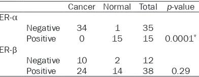
Table 2. Expression of ER subtypes in human cervical tissues
Cancer Normal Total p-value ER-α
Negative 34 1 35
Positive 0 15 15 0.0001*
ER-β
Negative 10 2 12
Positive 24 14 38 0.29
Data analyses were performed with Fisher’s exact test.
[image:3.612.91.287.196.272.2]the epithelial basal and parabasal cells in 14/16 (87.5%) normal tissues (Table 2, Figure 1C). Some normal tissues showed nuclear and cytoplasmic immunoreaction in cells through-out the all squamous epithelium (data not shown). Few stromal cells showed exclusively
nuclear immunoreaction for ER-β. To confirm
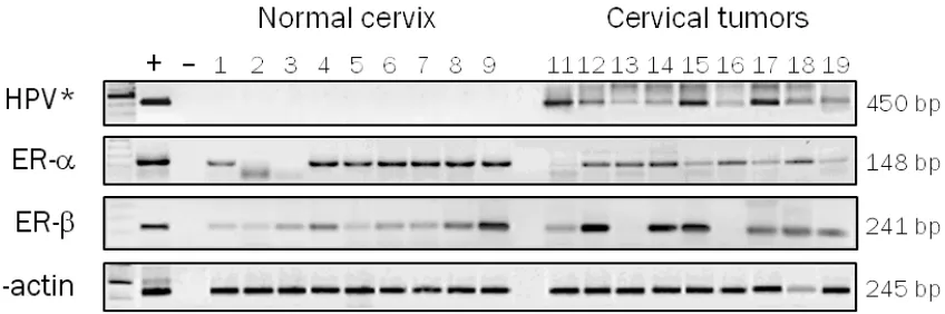
the results obtained by immunohistochemistry, analyses by RT-PCR were made in normal tis-sues chosen randomly. We detected mRNA
expression of ER-α and ER-β genes in 7/9
(77.8%) and 9/9 (100%) normal tissues, respec-tively (Figure 2). These results demonstrate
that ER-α and ER-β are expressed in the epithe -lium of adult cervical tissue and suggest
spe-cific roles in the human cervical biology.
Expression of ER-β in invasive cervical carci-noma
According to our results, ER-α expression was
not detected in any of the analyzed invasive cervical carcinomas (Table 2, Figure 1B). These
results show a statistically significant correla
-tion between the specific loss of expression of ER-α subtype and the neoplastic cervical phe
-notype (p≤0.0001) (Table 2). To confirm our
results obtained by immunohistochemistry, analyses by RT-PCR were made in invasive car-cinomas chosen randomly. We detected mRNA
transcripts of ER-α in 8 of 9 (90%) invasive car -cinomas (Figure 2) but not its protein (Figure 1B). These results suggest that a regulation
mechanism of the ER-α gene could be involved
in cervical cancer. Interestingly,
immunopositiv-ity to ER-β was observed in 24/34 (70.6%) inva -sive cervical carcinomas (Table 2). Figure 1D shows an invasive cervical carcinoma where can be observed nuclear and cytoplasmic
immunoreaction for ER-β in neoplastic cells, besides, ER-β mRNA transcripts were found in
7/9 invasive cervical tumors, a similar propor-tion of positive cases (77.6%) (Figure 2). We
found not a statistically significant correlation between the ER-β expression and the neoplas -tic phenotype (p=0.29), however, these results
demonstrate that ER-β is expressed in the cer -vical neoplastic cells and could be an important role in cervical carcinogenesis.
Discussion
The discovery of a second estrogen receptor,
ERβ, has led to a reevaluation of estrogen
action on their target tissues and its possible role in human carcinogenesis. In this study we
analyzed the expression of ER-α and ER-β in
normal and neoplastic human cervical tissues in order to determine their potential roles in cervical cancer development. Our results show Figure 1. Immunostaining of ER-α and ER-β receptors in human cervical tissues. A. Immunopositivity of ER-α in the
normal cervical epithelium showing nuclear reaction mainly in the parabasal layers (X40). B. Immunonegativity of
ER-α in invasive carcinomas (X10). C. Immunopositivity of ER-β in normal cervical epithelium showing nuclear and cytoplasmic reaction in the cells of basal and parabasal layers (X40). D. Immunopositivity of ER-β in cervical invasive
carcinoma with nuclear and cytoplasmic reaction in epithelial invasion zones (X10). E. A negative control in normal
cervical tissue consisted of the omission of the primary antibody (X40). F. Positive control of ER-α in normal breast
strong nuclear expression of ER-α in the cervi -cal squamous epithelium parabasal cells main-ly, and to a lesser extent in the basal cells and in some cells of the intermediate layers. These results agree with previous data that reported nuclear expression of ER mainly in the parabas-al cells of the normparabas-al cervicparabas-al squamous epi-thelium [12, 13]. It has been reported the expression of ER between 9% [13, 14] and 18% [12] of invasive cervical carcinomas, however,
we did not detect immunopositivity of ER-α in
the neoplastic cells on practically any of inva-sive carcinomas analyzed in this study, accord-ing with other groups [13, 15, 16].
The inconsistency between our results and those could be due to the preparation and han-dling of tissues or to the primary antibody used;
we used antibodies specific for the human ER-α and ER-β which fails to cross between them,
however other groups could have used antibod-ies that identify more than one type of ER. Zhai
et al [17] have suggested that this loss of ER-α
has a major role in the invasion and progres-sion of cervical cancer, as demonstrated by in vitro assays in cell lines derived from cervical
cancer where the inhibition of ER-α with short
hairpin RNA increased the cellular
invasive-ness, while restoring of ER-α in ER-α negative
cell lines reduced its invasiveness. Additionally,
it has been suggested that ER-α plays an
impor-tant role in the early stages of cervical
carcino-genesis in transgenic mice K14E7/ERα-/- [18].
The invasive cervical carcinoma included in our
work were negative for the ER-α protein, and
positive for HPV infection, suggesting an asso
-ciation between loss of expression specifically of ER-α and the presence of high-risk HPV,
according to previous reports [13, 14, 16].
Interestingly, we were able to detect significant levels of mRNA ER-α by RT-PCR in invasive cer -vical carcinomas. These results contrast with those found by immunohistochemistry, but a similar inconsistency between mRNA and pro-tein ER expression have also been reported in mammary gland carcinomas [19] where differ-ences in handling, preservation and prepara-tion time of tissues, as well as in the detecprepara-tion
sensitivity of the methods may have influenced
the results [20, 21].
Previous data indicate that ER-α is probably
regulated by multiple mechanisms, including transcriptional level, post-transcriptional and post-translational in breast cancer [22] and during the progression of cervical cancer [17].
ER-α have typical CpG islands within its pro
-moter and first exon, and though bisulfite
sequence analysis demonstrated promoter methylation in different cervical cancer cell
lines, ER-α is not frequently methylated in pri -mary tumors of cervical cancer [23]. Thus,
although ER-α methylation is associated with a reduced or absent expression of ER-α in some
cell lines of cervical cancer, it is probably not
involved in the negative status of ER-α in the
cervical cancer tissues without protein expres-sion [17]. According to these reports, in our study the presence of the messenger RNA of
[image:5.612.98.522.72.213.2]ER-α suggests that a regulatory mechanism at the level of ER-α promoter methylation would Figure 2. ER-α and ER-β analyses expression in human cervical tissues by RT-PCR. Invasive carcinomas and normal tissues were chosen randomly. (+) = positive control (RNA from uterus), (-) = negative control (H2O), 1-9 = normal
not be involved; so our findings suggest that the down regulation of ER-α in CC could be due to a
mechanism of post-transcriptional regulation,
as has been suggested for the ER-β subtype [24, 25]. Additionally, the loss of ER-α could be
due to the dedifferentiation progressive of the cancer cells caused by the ubiquitination and proteolysis processes or by a mechanism of post-translational regulation as has been sug-gested previously in breast cancer [25]. Thus, down regulation of ER expression may be the
first alteration to take place in normal epitheli -um during the development of cervical
dyspla-sia in women infected with high-risk HPV [26].
On the other hand, there is limited and contra-dictory information about the expression of
ER-β in the human uterine cervix [8, 9, 18] and
its role in human cancer is not completely
understood. Beside the presence of ER-α, we identified nuclear expression of ER-β in the nor -mal cervical tissue with an epithelial
expres-sion pattern similar to the ER-α, and it has been
speculated that the biology effect of estrogens might be governed by the relative expression of both estrogen receptors on the same tissue
[27], so, the simultaneous expression of ER-α and ER-β in the normal cervix suggest that each subtype has specific physiological functions in
the human cervical epithelium. Furthermore,
ER-β can alter gene expression profiles in the
presence of estradiol [28] and regulate the expression of genes associated with cell cycle
and apoptosis [28], whereby ER-β could consti -tute a tumor suppressor gene with antiprolifer-ative capacities [29].
We found immunopositivity for the ER-β protein
in the most (70.6%) of invasive cervical carci-noma in our study. These results demonstrate
that cervical neoplastic cells express ER-β and
suggest that estrogens could be regulating
some cervical tumors functions via the ER-β. Furthermore, a high expression of ER-β in endo -metrial cancer samples with severe myometri-um invasion has been reported [30]. Some of the proteins involved in invasion and metasta-sis as metalloproteinase MMP2 and MMP9, are activated by the estrogen effects [31], over-regulated in cervical cancer [32] and maybe activated in these neoplastic cervical cells
through ER-β [33].
Previous results published by our group have shown overexpression of MMP9, 10, 11 and 12
in invasive cervical carcinomas [34], so that some of the mechanisms of invasion in cervical cancer could be also governed in part by the
activation of MMPs via the ER-β subtype. Additionally, ER-β might have a role in transcrip -tional activation of MMP9 in the invasive cervi-cal carcinomas by the no canonicervi-cal (genomic) way through to the activation of theirs sites AP-1 [35].
We detected significant levels of ER-β in the
invasive cervical carcinomas, and interestingly, almost exclusively cytoplasmic staining was found in the neoplastic cells in these tumors.
This pattern of cytoplasmic expression of ER-β
has also been reported in vulvar squamous cell carcinoma [36], breast cancer [37] and colorec-tal cancer [38].
Although the ER-β is a transcription factor, changing ER-β nuclear expression in healthy
tissue to cytoplasmic expression in neoplastic
cells was an interesting finding; the presence of ER-β in the cytoplasm of the neoplastic cells may indicate well-defined roles on the gene activation. The presence of ER-β in cytoplasm
could be due to the transcriptional activation of
ER-β that shows preferentially by the
non-genomic way rather than the classic way [39], modulating the transcriptional activity of AP-1 sites by the non-genomic way. Additionally, the cytoplasmic immunoreaction in the neoplastic
cervical cells may be due in part to the specific expression of some isoforms of ER-β which can
present nuclear or cytoplasmic expression that has been associated with different prognoses in human cancer depending on the isoform involved and its subcellular distribution [22, 40, 41], and apparently could be related to neo-plastic progression with a high incidence of car-cinoma in situ/invasive compared with healthy tissue [42].
The specific cytoplasmic expression of ER-β2 and ER-β5 isoforms has been associated with
an overall survival poor and metastasis in breast and prostate cancers [40, 43]. In our study, the neoplastic cells presented
cytoplas-mic staining of ER-β in most of the cervical
tumors, but we were unable to discriminate any
specific receptor isoform since our primary anti -body recognizes the N-terminal domain present
invasive carcinomas, and if these could play a role in the cervical neoplastic tissue. We are
currently investigating the specific expression of ER-β isoforms in invasive tumors and precan -cerous lesions of the uterine cervix and its association with clinical variables in an attempt to determine if the expression of any isoform may be relevant as a diagnostic marker and/or important to the prognosis in cancer patients cervical or as a target for the development of new therapies in this neoplasm.
In conclusion, our results demonstrate that
ER-α and ER-β are expressed in the normal cer -vical epithelium, but only ER-β is expressed en
the invasive cervical tumors. The loss of ER-α
could be due to mechanisms of post-transcrip-tional and/or post-translapost-transcrip-tional regulation in
the ER-α gene. The loss of ER-α is likely one of
the factors involved in the onset of
carcinogen-esis process while that ER-β expression might
be involved in the subsequent stages in this
malignancy. Furthermore, our findings suggest that the loss of expression specifically of ER-α
is that associated with infection with high-risk
HPV, mainly with the viral types HPV16 and 18.
Our results are not conclusive, but suggest that
cytoplasmic expression of ER-β can be impor -tant in cervical cancer. Since tumor cells do not
express ER-α, any effect of estrogens on these cells could be mediated through ER-β in cervi -cal cancer.
Acknowledgments
This work was partially supported by grants from CONACyT-Mexico No. 87244 and IMSS- FIS/478.
Disclosure of conflict of interest
We have no conflict of interest in association
with this work.
Address correspondence to: Dr. Mauricio Salcedo, Laboratorio de Oncología Genómica, Unidad de Investigación Médica en Enfermedades Oncológicas,
Hospital de Oncología, CMN SXXI-IMSS. Av.
Cuauhtémoc 330, Col. Doctores, México, Distrito Federal 06720, México. Phone: 55-56276900 Ext. 22706; E-mail: [email protected]
References
[1] Nair HB, Luthra R, Kirma N, Liu YG, Flowers L,
Evans D, Tekmal RR. Induction of aromatase
expression in cervical carcinomas: effects of endogenous estrogen on cervical cancer cell proliferation. Cancer Res 2005 Dec 1; 65: 11164-73.
[2] Harris HA, Bapat AR, Gonder DS, Frail DE. The ligand binding profiles of estrogen receptors
alpha and beta are species dependent. Ste-roids 2002 Apr; 67: 379-84.
[3] Mosselman S, Polman J and Dijkema R. ERβ: Identification and characterization of a novel
human estrogen receptor. FEBS Lett 1996; 392: 49-53.
[4] Kuiper G and Gustafsson A. The novel estro-gen receptor-b subtype: potential role in the
cell- and promoter-specific actions of estro -gens and anti-estro-gens. FEBS Lett 1997; 410: 87-90.
[5] Pelletier G and El-Alfy M. Immunocytochemical
localization of estrogen receptors α and β in
the human reproductives organs. J Clin Endo-crinol Metabol 2000; 85: 4835-4840. [6] Younes M, Honma N. Estrogen receptor β. Arch
Pathol Lab Med 2011 Jan; 135: 63-6. [7] Cavaillès V. Estrogens and receptors: an evolv
-ing concept. Climacteric 2002 Jun; 5 Suppl 2: 20-6.
[8] Stygar D, Wang H, Vladic YS, Ekman G, Eriks
-son H, Sahlin L. Co-localization of oestrogen
recptor β and leukocyte markers in the human cervix. Mol Hum Reprod 2001; 7: 881-886.
[9] Wang H, Stjernholm Y, Ekman G, Eriksson H,
Sahlin L. Different regulation of oestrogen re-ceptors alpha and beta in the human cervix at
term pregnancy. Mol Hum Reprod 2001 Mar;
7: 293-300.
[10] Manos MM, Ting Y, Wright DK. Use of Poly
-merase Chain Reaction amplification for the detection of genital Human Papillomaviruses.
In: Furth M, Greaves M, editors. Cancer cells
7/Molecular Diagnostics of Human Cancer. U.S.A.: Cold Spring Harbor Lab Press 1989; pp:
209-214.
[11] Leygue E, Dotzlaw H, Watson PH and Murphy LC. Altered estrogen receptor α and β Messen -ger RNA expresión during human breast tu-morigenesis. Cancer Res 1998; 58: 3197-3201.
[12] Kanai M, Shiozawa T, Xin L, Nikaido T, Fujii S. Immunohistochemical detection of sex ster-poid receptor, cyclins, and cyclin-dependent kinases in the normal snd neoplastic squa-mous epithelia of the uterini cervix. Cancer 1998; 82: 1709-1719.
[14] Konishi I, Fujii S, Nonogaki H, Nanbu Y, Iwai T,
Mori T. Immnunohistochemical analisys of es-trogen receptor, progesterone receptors, Ki-67 antigen, and normal papillomavirus DNA in normal and neoplastic epithelium of the uter-ine cervix. Cancer 1991; 68: 1340-1350. [15] Mosny DS, Herholz J, Degen W, Bender HG. Im
-munohistochemical investigations of steroid receptors in normal and neoplastic squamous epithelium of the uterine cervix. Gynecol Oncol 1989 Dec; 35: 373-7.
[16] Monsonego J, Magdelenat H, Catalan F, Cos
-cas Y, Sastre X. Estrogen and progesterone re -ceptors in cervical human papillomavirus re-lated lesions. Int J Cancer 1991; 48: 533-539. [17] Zhai Y, Bommer GT, Feng Y, Wiese AB, Fearon
ER, Cho KR. Loss of estrogen receptor 1 en-hances cervical cancer invasion. Am J Pathol 2010 Aug; 177: 884-95.
[18] Chung SH, Wiedmeyer K, Shai A, Korach KS,
Lambert PF. Requirement for estrogen recep-tor alpha in a mouse model for human papillo-mavirus-associated cervical cancer. Cancer Res 2008 Dec 1; 68: 9928-34.
[19] Graham DM, Jin L, Lloyd RV. Detection of estro
-gen receptor in paraffin-embedded sections of
breast carcinoma by immunocytochemistry and in situ hibridization. Am J Surg Pathol 1991; 15: 475-485.
[20] Wu X, Subramaniam M, Negron V, Cicek M,
Reynolds C, Lingle WL, Goetz MP, Ingle JN,
Spelsberg TC, Hawse JR. Development, char -acterization, and applications of a novel estro-gen receptor beta monoclonal antibody. J Cell Biochem 2012 Feb; 113: 711-23.
[21] Carder PJ, Murphy CE, Dervan P, Kennedy M, McCann A, Saunders PT, Shaaban AM, Foster
CS, Witton CJ, Bartlett JM, Walker RA, Speirs V.
A multi-centre investigation towards reaching a consensus on the immunohistochemical
de-tection of erbeta in archival formalinfixed par
-affin embedded human breast tissue. Breast
Cancer Res Treat 2005; 92: 287-293. [22] Parl FF. Multiple mechanisms of estrogen
re-ceptor gene repression contribute to ER-nega-tive breast cancer. Pharmacogenomics J 2003; 3: 251-253.
[23] Wentzensen N, Sherman ME, Schiffman M, Wang SS. Utility of methylation markers in cer-vical cancer early detection: appraisal of the state-of-the-science. Gynecol Oncol 2009; 112: 293-299.
[24] Jarzabek K, Koda M, Kozlowski L, Mittre H,
Sulkowski S, Kottler ML, Wolczynski S. Distinct mRNA, protein expression patterns and
distri-bution of oestrogen receptors α and β in hu -man primary breast cancer: Correlation with proliferation marker Ki-67 and
clinicopatho-logical factors. Eur J Cancer 2005; 41: 2924-2934.
[25] Smith L, Coleman LJ, Cummings M, Satheesha
S, Shaw SO, Speirs V, Hughes TA. Expression of
oestrogen receptor beta isoforms is regulated by transcriptional and post-transcriptional mechanisms. Biochem J 2010 Jul 15; 429: 283-90.
[26] Bekkers RL, van der Avoort IA, Melchers WJ, Bulten J, de Wilde PC, Massuger LF. Down reg-ulation of estrogen receptor expression is an early event in human papillomavirus infected cervical dysplasia. Eur J Gynaecol Oncol 2005; 26: 376-82.
[27] Böttner M, Thelen P, Jarry H. Estrogen receptor
beta: Tissue distribution and the still largely enigmatic physiological function. J Steroid Bio-chem Mol Biol 2013 Mar 20; pii: S0960-0760(13)00052-6.
[28] Chang EC, Frasor J, Komm B, Katzenellenbo-gen BS. Impact of estroKatzenellenbo-gen receptor beta on gene networks regulated by estrogen receptor alpha in breast cancer cells. Endocrinology 2006 Oct; 147: 4831-42.
[29] Morani A, Warner M, Gustafsson JA. Biological functions and clinical implications of oestro-gen receptors alfa and beta in epithelial tis-sues. J Intern Med 2008 Aug; 264: 128-42. [30] Takama F, Kanuma T, Wang D, Kagami I,
Mizunuma H. Oestrogen receptor β expression
and depth of myometrial invasion in human endometrial cancer. Br J Cancer 2001; 84: 545-549.
[31] Merlo S, Sortino MA. Estrogen activates matrix metalloproteinases-2 and -9 to increase beta amyloid degradation. Mol Cell Neurosci 2012 Apr; 49: 423-9.
[32] Rouyer N, Wolf C, Chenard MP, Rio MC, Cham-bon P, Bellocq JP, Basset P. Stromelysin-3 gene expression in human cancer: an overview. In-vasion Metastasis 1994-1995; 14: 269-75. [33] Lu T, Achari Y, Rattner JB, Hart DA. Evidence
that estrogen receptor beta enhances
MMP-13 promoter activity in HIG-82 cells and that this enhancement can be influenced by li
-gands and involves specific promoter sites.
Biochem Cell Biol 2007 Jun; 85: 326-36. [34] Vazquez-Ortiz G, Pina-Sanchez P, Vazquez K,
Duenas A, Taja L, Mendoza P, Garcia JA, Sal-cedo M. Overexpression of cathepsin F, matrix metalloproteinases 11 and 12 in cervical can-cer. BMC Cancer 2005 Jun 30; 5: 68. [35] Yoshizaki T, Sato H, Murono S, Pagano JS, Fu
-rukawa M. Matrix metalloproteinase 9 is in-duced by the Epstein-Barr virus BZLF1 transac-tivator. Clin Exp Metastasis 1999 Jul; 17: 431-6.
[36] Stefano I, Vizzielli G, Tortorella L, Fagotti A,
oestrogen receptor beta (ERβ) as a prognostic
factor in vulvar squamous cell carcinoma in
el-derly women. Histopathology 2011 Nov; 59:
909-17.
[37] Fuqua SA, Schiff R, Parra I, Moore JT, Mohsin SK, Osborne CK, Clark GM, Allred DC. Estrogen receptor beta protein in human breast cancer: correlation with clinical tumor parameters. Cancer Res 2003 May 15; 63: 2434-9. [38] Xie LQ, Yu JP, Luo HS. Expression of estrogen
receptor beta in human colorectal cancer. World J Gastroenterol 2004 Jan 15; 10: 214-7. [39] Björnström L, Sjöberg M. Estrogen receptor-dependent activation of AP-1 via non-genomic signalling. Nucl Recept 2004 Jun 14; 2: 3. [40] Shaaban AM, Green AR, Karthik S, Alizadeh Y,
Hughes TA, Harkins L, Ellis IO, Robertson JF, Paish EC, Saunders PT, Groome NP, Speirs V.
Nuclear and Cytoplasmic Expression of ERB1,
ERB2, and ERB5 Identifies Distinct Prognostic
Outcome for Breast Cancer Patients. Clin Can-cer Res 2008 Aug 15; 14: 5228-35.
[41] Shaaban AM, O’Neill PA, Davies MP, Sibson R,
West CR, Smith PH, Foster CS. Declining estro
-gen receptor-h expression defines malignant
progression of human breast neoplasia. Am J Surg Pathol 2003; 27: 1502-12.
[42] Shaaban AM, Jarvis C, Moore F. Prognostic
sig-nificance of estrogen receptor h in epithelial
hyperplasia of usual type with known outcome. Am J Surg Pathol 2005; 29: 1593-9.