Original Article
A RTK-based functional RNAi screen reveals
determinants of PTX-3 expression
Hua Liu*, Xin-Kai Qu*, Fang Yuan, Min Zhang, Wei-Yi Fang
Department of Cardiology, Shanghai Chest Hospital affiliated to Shanghai JiaoTong University, Shanghai, China. *These authors contributed equally to this work.
Received January 30, 2013;Accepted February 15, 2013; Epub March 15, 2013; Published April 1, 2013
Abstract: Aim: The aim of the present study was to explore the role of receptor tyrosine kinases (RTKs) in the regu-lation of expression of PTX-3, a protector in atherosclerosis. Methods: Human monocytic U937 cells were infected with a shRNA lentiviral vector library targeting human RTKs upon LPS stimuli and PTX-3 expression was determined by ELISA analysis. The involvement of downstream signaling in the regulation of PTX-3 expression was analyzed by both Western blotting and ELISA assay. Results: We found that knocking down of ERBB2/3, EPHA7, FGFR3 and RET impaired PTX-3 expression without effects on cell growth or viability. Moreover, inhibition of AKT, the downstream effector of ERBB2/3, also reduced PTX-3 expression. Furthermore, we showed that FGFR3 inhibition by anti-cancer drugs attenuated p38 activity, in turn induced a reduction of PTX-3 expression. Conclusion: Altogether, our study demonstrates the role of RTKs in the regulation of PTX-3 expression and uncovers a potential cardiotoxicity effect of RTK inhibitor treatments in cancer patients who have symptoms of atherosclerosis or are at the risk of athero-sclerosis.
Keywords: PTX-3, RNAi screening, RTKs, target therapy, cardiotoxicity, atherosclerosis
Introduction
Treatment of patients with cancer has changed radically over the last several years with the advent of “targeted therapeutics.” This target-ed approach, prtarget-edominantly via inhibition of tyrosine kinase activity, has markedly improved the management of cancers. Given the initial success of this approach, the number of tar-geted therapy drugs entering into development in the last 5 years has increased dramatically. And so far, there are hundreds of kinase inhibi-tors somewhere in between discovery and mar-ket, with 80% of drug development being in cancer [1].
Although target therapy has an improve antitu-mor activity with fewer toxic side effects than traditional anticancer therapies including radia-tion therapies and chemotherapies, many approved RTK inhibitors have been found to evoke cardiac dysfunction in some cancer patients [2-4]. Furthermore, there is a strong link between kinase pathway inhibition and car-diotoxicity as compared with other organ
toxici-ties [5-7]. However, in many cases, adverse car-diac events in the clinic were not anticipated based on the fact that predefined cardiac end-points was not included in early clinical trials and the difficulties to diagnose heart failure in patients with cancer. Since kinase inhibition has revolutionized the treatment of cancer, which can be managed effectively for years and could be eventually regarded as a chronic dis-ease, one could except that the rate of cardio-toxicity in cancer patients treated with kinase inhibitors would increase in future.
kinase inhibitors on atherosclerosis in cancer patients have not been fully investigated due to lack of appropriate preclinical model.
Given the critical role of inflammation in athero-sclerosis, circulating factors related to inflam-mation and atherosclerosis therefore attracted attention [11]. Of interest, pentraxin protein family is highly associated with CVD [11]. Pentraxin protein family consists of C-reactive protein (CRP), serum amyloid P component (SAP) and pentraxin-3 (PTX-3) [11-13], all of which have the features of pattern recognition receptors and are involved in human humoral immune response [14, 15]. In contrast to CRP, PTX-3 demonstrates to be more specifically associated with advanced atherosclerosis [15-17]. PTX-3 is highly expressed in advanced ath-erosclerosis tissues [18, 19], including macro-phages, surviving endothelial cells, activated monocytes and infiltrating neutrophils [19]. Amounts of evidence suggest the possibility that the increased levels of PTX-3 in subjects with CVD may reflect a protective physiologic response that correlates with the severity of the disease [20-22]. More importantly, defi-ciency of the long pentraxin PTX-3 promotes vascular inflammation and atherosclerosis [23].
To systematically investigate the function of receptor tyrosine kinases (RTKs), the main tar-gets in target cancer therapies, on atheroscle-rosis, we set out to screen a short hairpin RNA (shRNA) library representing the full comple-ment of 56 human RTKs (Table 1) for genes whose inhibition could impair PTX-3 expression in U937 cells. Our results revealed multiple functionally important pathways, particularly the FGFR/p38 signaling in the regulation of PTX-3 expression. Our study suggests that clini-cally used kinase inhibitor might contribute to atherosclerosis, in turn inducing cardiotoxicity, by interfering with PTX-3 expression. These findings could provide a preclinical model for studying the side effects of kinase inhibitors on atherosclerosis.
Materials and methods
Cell culture
293T cells (ATCC) were cultured in DMEM medi-um supplemented with 10% FBS (Hyclone). U937 cells (ATCC, CRL-1593.2) were grown in
RPMI 1640 medium supplemented with 10% FBS (Hyclone). All media were supplemented with penicillin (100 IU·ml-1) and Streptomycin (100 μg·ml-1) (Life Technologies).
Induction of PTX-3 expression by LPS stimuli
The cells were washed and incubated in endo-toxin-free RPMI 1640 (10% FBS) at 5 X 10⁴ cells each well in 24-well plate, with or without LPS (lipopolysaccharide, Escherichia coli 0127:B8, Sigma, L4516 ) stimuli for 6 hrs at 37°C in the presence of 5% CO2. The optimized concentra-tion of LPS to induce PTX-3 expression was tested with 0, 20, 50 and 100 ng· ml-1.
U937 cells induced by series of concentration of LPS were homogenized with 0.5 ml Trizol (life technologies) and total RNA was isolated as instruction manual (life technologies). Reverse transcription was carried out at 42°C for 1 hour with oligo dT. Real-time PCR primers were designed as follows: forward primer: tgtatgt-gaatttggacaacgaa; reverse primer: cattccgagt-gctcctgac. Reaction program was 2 min at 95°C, 40 cycles of 95°C for 30 s, 60°C for 30 s and 72°C for 30 s. The concentration of LPS induced the maximal expression of PTX-3 was selected to carry out RNAi screening.
RNAi screening
U937 cells were incubated in endotoxin-free RPMI 1640 (10% FBS) at 1 × 10⁴ cells each well in 96-well plates, and then infected respective-ly with 55 RTK lentiviral shRNAs in the pres-ence of polybrene to the final concentration of 8 μg· ml-1, each gene in the library was duplicate in two plates. LPS was added to cell culture to final concentration 100 ng· ml-1 120 h after infection for 6 hrs. After the treatment, cells were pelleted and divided into two parts: the supernate was collected to measure PTX-3 lev-els by ELISA, and the pellets were used to detect cell viability by MTS assay (Promega, G3580) according to the operation manual.
Determination of PTX-3 expression by ELISA assay
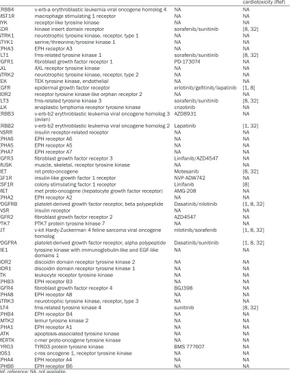
Table 1. RTKs genes for screen
Gene symbol Official Full Name Inhibitor Associated with
cardiotoxicity (Ref)
ERBB4 v-erb-a erythroblastic leukemia viral oncogene homolog 4 NA NA
MST1R macrophage stimulating 1 receptor NA NA
RYK receptor-like tyrosine kinase NA NA
KDR kinase insert domain receptor sorafenib/sunitinib [8, 32]
NTRK1 neurotrophic tyrosine kinase, receptor, type 1 NA NA
STYK1 serine/threonine/tyrosine kinase 1 NA NA
EPHA3 EPH receptor A3 NA NA
FLT1 fms-related tyrosine kinase 1 sorafenib/sunitinib [8, 32]
FGFR1 fibroblast growth factor receptor 1 PD-173074 NA
AXL AXL receptor tyrosine kinase NA NA
NTRK2 neurotrophic tyrosine kinase, receptor, type 2 NA NA
TEK TEK tyrosine kinase, endothelial NA NA
EGFR epidermal growth factor receptor erlotinib/gefitinib/lapatinib [1, 8]
ROR2 receptor tyrosine kinase-like orphan receptor 2 NA NA
FLT3 fms-related tyrosine kinase 3 sorafenib/sunitinib [8, 32]
ALK anaplastic lymphoma receptor tyrosine kinase crizotinib NA
ERBB3 v-erb-b2 erythroblastic leukemia viral oncogene homolog 3
(avian) AZD8931 NA
ERBB2 v-erb-b2 erythroblastic leukemia viral oncogene homolog 2 Lapatinib [1, 32]
INSRR insulin receptor-related receptor NA NA
EPHA6 EPH receptor A6 NA NA
EPHA5 EPH receptor A5 NA NA
EPHA7 EPH receptor A7 NA NA
FGFR3 fibroblast growth factor receptor 3 Linifanib/AZD4547 NA
MUSK muscle, skeletal, receptor tyrosine kinase NA NA
RET ret proto-oncogene Motesanib [8, 32]
IGF1R insulin-like growth factor 1 receptor NVP-ADW742 NA
CSF1R colony stimulating factor 1 receptor Linifanib [8]
MET met proto-oncogene (hepatocyte growth factor receptor) AMG-208 NA
EPHA2 EPH receptor A2 NA NA
PDGFRB platelet-derived growth factor receptor, beta polypeptide Dasatinib/nilotinib [1, 8, 32]
INSR insulin receptor NA NA
FGFR2 fibroblast growth factor receptor 2 AZD4547 NA
PTK7 PTK7 protein tyrosine kinase 7 NA NA
KIT v-kit Hardy-Zuckerman 4 feline sarcoma viral oncogene
homolog nilotinib/sorafenib [1, 8, 32]
PDGFRA platelet-derived growth factor receptor, alpha polypeptide Dasatinib/sunitinib [1, 8, 32] TIE1 tyrosine kinase with immunoglobulin-like and EGF-like
domains 1 NA NA
DDR2 discoidin domain receptor tyrosine kinase 2 NA NA
DDR1 discoidin domain receptor tyrosine kinase 1 NA NA
LTK leukocyte receptor tyrosine kinase NA NA
EPHB3 EPH receptor B3 NA NA
FGFR4 fibroblast growth factor receptor 4 BGJ398 NA
EPHA8 EPH receptor A8 NA NA
NTRK3 neurotrophic tyrosine kinase, receptor, type 3 NA NA
FLT4 fms-related tyrosine kinase 4 sunitinib [8, 32]
EPHB4 EPH receptor B4 NA NA
LMTK2 lemur tyrosine kinase 2 NA NA
EPHA1 EPH receptor A1 NA NA
AATK apoptosis-associated tyrosine kinase NA NA
MERTK c-mer proto-oncogene tyrosine kinase NA NA
TYRO3 TYRO3 protein tyrosine kinase BMS 777607 NA
ROS1 c-ros oncogene 1, receptor tyrosine kinase NA NA
EPHA4 EPH receptor A4 NA NA
EPHB6 EPH receptor B6 NA NA
Ref, reference; NA, not available.
used to wash plates thoroughly after each step. Non-specific binding to the plates was blocked with 2% BSA in PBS for 2 h at room tempera-ture before adding unknown samples. After incubation for 2 h at room temperature, 10 ng each well of biotin conjugated anti-PTX-3 goat
IgG were then added (1 h at room temperature) followed by the addition of 50 μl of streptavi-din-peroxidase. Finally, 100 μl of Substrate
Solution (1:1 mixture of Color Reagent A (H2O2)
and Color Reagent B (Tetramethylbenzidine),
and absorbance values were read at 450 nm in an automatic ELISA reader.
Protein Isolation and Western Blotting
Cell pellets were resuspended in 1×SDS load-ing buffer (1 mmol·L-1 Na
3VO4, 10 mmol·L-1 NaF, 1 mmol·L-1 PMSF) containing protease inhibi-tors. Lysates (20 μg each lane) were applied to SDS-PAGE. Immunoblotting of Abs specific for GAPDH (Abmart, 080922), p38 (A-12, Santa Cruz, sc-7972) and p-p38 (D-8, Santa Cruz, SC-7973) were detected using HRP-conjugated anti-mouse (Promega) and visualized by chemi-luminescence detection system (Millipore, WBKLS0500).
Results
Loss-of-function Screen for RTKs regulating PTX-3 expression
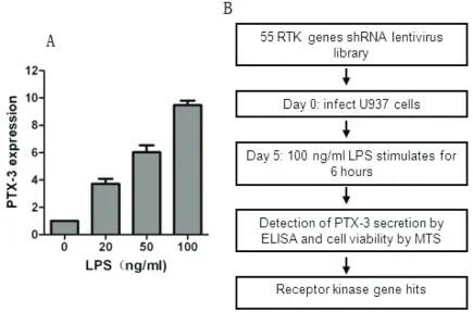
PTX-3 is produced by macrophages and a vari-ety of tissue cells upon exposure to LPS, which
has been commonly used as a reagent to induce inflammatory response [15]. Consistent with previous reports, we found a dose-depen-dent induction in PTX-3 expression after LPS stimulation in monoblastic U937 cells (Figure 1A). We therefore analyze PTX-3 expression in U937 cells after treatment with LPS at 100 ng· ml-1 for 6 hrs with or without other treatment in our experiments.
[image:4.612.89.524.75.362.2]Given that RTKs have been widely demonstrat-ed as most important driver genes for tumor development, many small molecular inhibitors are designed to inhibit RTKs and a bunch of RTK inhibitors have been extensively used in both cancer treatment and clinical trials. Hence, the lost-of-function screen used a shRNA library targeting human RTKs involved in cancer, in which each shRNA induces strong and specific suppression of gene expression over prolonged period of time. We configured the screen to allow the identification of RTKs that regulate the expression of PTX-3 in U937
Figure 1. Schematic of RNAi screen. A. U937 cells were treated with 100 ng·ml-1 of LPS for 6 hrs and PTX-3
expres-sion was determined by qPCR analysis. B. Schematic outline of high-throughput RNAi screen. The human RTKs
lentiviral vector library was used to infect U937 cells for 4 days to allow efficient knocking down of RTKs and was
then treated with LPS at 100 ng·ml-1 for 6 hrs. The supernatant media and U937 cells were collected by centrifuged
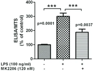
Figure 3. The downstream signaling involved in RTKs regulating PTX-3 secretion induced by LPS. U937 cells were pretreated with AKT inhibitor MK2206 for 24 hrs and then added with LPS for another 6 hrs. U937 cells were centrifuged and analyzed by MTS assay. The supernatant were collected and PTX-3 se-cretion was determined by ELISA assay. The ELISA/ MTS value was calculated as described in Materials and methods.
cells (Figure 1B). The output of the screen is described in Table S1 available online. The screen was carried out in duplicate and the expression of PTX-3 was detected by ELISA as described in Material and Methods. Genes that could affect survival and proliferation of U937 cells were also obtained by MTS assay (Table S1). The ratio of the ELISA to MTS value after infection with RTKs shRNA lentiviral libraries and the mean value of the reduction of ELISA/ MTS was calculated (Table S1 and Figure 2A). We set a cutoff for gene with > 30% of reduc-tion in the value of ELISA/MTS to be considered potential candidates, and 5 genes were obtained (Figure 2B).
Characterization of RTKs with respect to PTX-3 expression
Several of the positive regulators of PTX-3 expression we identified were already shown to have cardiotoxicity. Of particular interest were ERBB2 and ERBB3, inhibitors of which are widely used in breast cancer target therapy
[24]. Expression of both ERBB2 and ERBB3 is usually enhanced during tumorigenesis, result-ing in promotion of cell proliferation and inhibi-tion of apoptosis [25]. We found a 30-40% of reduction in PTX-3 expression after either ERBB2 or ERBB3 inhibition (Table 1), indicating the potential toxicity of ERBB2/3 signaling inhi-bition by influencing atherosclerosis regulation. Additionally, we identified RET as a regulator of PTX-3 expression (Figure 2B). Knocking down of RET expression induced a 40% of reduction in PTX-3 expression (Table 1). Interestingly, RET inhibitor, sunitinib, has also been widely recog-nized as an inducer of cardiotoxicity [3, 26]. Since AKT is a canonical downstream signaling of ERBB2/3 signaling and AKT inhibitor is a promising anti-cancer drug on clinical trials, we examine whether inhibition of AKT signaling could also impair PTX-3 expression. We treated U937 cells with AKT inhibitor, MK2206, which is on clinical trials and found that, similar with inhibition of ERBB2/3 signaling, AKT signaling inhibition reduced PTX-3 expression (Figure 3), suggesting that ERBB2/3 might regulate PTX-3 expression via AKT signaling and could in turn influence atherosclerosis.
Identification of p38 signaling in the regulation of atherosclerosis dysfunction
FGFR3 has been shown to drive oncogenesis in a subset of patients with multiple myeloma and
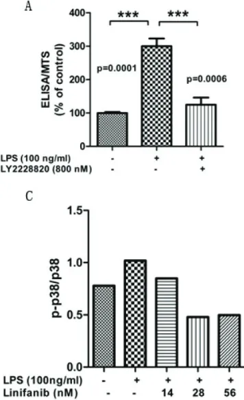
[image:5.612.329.518.76.220.2]some epithelial cancers. We found that knock-ing down of FGFR3 expression induced a 30% of reduction in PTX-3 expression (Figure 2B). Notably, FGFR3 inhibition had no effect on cell viability (Figure S1), indicating that the inhibi-tion of PTX-3 expression is not due to cytotoxicity.
Next, we explored the mechanism by which FGFR3 regulates PTX-3 expression. Since p38 signaling has an important role in the regula-tion of atherosclerosis [27], we examined p38 signaling activity after FGFR3 inhibition. We found that treatment of Linifanib, a potent FGFR3 inhibitor, induced a dose-dependent reduction of phosphorylation of p38 (Figure 4B, C). Consistently, p38 inhibitor treatment impaired LPS-induced PTX-3 expression (Figure 4A). Collectively, these data demonstrated a link between FGFR3-p38 signaling and the reg-ulation of PTX-3 expression.
Discussion
Cardiovascular disease, a leading cause of mortality in developed countries, is mainly caused by atherosclerosis, a chronic
inflamma-tory disease. Atherosclerosis is referred to as a hardening or furring of the arteries, in which an artery wall thickens as a result of the accumula-tion of fatty materials such as cholesterol. PTX-3, a molecule acting as the humor arm of innate immunity, is produced by the major cell types in atherosclerotic lesion in response to inflamma-tory stimuli. Previous reports points out a potential protective effect of PTX-3 in the ath-erosclerotic, which PTX-3 deficiency is associ-ated with increased atherosclerosis in apolipo-protein-E-deficient mice and increased macrophage accumulation in the atheroscle-rotic lesions.
[image:6.612.94.272.79.370.2]Although growing evidences show a cardiotoxic-ity induced by target therapies with kinase inhibitors, little is known about the side effects on atherosclerosis. In this study, we demon-strate the utility of an RNAi-based screen to identify molecules required for the regulation of PTX-3 expression, which is critical for the pro-tection of atherosclerosis. The results present-ed here raise the possibility that treatment with multiple RTK inhibitors contributes to the accel-erating process of atherosclerosis because the expression of PTX-3, an important protector of
atherosclerosis, is impaired. To our knowledge, it is the first time to systematically explore the side effect of RTK inhibition on atherosclerosis.
Previous report which studied FGFR3 expres-sion between normal and athermanous human arteries show that FGFR3 exhibited more restricted patterns of distribution within the plaque [28], suggesting an important role of FGFR3 in the regulation of atherogenesis. Consistent with this, we demonstrate that depleting FGFR3 expression induced a reduc-tion of PTX-3 expression via p38 signaling. While FGFR3 functions in tumor development and therefore is a promising drug target for can-cer therapy [29, 30], our evidence suggests a possibility of cardiotoxicity side effect when treating cancer patients with FGFR3 inhibitor. Further analysis about the markers associated with atherosclerosis in either animal models or target therapy treated cancer patients who have less severe atherosclerosis are needed to demonstrate in vivo effects.
Moreover, we provide a novel explanation about the cardiotoxicity effect of cancer therapies tar-geting ERBB2/3. Retrospective studies have reported incidences of symptoms of heart fail-ure or LV dysfunction as high as 35% causing discontinuation of therapy in 20% of cancer patients with ERBB2/3 inhibition treatment [31]. Here, we found that ERBB2/3 inhibition impairs PTX-3 expression, indicating a highly possibility that ERBB2/3 inhibition therapy might accelerate the risk of atherosclerosis and in turn induce cardiotoxicity. Importantly, we also explored the potential mechanism by which ERBB2/3 inhibition impairs PTX-3 expression and found the involvement of AKT signaling in this side effect. It is worth noting that our results suggest the therapy involving an AKT inhibitor need to be careful when can-cer patients either have symptoms of athero-sclerosis or are at high risk of atheroathero-sclerosis. In conclusion, we have used a RTKs-based loss-of-function screening approach to system-atically explore the link between RTKs, com-mon cancer drug targets, and atherosclerosis, one of the most common cardiovascular dis-eases. Our study has unearthed a novel mech-anism by which multiple RTK inhibition-based cancer therapies could induce cardiotoxicity via accelerating atherosclerosis.
Acknowledgments
We gratefully acknowledge Lei Xiong, Feng-Qing Li, Lin Shi and Li Zhang for helpful discussion, Tao Zhou for the technical supports of western blot analysis. This work was supported by grants of The National Natural Science Foundation of China (81000087), Basic Research of Science and Technology Commission of Shanghai Municipality (10JC1414000), Scientific Research Project of City Health Bureau of Shanghai (20114Y092) and The Development of Science and Technology Fund Project of Shanghai Chest Hospital (YZ11-07).
Address correspondence to: Dr. Wei-Yi Fang, Department of Cardiology, Shanghai Chest Hospital
affiliated to Shanghai JiaoTong University, Shanghai,
China. E-mail: [email protected]
References
[1] Force T, Kerkela R. Cardiotoxicity of the new cancer therapeutics--mechanisms of, and ap-proaches to, the problem. Drug discovery to-day2008; 13: 778-784.
[2] Seidman A, Hudis C, Pierri MK, Shak S, Paton V, Ashby M, Murphy M, Stewart SJ, Keefe D. Cardiac dysfunction in the trastuzumab clini-cal trials experience. J Clin Oncol2002; 20: 1215-1221.
[3] Chu TF, Rupnick MA, Kerkela R, Dallabrida SM, Zurakowski D, Nguyen L, Woulfe K, Pravda E, Cassiola F, Desai J, George S, Morgan JA, Har-ris DM, Ismail NS, Chen JH, Schoen FJ, Van den Abbeele AD, Demetri GD, Force T, Chen MH. Cardiotoxicity associated with tyrosine kinase inhibitor sunitinib. Lancet 2007; 370: 2011-2019.
[4] Escudier B, Eisen T, Stadler WM, Szczylik C, Oudard S, Siebels M, Negrier S, Chevreau C, Solska E, Desai AA, Rolland F, Demkow T, Hut-son TE, Gore M, Freeman S, Schwartz B, Shan M, Simantov R, Bukowski RM; TARGET Study Group. Sorafenib in advanced clear-cell renal-cell carcinoma. N Engl J Med2007; 356: 125-134.
[5] De Keulenaer GW, Doggen K, Lemmens K. The vulnerability of the heart as a pluricellular paracrine organ: lessons from unexpected trig-gers of heart failure in targeted ErbB2 antican-cer therapy. Circ Res2010; 106: 35-46. [6] Slamon DJ, Leyland-Jones B, Shak S, Fuchs H,
that overexpresses HER2. N Engl J Med2001; 344: 783-792.
[7] Zuppinger C, Suter TM. Cancer therapy-associ-ated cardiotoxicity and signaling in the myocar-dium. J Cardiovasc Pharmacol 2010; 56: 141-146.
[8] Chen MH, Kerkela R, Force T. Mechanisms of cardiac dysfunction associated with tyrosine kinase inhibitor cancer therapeutics. Circula-tion 2008; 118: 84-95.
[9] Stone GW, Maehara A, Lansky AJ, de Bruyne B, Cristea E, Mintz GS, Mehran R, McPherson J, Farhat N, Marso SP, Parise H, Templin B, White R, Zhang Z, Serruys PW; PROSPECT Investiga-tors. A prospective natural-history study of coronary atherosclerosis. N Engl J Med 2011; 364: 226-235.
[10] Libby P, Ridker PM, Hansson GK. Progress and challenges in translating the biology of athero-sclerosis. Nature 2011; 473: 317-325. [11] Garlanda C, Bottazzi B, Bastone A, Mantovani
A. Pentraxins at the crossroads between
in-nate immunity, inflammation, matrix deposi -tion, and female fertility. Annu Rev Immunol 2005; 23: 337-366.
[12] Breviario F, d’Aniello EM, Golay J, Peri G, Bot-tazzi B, Bairoch A, Saccone S, Marzella R, Pre-dazzi V, Rocchi M, et al. Interleukin-1-inducible genes in endothelial cells. Cloning of a new gene related to C-reactive protein and serum amyloid P component. J Biol Chem 1992; 267: 22190-22197.
[13] Lee GW, Lee TH, Vilcek J. TSG-14, a tumor ne-crosis factor- and IL-1-inducible protein, is a novel member of the pentaxin family of acute phase proteins. J Immunol 1993; 150: 1804-1812.
[14] Bottazzi B, Doni A, Garlanda C, Mantovani A. An integrated view of humoral innate immuni-ty: pentraxins as a paradigm. Annu Rev Immu-nol 2010; 28: 157-183.
[15] Mantovani A, Garlanda C, Doni A, Bottazzi B. Pentraxins in innate immunity: from C-reactive protein to the long pentraxin PTX3. J Clin Im-munol 2008; 28: 1-13.
[16] Alles VV, Bottazzi B, Peri G, Golay J, Introna M, Mantovani A. Inducible expression of PTX3, a new member of the pentraxin family, in human mononuclear phagocytes. Blood 1994; 84: 3483-3493.
[17] Luchetti MM, Piccinini G, Mantovani A, Peri G, Matteucci C, Pomponio G, Fratini M, Fraticelli P, Sambo P, Di Loreto C, Doni A, Introna M, Ga-brielli A. Expression and production of the long pentraxin PTX3 in rheumatoid arthritis (RA). Clin Exp Immunol 2000; 119: 196-202. [18] Rolph MS, Zimmer S, Bottazzi B, Garlanda C,
Mantovani A, Hansson GK. Production of the long pentraxin PTX3 in advanced atheroscle-rotic plaques. Arterioscler Thromb Vasc Biol 2002; 22: e10-4.
[19] Savchenko A, Imamura M, Ohashi R, Jiang S, Kawasaki T, Hasegawa G, Emura I, Iwanari H, Sagara M, Tanaka T, Hamakubo T, Kodama T, Naito M. Expression of pentraxin 3 (PTX3) in human atherosclerotic lesions. J Pathol 2008; 215: 48-55.
[20] Norata GD, Garlanda C, Catapano AL. The long pentraxin PTX3: a modulator of the
immunoin-flammatory response in atherosclerosis and
cardiovascular diseases. Trends Cardiovasc Med 2010; 20: 35-40.
[21] Garlanda C, Bottazzi B, Moalli F, Deban L, Mol-la F, Latini R, Mantovani A. Pentraxins and ath-erosclerosis: the role of PTX3. Curr Pharm Des 2011; 17: 38-46.
[22] Kotooka N, Inoue T, Fujimatsu D, Morooka T, Hashimoto S, Hikichi Y, Uchida T, Sugiyama A, Node K. Pentraxin3 is a novel marker for
stent-induced inflammation and neointimal thicken -ing. Atherosclerosis 2008; 197: 368-374. [23] Norata GD, Marchesi P, Pulakazhi Venu VK,
Pasqualini F, Anselmo A, Moalli F, Pizzitola I,
Garlanda C, Mantovani A, Catapano AL. Defi -ciency of the long pentraxin PTX3 promotes
vascular inflammation and atherosclerosis.
Circulation 2009; 120: 699-708.
[24] Alvarez RH, Valero V, Hortobagyi GN. Emerging targeted therapies for breast cancer. J Clin On-col2010; 28: 3366-3379.
[25] Vaught DB, Stanford JC, Young C, Hicks DJ, Wheeler F, Rinehart C, Sánchez V, Koland J, Muller WJ, Arteaga CL, Cook RS. HER3 is re-quired for HER2-induced preneoplastic chang-es to the breast epithelium and tumor forma-tion. Cancer Res 2012; 72: 2672-2682. [26] Telli ML, Witteles RM, Fisher GA, Srinivas S.
Cardiotoxicity associated with the cancer ther-apeutic agent sunitinib malate. Ann Oncol 2008; 19: 1613-1618.
[27] Seeger FH, Sedding D, Langheinrich AC, Haendeler J, Zeiher AM, Dimmeler S. Inhibition of the p38 MAP kinase in vivo improves num-ber and functional activity of vasculogenic cells and reduces atherosclerotic disease pro-gression. Basic Res Cardiol 2010; 105: 389-397.
[28] Hughes SE. Localisation and differential
ex-pression of the fibroblast growth factor recep -tor (FGFR) multigene family in normal and ath-erosclerotic human arteries. Cardiovasc Res 1996; 32: 557-569.
[30] Chen J, Lee BH, Williams IR, Kutok JL, Mitsia-des CS, Duclos N, Cohen S, Adelsperger J, Ok-abe R, Coburn A, Moore S, Huntly BJ, Fabbro D,
Anderson KC, Griffin JD, Gilliland DG. FGFR3 as
a therapeutic target of the small molecule in-hibitor PKC412 in hematopoietic malignan-cies. Oncogene 2005; 24: 8259-8267. [31] Guglin M, Hartlage G, Reynolds C, Chen R,
Pa-tel V. Trastuzumab-induced cardiomyopathy:
not as benign as it looks? A retrospective study. J Card Fail 2009; 15: 651-657.
Supplementary information
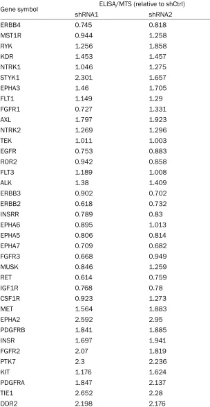
Gene symbol ELISA/MTS (relative to shCtrl)
shRNA1 shRNA2
ERBB4 0.745 0.818
MST1R 0.944 1.258
RYK 1.256 1.858
KDR 1.453 1.457
NTRK1 1.046 1.275
STYK1 2.301 1.657
EPHA3 1.46 1.705
FLT1 1.149 1.29
FGFR1 0.727 1.331
AXL 1.797 1.923
NTRK2 1.269 1.296
TEK 1.011 1.003
EGFR 0.753 0.883
ROR2 0.942 0.858
FLT3 1.189 1.008
ALK 1.38 1.409
ERBB3 0.902 0.702
ERBB2 0.618 0.732
INSRR 0.789 0.83
EPHA6 0.895 1.013
EPHA5 0.806 0.814
EPHA7 0.709 0.682
FGFR3 0.668 0.949
MUSK 0.846 1.259
RET 0.614 0.759
IGF1R 0.768 0.78
CSF1R 0.923 1.273
MET 1.564 1.883
EPHA2 2.592 2.95
PDGFRB 1.841 1.885
INSR 1.697 1.941
FGFR2 2.07 1.819
PTK7 2.3 2.236
KIT 1.176 1.624
PDGFRA 1.847 2.137
TIE1 2.652 2.28
[image:10.612.152.456.121.708.2]DDR2 2.198 2.176
DDR1 1.146 1.357
LTK 1.381 1.702
EPHB3 1.128 1.326
FGFR4 1.267 1.426
EPHA8 1.046 1.534
NTRK3 1.377 1.128
FLT4 0.964 1.258
EPHB4 1.516 0.986
LMTK2 1.261 1.738
EPHA1 1.189 1.012
AATK 0.884 1.052
MERTK 1.506 1.05
TYRO3 1.256 1.553
ROS1 1.064 1.006
EPHA4 1.156 1.132
[image:11.612.148.437.59.530.2]EPHB6 1.247 1.444