Original Article
Lysosomal integral membrane protein Sidt2 plays a
vital role in insulin secretion
Jialin Gao1,2, Cui Yu1,2, Qianyin Xiong1,2, Yao Zhang2,3, Lizhuo Wang2,3
1Department of Endocrinology and Genetic Metabolism, Yijishan Hospital of Wannan Medical College, Wuhu,
China; 2Anhui Province Key Laboratory of Biological Macro-Molecules Research, Wannan Medical College, Wuhu,
China; 3Department of Biochemistry and Molecular Biology, Wannan Medical Collage, Wuhu, China
Received October 12, 2015; Accepted November 25, 2015; Epub December 1, 2015; Published December 15, 2015
Abstract: Abnormal insulin secretion results in impaired glucose tolerance and is one of the causal factors in the
etiology of type 2 diabetes mellitus. Sidt2, a lysosomal integral membrane protein, plays a critical role in insulin se-cretion. Here, we further investigate its regulation in insulin sese-cretion. We show that Sidt2-/- mice exhibit weight loss, decreased postnatal survival rate with aging, increased fasting glucose and impaired glucose tolerance. After load-ing high levels of glucose in their diet, Sidt2-/- mice produce notably lower insulin levels at the first-phase secretion compared with Sidt2+/+ mice. Consistent with the in vivo study, INS-1 cells treated with Sidt2 siRNA produced less insulin when loaded with 16.7 mM of glucose. Only 2 of the 13 genes, synap1 and synap3 which encode soluble N-ethylmaleimide-sensitive factor attachment receptor (SNARE) proteins, showed significantly decreased expres-sion in Sidt2-/- mice. In conclusion, Sdit2 may play a vital role in the regulation of insulin secretion via two SNARE proteins synap1 and syanp3.
Keywords: Lysosome membrane protein, Sidt2, insulin, secretion, diabetes
Introduction
Insulin is released by pancreatic-islet β cells in response to elevated blood glucose levels in order to maintain normal glucose homeostasis, and more than 99% of the insulin released from β cells is regulated via a secretory pathway [1]. When extracellular glucose levels rise, regulat-ed insulin-granule exocytosis ensues and plas-ma glucose enters the β cells via the glucose transporter GLUT2. Subsequently, a series of metabolic processes occur, including a net increase in the ATP: ADP ratio [2, 3], increased intracellular Ca2+ concentration via the opening of voltage-gated Ca2+ channels [4, 5], and insu-lin vesicle fusion with plasma membrane [4]. An impaired secretory response is one of the caus-al factors in the etiology of type 2 diabetes mel-litus (T2DM), hallmarked with hyperglycemia [6].
This type of exocytosis requires synergistic molecular handshakes between three different soluble N-ethylmaleimide-sensitive factor atta-
They include vesicle-associated membrane pro-tein (VAMP) which is associated with the vesicle (a v-SNARE), synaptosomal-associated protein of 25 kDa (SNAP-25) and syntaxin which are associated with the plasma membrane (t-SNAREs) [7]. Neuroendocrine cells, such as pancreatic islet β cells, express SNAREs: VAMP2, SNAP-25b, and syntaxin-1 [8, 9]. Studies have demonstrated that patients with T2DM and animal models of the disease showed reduced expression of all three of these SNAREs [10, 11].
In the last few years, scientists have revealed some of the regulatory processes that control insulin exocytosis. For example, Rab27a, a small GTPase, is involved in insulin secretion by modulating the transport and docking steps of insulin granules via its effector proteins [12-16]. Rab3a, another small GTPase, also regu-lates insulin exocytotic release [17]. However, the ways in which these proteins regulate insu-lin secretion remains unclear.
Sidt2 is involved in insulin secretion
(KRB). The cells were incubated with 1 ml of KRB at 37°C for 1 h, and then incubated with KRB containing two different glucose concen-trations (4 and 16.7 mM) for 1 h at 37°C. The supernatant was collected and the insulin con-tent was assayed by ELISA (Millipore. Charles, MO). Cells were then treated with acidified eth-anol (75% etheth-anol, 1.5% HCl), and the total pro-tein was measured using the Bio-Rad Dc Protein assay (Hercules, CA, USA). The amount of insulin secretion was corrected by compar-ing to the amount of total protein in each well.
Sidt2 siRNA transfection
Cells were transfected with two siRNAs: a nega-tive control and Sidt2 siRNA (siRNA transfec-tion kit, sc-45064, from Santa Cruz Biotech- nology, CA, USA). Sidt2 siRNA or non-targeting negative control duplex (sc-37007, Santa Cruz tion [18, 19]. Dysfunction of the islet lysosomal
system impaired glucose-induced insulin secre-tion in diabetic Goto-Kakizaki rat [20]. Pre- viously, we described a novel lysosomal inte-gral membrane protein, Sidt2, which belongs to the SID1 transmembrane family [21] and showed that Sidt2 deficiency impaired glucose metabolism [22]. Hypothetically, Sidt2 could promote insulin-secretory exocytosis by affect-ing some SNARE proteins. Therefore, we explored the role of Sidt2 in insulin secretion in vivo and in vitro.
Materials and methods
Animals
Sidt2-/- mice were generated using the Cre-LoxP system and described in our previous study [22]. All animals were bred under conventional pathogen-free conditions. Animals were fed
with food and water ad libitum. All animal experimental protocols were approved by the Animal Ethics Committee of Yijishan Hospital of Wannan Medical College and performed according to their recommendations.
Cell culture
The insulin-secreting cell line INS-1 was purchased from the Shanghai Institute of Biochemistry and Cell Biology, Academy of Sciences of China. INS-1 cells were cul-tured in RPMI-1640 media (Gibco®, Invitrogen, Shanghai, China) supplement-ed with 10% fetal bovine serum (FBS) and routinely grown at 5% CO2/95% air at 37°C. The cells were passaged weekly by performing trypsin-EDTA detachment. All of the studies were performed in INS-1 between passages 5 and 20.
Insulin secretion assay
[image:2.629.100.334.92.480.2]Insulin secretion was measured as previ-ously described [23]. Briefly, 5×10⁵ insuli-noma cells were placed in 12-well plates in growing medium. The cell lines were treated with siRNA (1 nM or 10 nM, respectively) for 48 h. Subsequently, fresh medium containing 4.0 mM glucose was added after 24 h. And then, cells were cul-tured for an additional 48 h. The medium was removed on the day of the experi-ment, and the cell lines washed three times with warm Krebs-Ringer buffer
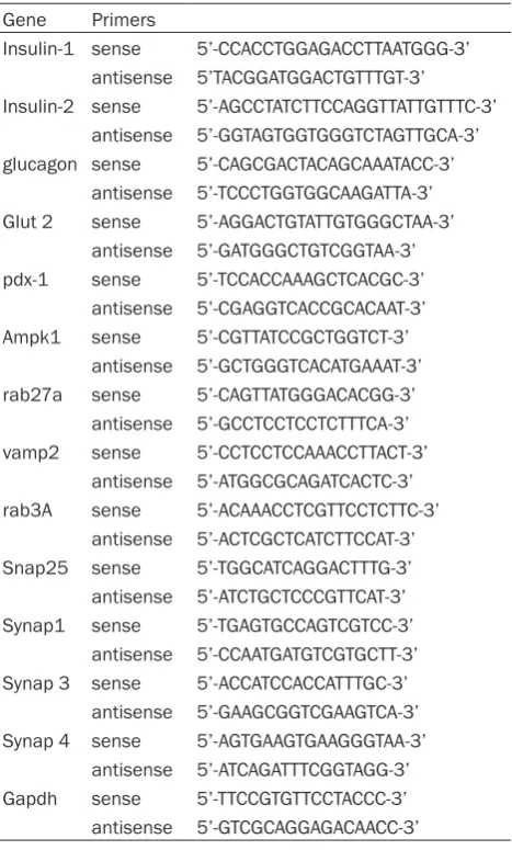
Table 1. The primers used in this project Gene Primers
Insulin-1 sense 5’-CCACCTGGAGACCTTAATGGG-3’ antisense 5’TACGGATGGACTGTTTGT-3’ Insulin-2 sense 5’-AGCCTATCTTCCAGGTTATTGTTTC-3’
antisense 5’-GGTAGTGGTGGGTCTAGTTGCA-3’ glucagon sense 5’-CAGCGACTACAGCAAATACC-3’
antisense 5’-TCCCTGGTGGCAAGATTA-3’ Glut 2 sense 5’-AGGACTGTATTGTGGGCTAA-3’
antisense 5’-GATGGGCTGTCGGTAA-3’ pdx-1 sense 5’-TCCACCAAAGCTCACGC-3’
antisense 5’-CGAGGTCACCGCACAAT-3’ Ampk1 sense 5’-CGTTATCCGCTGGTCT-3’
antisense 5’-GCTGGGTCACATGAAAT-3’ rab27a sense 5’-CAGTTATGGGACACGG-3’
antisense 5’-GCCTCCTCCTCTTTCA-3’ vamp2 sense 5’-CCTCCTCCAAACCTTACT-3’
antisense 5’-ATGGCGCAGATCACTC-3’ rab3A sense 5’-ACAAACCTCGTTCCTCTTC-3’
antisense 5’-ACTCGCTCATCTTCCAT-3’ Snap25 sense 5’-TGGCATCAGGACTTTG-3’ antisense 5’-ATCTGCTCCCGTTCAT-3’ Synap1 sense 5’-TGAGTGCCAGTCGTCC-3’
antisense 5’-CCAATGATGTCGTGCTT-3’ Synap 3 sense 5’-ACCATCCACCATTTGC-3’
antisense 5’-GAAGCGGTCGAAGTCA-3’ Synap 4 sense 5’-AGTGAAGTGAAGGGTAA-3’
Reverse transcription polymerase chain reac-tion (RT-PCR) analysis
Total RNA was prepared using an RNA extrac-tion kit (Sangon Biotech Co. LTD) according to the manufacturer’s instructions. RT-PCR was performed as previously described in our litera-ture [21]. The primers used in this study were designed by Primer 5.0 software and synthe-sized by Sangon Biotech Co. LTD. The primers sequences are listed in Table 1.
Western blotting analysis
For protein expression studies, tissues or insu-linoma cells were homogenized in lysis buffer (Beyotime, Beijing, China). Homogenates were centrifuged at 12,000×g at 4°C for 10-20 min. Western blot analysis was performed as described previously [24]. Briefly, 5 and 10 µg of total soluble proteins were separated on 12.5% SDS-PAGE and transferred onto PVDF Biotechnology) and transfection reagent were
mixed with transfection medium. After 6 h of transfection, the culture media was replen-ished with fresh medium containing 10% fetal bovine serum (FBS). After an additional 48 h in culture, the cells were harvested for further analysis.
Glucose tolerance test
[image:3.629.102.529.81.397.2]Intraperitoneal glucose tolerance tests (IPGTTs) were performed as described in our precious study [22]. Briefly, after a 12 h fasting period, a blood sample from Sidt2-/- or Sidt2+/+ mice was collected from the tail vein at time 0, 15, 30, 60 and 120 min after intraperitoneal injection (i.p.) of 1.5 g/kg body weight glucose. Blood glucose concentrations were measured using a blood glucose meter (Johnson & Johnson). Insulin ELISA Kit (Millipore) was used to analyze insulin levels according to the manufacturer’s instru- ctions.
Sidt2 is involved in insulin secretion
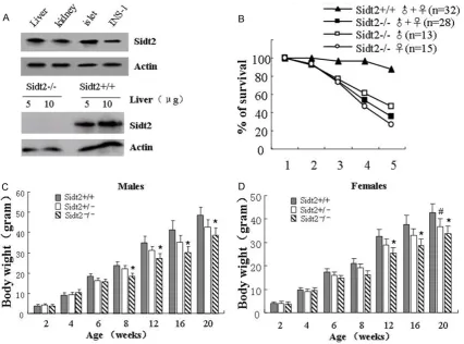
Sidt2+/+ littermates (Figure 1B). Moreover, the Sidt2-/- mice had thinner and smaller physiques (data not shown). After assessing body mass, we found that the male Sidt2-/- mice lost the majority of their weight at 8 to 20 weeks of age, compared with those of Sidt2+/+ litter-mates (Figure 1C). However, the female Sidt2 -/-mice lost their weights at 12 to 20 weeks of age, compared with their Sidt2+/+ littermates (Figure 1D).
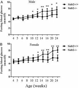
Sidt2-/- mice have a high level of fasting
glu-cose (FG) during aging
In order to investigate the effect of Sidt2 defi-ciency on glucose metabolism, the levels of fasting glucose (FG) were assessed in Sidt2 -/-mice and Sidt2+/+ mice (as control) from 4 to 24 weeks after birth. The results showed that the levels of FG in male Sidt2-/- mice were signifi-cantly higher from 10 to 24 weeks-old than those in male wild type littermates (Figure 2A). membranes. Membranes were incubated with
rabbit anti-Sidt2 specific antibody (Sigma-Aldrich® Co. LLC., St. Louis, MO, USA) and then with a horseradish peroxidase-conjugated goat anti-rabbit IgG as secondary antibody (Sangon Biotech Co. LTD, Shanghai, China). Antibodies specific to actin (Sangon Biotech Co. LTD) served as an internal control. The target pro-teins were visualized using an enhanced chemi-luminescence detection system (Sangon Biotech Co. LTD).
Statistical analysis
Generally, data from independent experiments were averaged. Error bars represent standard deviations (SD). The data was analyzed using SPSS for Windows, version 16.0 (SPSS, Chica- go, IL, USA). Differences between groups were performed using unpaired Student t-test or one-way analysis of variance (ANOVA) with a significance limit set at P<0.05.
Results
Sidt2-/- mouse shows weight
loss and lower postnatal sur-vival rate
Previously, we used RT-PCR to show that the Sidt2 gene was widely expressed in vari-ous organs, including heart, liver, kidney, pancreas and brain [22]. In the present study, the expression of the Sidt2 protein was confirmed in liver, kidney, pancreatic islet, as well as in the ISN-1 cell line using western blot analysis (Figure 1A, on the top). Furthermore, in the Sidt2-/- mouse, the pro-tein was not detected by west-ern blot (Figure 1A, on the bottom).
[image:4.629.103.380.84.390.2]In order to determine the effect of Sidt2 on the postnatal sur-vival rate and weight, we mea-sured the survival rates and weights of the Sidt2-/- mice. The results showed that the survival rates of the Sidt2 -/-mice declined significantly from 3 weeks to 4 weeks of age, compared with their
A similar trend was also observed in female sidt2-/- mice (Figure 2B). These findings sug-gested that the KO mice had a diminished abil-ity to use the lower levels of insulin for glucose uptake or were less insulin-sensitive.
Sidt2-/- mice exhibit impaired glucose
toler-ance
To determine whether Sidt2 knockout causes insulin insensitivity, glucose tolerance tests were performed. Animals were fasted over-night, injected i.p. with 2 g/kg (body weight) glucose and their blood glucose was assessed. In two month-old mice, the levels of blood glu-cose in both male and female Sidt2-/- mice were significantly higher than those in the wild-type littermates, 30 and 60 min after challenge with glucose (Figure 3A and 3D). Furthermore, in aged mice that were 4 and 6 months old, after loading glucose, both male (Figure 3B and 3D) and female (Figure 3C and 3F) KO mice at dif-ferent points in time 0, 15, 30, 60 and 120 min cleared the glucose significantly slower than
the controls, respectively. Moreover, the glu-cose tolerance was progressively attenuated with aging (4-month-old mice versus 6-month-old mice, Figure 3B-F).
Dysfunction of insulin secretion in Sidt2 knock-out mice
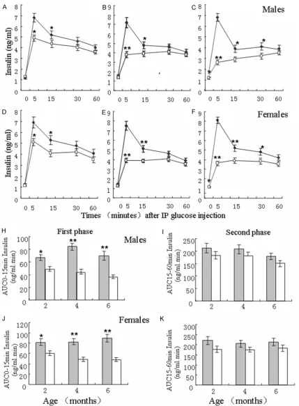
[image:5.629.102.529.80.354.2]To investigate the role of Sidt2 in insulin secre-tion, blood insulin were measured in Sidt2 -/-mice at 2-, 4- and 6-months old; as well as in Sidt2+/+ mice. Compared with the male litter-mate controls at 2-months old, male Sidt2 -/-mice had much lower blood levels of insulin in the first-phase (0-15 min; Figure 4A). Moreover, the peak of insulin secretion occurred 5 min after injection and then decreased until the 60 min time point. However, insulin blood levels were not evident at the first- and second-phase (15-60 min) in 4 and 6 month-old Sidt2-/- mice; although the difference was also observed in which case insulin levels were significantly lower than those in the controls, respectively (Figure 4B and 4C). A similar trend was also
Figure 3. Sidt2-/- mice characterized with a progressively impaired glucose tolerance with aging. (A-C) Glucose
Sidt2 is involved in insulin secretion
Figure 4. Changes in insulin secretion in the first- and second-phases in 2-, 4-, and 6-month-old Sidt2+/+ and Sidt2
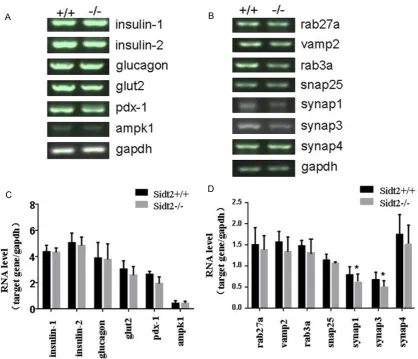
were no differences in the expression of 6 genes related to glucose metabolism between Sidt2-/- and Sidt2+/+ mice, including insulin-1,
insulin-2, glucagon, glut2, pdk-1 and ampk1
(Figure 6A and 6C). However, 2 of the 7 genes involved in insulin secretion, including synap1
and synap3, were significantly decreased in
Sidt2-/- mice, compared with controls (p<0.05,
Figure 6B and 6D). The expression of the other SNARE proteins, such as rab27a, vamp2, snap25 and synap4, showed no significant changes compared with controls.
Discussion
In this study, we show that Sidt2 is expressed in the liver and kidneys, tissues that have abun-dant lysosomes. Gluconeogenesis in these tis-sues contributes to an increase in plasma glu-cose level after overnight fasting [25, 26]. Sidt2 expression was also detected in islets and INS-1 cells. However, there was no expression of Sidt2 in the liver from Sidt2-deficient mice. It has been postulated that the liver is the main source of glucose under physiologic circum-noted in female littermate Sidt2-/- mice (Figure
4D-F).
After analyzing the Area Under the Curve (AUC) s of the first-and second-phases, respectively, the results showed that the AUCs of the first-phase of insulin secretion in male Sidt2-/- mice were significantly decreased compared with the male littermate controls (Figure 4H). However, the AUCs at the second-phase of insulin secretion were not markedly changed between male Sidt2-/- and Sidt2+/+ mice (Figure
4I). Similar trends were also observed in female Sidt2-/- and Sidt2+/+ mice (Figure 4J and 4K).
Sidt2 drives glucose-induced insulin secretion in cultured cells
In order to address the role of Sidt2 of insulin secretion in vitro, the pancreatic β-cell line INS-1 was employed. INS-1 cells were grown in culture (Figure 5A). INS-1 cells were transfect-ed with Sidt2 siRNA or a negative control for 48 h, and the western blot analysis showed that the expression level of Sidt2 protein in
Sidt2-siRNA group was notably decreased compared to the control (Figure 5B). The cells were treated with glucose (4.0 mM or 16.7 mM) for 48 h, and analysis indicated that the control cells showed robust induction of insulin secretion by these two glu-cose concentrations (Figure 5C). However, the Sidt2-siRNA-treated cells displayed a significantly reduced insulin secretion after treatment with 16.7 mM glucose, compared with the control (Figure 5C).
Impact of Sidt2 expression levels on islet and β-cell gene expression
To elucidate the possible mechanisms of Sidt2 effects on insulin secretion, we exam-ined changes in the expres-sion of 13 genes involved in glucose-stimulated insulin exocytosis and glucose meta- bolism by RT-PCR analysis. The results showed there
Figure 5. Impact of Sdit2 deficiency for insulin secretion in INS-1 cells. A:
Sidt2 is involved in insulin secretion
stances [25]. Hepatic abnormalities contribute to impaired glucose tolerance [27]. Our results showed that the postnatal survival rate of Sidt2 KO mice was significantly decreased compared with controls. Both the male and female Sidt2 -/- mice also showed increased weight loss with age, suggesting that Sidt2 might be involved in glucose metabolism.
In order to determine the role of Sidt2 in glu-cose metabolism, we measured the levels of FG in Sidt2-/- mice. The results showed that Sidt2-/- mice had a higher level of FG compared with the controls. In other words, the depletion of Sidt2 might cause impaired glucose toler-ance and this may be aggravated with age.
Glucose tolerance tests further confirmed that Sidt2 was involved in normal glucose tole- rance.
[image:8.629.107.526.82.441.2]Rapid insulin exocytosis stimulated by elevated blood glucose levels occurs within the initial 5-10 min after stimulation, referred as the first phase. Subsequently, the second-phase secre-tion is sustained for up to several hours when increased plasma glucose levels persist [28, 29]. To demonstrate that Sidt2 exerts an effect on insulin secretion, blood insulin was mea-sured in Sidt2-/- mice. The Sidt2-/- mice released lower blood levels of insulin during the first and second phases compared with the controls. However, the AUC analysis confirmed that there
Figure 6. Changes in the expression of 13 genes involved in glucose-stimulated insulin exocytosis and glucose
was no significant difference during the second phase of insulin release between Sidt2-/- and Sidt2+/+ mice. Our data shows that Sidt2 plays a vital role on the insulin secretion during the first-phase.
To address the role of Sidt2 on insulin secre-tion, Sidt2 siRNA was transfected into INS-1 cells. The results showed that Sidt2-siRNA-treated cells released lower insulin levels after injection with 16.7 mM glucose, compared with the control untreated cells. In order to further analyze the deficit in insulin secretion in the Sidt2 KO mice, we analyzed the expression of 13 genes involved in insulin release and glu-cose metabolism. Insulin, encoded by
nonalle-lic insulin-1 and -2 genes, plays a vital role in
glucose homeostasis and is a negative regula-tor of islet cell growth [30]. Pdx1, also known as IPF1 in human, is thought to play essential roles in insulin gene expression [31, 32]. AMP-activated protein kinase (AMPK) in the liver is a master regulator of glucose homeostasis main-ly through the inhibition of gluconeogenic gene expression and hepatic glucose production [33]. Our results showed that the expression levels of these genes have no statistical chang-es between Sidt2-/- and Sidt2+/+ mice. The data indicates that Sidt2 is not related to the pro-cess of glucose metabolism. The expression two insulin-exocytosis related SNARE genes,
synap1 and synap3, was decreased in Sidt2 KO
mice. It implies that insulin secretion is regu-lated by Sidt2 via synap1 and synap3 proteins instead of the other SNARE proteins and regu-lators, such as synap 4, VAMP2, SNAP-25, rab3a and rab27a.
In general, our study confirms that Sidt2, a lyso-somal integral membrane protein, plays a key role in insulin exocytosis via two SNARE pro-teins, synap1 and synap3. Depletion of Sidt2 results in weight loss, elevated postnatal sur-vival rate, and impaired glucose tolerance. However, the precise mechanism of insulin exo-cytosis regulation by Sidt2 needs further investigation.
Acknowledgements
This study was supported by the National Natural Science Foundation of China (Nos. 81200632, 81471002), the Natural Science
Foundation of Anhui, China (No. 1308085- QH134) and the Introduction of Talents Foundation of Yijishan Hospital (No. YR201104).
Disclosure of conflict of interest
None.
Address correspondence to: Jialin Gao, Department
of Endocrinology and Genetic Metabolism, Yijishan Hospital of Wannan Medical College, 2, Zheshan Road, Wuhu 241002, China. E-mail: jialing.gao@ yahoo.com; Lizhuo Wang, Anhui Province Key Laboratory of Biological Macro-Molecules Research, Wannan Medical College, 22 Wenchang Road, Wuhu 242001, China. E-mail: [email protected]
References
[1] Wang Z and Thurmond DC. Mechanisms of bi-phasic insulin-granule exocytosis-roles of the cytoskeleton, small GTPases and SNARE pro-teins. J Cell Sci 2009; 122: 893-903.
[2] Kennedy HJ, Pouli AE, Ainscow EK, Jouaville LS, Rizzuto R and Rutter GA. Glucose gener-ates sub-plasma membrane ATP microdo-mains in single islet beta-cells. Potential role for strategically located mitochondria. J Biol Chem 1999; 274: 13281-13291.
[3] Malaisse WJ and Sener A. Glucose-induced changes in cytosolic ATP content in pancreatic islets. Biochim Biophys Acta 1987; 927: 190-195.
[4] Rorsman P, Eliasson L, Renstrom E, Gromada J, Barg S and Gopel S. The Cell Physiology of Biphasic Insulin Secretion. News Physiol Sci 2000; 15: 72-77.
[5] Satin LS and Cook DL. Voltage-gated Ca2+ cur-rent in pancreatic B-cells. Pflugers Arch 1985; 404: 385-387.
[6] D’Alessio D. The role of dysregulated glucagon secretion in type 2 diabetes. Diabetes Obes Metab 2011; 13 Suppl 1: 126-132.
[7] Takahashi N, Hatakeyama H, Okado H, Noguchi J, Ohno M and Kasai H. SNARE conformational changes that prepare vesicles for exocytosis. Cell Metab 2010; 12: 19-29.
[8] Lang J. Molecular mechanisms and regulation of insulin exocytosis as a paradigm of endo-crine secretion. Eur J Biochem 1999; 259: 3-17.
[9] Gerber SH and Sudhof TC. Molecular determi-nants of regulated exocytosis. Diabetes 2002; 51 Suppl 1: S3-11.
expres-Sidt2 is involved in insulin secretion
sion of exocytotic soluble N-ethylmaleimide at-tachment protein receptor complex proteins in pancreatic islets of type 2 diabetic patients. Diabetes 2006; 55: 435-440.
[11] Del Prato S, Marchetti P and Bonadonna RC. Phasic insulin release and metabolic regula-tion in type 2 diabetes. Diabetes 2002; 51 Suppl 1: S109-116.
[12] Kasai K, Ohara-Imaizumi M, Takahashi N, Mizutani S, Zhao S, Kikuta T, Kasai H, Nagamatsu S, Gomi H and Izumi T. Rab27a mediates the tight docking of insulin granules onto the plasma membrane during glucose stimulation. J Clin Invest 2005; 115: 388-396. [13] Izumi T. Physiological roles of Rab27 effectors
in regulated exocytosis. Endocr J 2007; 54: 649-657.
[14] Kimura T and Niki I. Rab27a in pancreatic be-ta-cells, a busy protein in membrane traffick-ing. Prog Biophys Mol Biol 2011; 107: 219-223.
[15] Yamaoka M, Ishizaki T and Kimura T. GTP- and GDP-Dependent Rab27a Effectors in Pancre- atic Beta-Cells. Biol Pharm Bull 2015; 38: 663-668.
[16] Yamaoka M, Ishizaki T and Kimura T. Interplay between Rab27a effectors in pancreatic beta-cells. World J Diabetes 2015; 6: 508-516. [17] Cazares VA, Subramani A, Saldate JJ, Hoerauf
W and Stuenkel EL. Distinct actions of Rab3 and Rab27 GTPases on late stages of exocyto-sis of insulin. Traffic 2014; 15: 997-1015. [18] Lundquist I and Lovdahl R. Effect of fasting on
islet lysosomal enzyme activities and the in vivo insulin response to different secreta-gogues. Horm Metab Res 1983; 15: 11-14. [19] Lundquist I, Panagiotidis G and Salehi A. Islet
acid glucan-1,4-alpha-glucosidase: a putative key enzyme in nutrient-stimulated insulin se-cretion. Endocrinology 1996; 137: 1219-1225. [20] Salehi A, Henningsson R, Mosen H, Ostenson
CG, Efendic S and Lundquist I. Dysfunction of the islet lysosomal system conveys impairment of glucose-induced insulin release in the dia-betic GK rat. Endocrinology 1999; 140: 3045-3053.
[21] Jialin G, Xuefan G and Huiwen Z. SID1 trans-membrane family, member 2 (Sidt2): a novel lysosomal membrane protein. Biochem Biophys Res Commun 2010; 402: 588-594. [22] Gao J, Gu X, Mahuran DJ, Wang Z and Zhang H.
Impaired glucose tolerance in a mouse model of sidt2 deficiency. PLoS One 2013; 8: e66139.
[23] Bordone L, Motta MC, Picard F, Robinson A, Jhala US, Apfeld J, McDonagh T, Lemieux M, McBurney M, Szilvasi A, Easlon EJ, Lin SJ and Guarente L. Sirt1 regulates insulin secretion by repressing UCP2 in pancreatic beta cells. PLoS Biol 2006; 4: e31.
[24] Thomas DM, Ferguson GD, Herschman HR and Elferink LA. Functional and biochemical analysis of the C2 domains of synaptotagmin IV. Mol Biol Cell 1999; 10: 2285-2295. [25] Meyer C, Woerle HJ, Dostou JM, Welle SL and
Gerich JE. Abnormal renal, hepatic, and mus-cle glucose metabolism following glucose in-gestion in type 2 diabetes. Am J Physiol Endocrinol Metab 2004; 287: E1049-1056. [26] Meyer C, Stumvoll M, Nadkarni V, Dostou J,
Mitrakou A and Gerich J. Abnormal renal and hepatic glucose metabolism in type 2 diabetes mellitus. J Clin Invest 1998; 102: 619-624. [27] Mitrakou A, Kelley D, Veneman T, Jenssen T,
Pangburn T, Reilly J and Gerich J. Contribution of abnormal muscle and liver glucose metabo-lism to postprandial hyperglycemia in NIDDM. Diabetes 1990; 39: 1381-1390.
[28] Henquin JC, Ishiyama N, Nenquin M, Ravier MA and Jonas JC. Signals and pools underlying biphasic insulin secretion. Diabetes 2002; 51 Suppl 1: S60-67.
[29] Henquin JC, Nenquin M, Stiernet P and Ahren B. In vivo and in vitro glucose-induced biphasic insulin secretion in the mouse: pattern and role of cytoplasmic Ca2+ and amplification sig-nals in beta-cells. Diabetes 2006; 55: 441-451.
[30] Duvillie B, Cordonnier N, Deltour L, Dandoy-Dron F, Itier JM, Monthioux E, Jami J, Joshi RL and Bucchini D. Phenotypic alterations in insu-lin-deficient mutant mice. Proc Natl Acad Sci U S A 1997; 94: 5137-5140.
[31] Ohlsson H, Karlsson K and Edlund T. IPF1, a homeodomain-containing transactivator of the insulin gene. EMBO J 1993; 12: 4251-4259. [32] Glick E, Leshkowitz D and Walker MD. Tran-
scription factor BETA2 acts cooperatively with E2A and PDX1 to activate the insulin gene pro-moter. J Biol Chem 2000; 275: 2199-2204. [33] Zhang BB, Zhou G and Li C. AMPK: an