Original Article
Analysis of whole genomic expression profiles and
screening of the key signaling pathways
associated with pancreatic cancer
Chengzhi He1,2, Hua Jiang1,*, Shasha Geng1, Haihui Sheng3, Xiaoying Shen3, Xiaoyan Zhang3, Shizhang Zhu3, Ximei Chen2, Changqing Yang2, HengJun Gao2, 3,*
1Department of Gastroenterology, Shanghai East Hospital, Tongji University School of Medicine, Shanghai
200120, China; 2Department of Gastroenterology, Institute of Digestive Diseases, Tongji Hospital, Tongji
Universi-ty School of Medicine, Shanghai 200065, China; 3National Engineering Center for Biochip at Shanghai, 201203,
China. *Authors contributed equally.
Received May 24, 2012; Accepted July 1, 2012; Epub July 29, 2012; Published August 15, 2012
Abstract: The tumorigenesis of pancreatic cancer is thought to be a complex process. Investigation of the molecular mechanism of pancreatic cancer and exploring the specific markers for early diagnosis and specific targets of therapy is a key point to prevent and treat pancreatic cancer effectively and to improve their prognosis. In this study, expression profiles experiment was performed using Agilent human whole genomic oligonucleotide microarrays with 41,000 genes. Differentially expressed genes related with pancreatic cancer were screened, and analyzed further by GO term analysis and KEGG Pathway analysis. Our results showed that there were 1276 differentially expressed genes associated with pancreatic cancer. 691 genes were up regulated and 585 were down regulated in pancreatic cancer group. The present study confirmed that the occurrence of pancreatic cancer was involved in multiple-gene interaction. In addition, our study found that pancreatic cancer was related to an activation of the mTOR signaling pathway and renal cell carcinoma pathway.
Keywords: Pancreatic cancer, microarray, gene expression profile, signaling pathway
Introduction
Pancreatic ductal adenocarcinoma (PDAC) is one of the most fatal cancers in the world with a 5-year survival rate of <5% [1], and the incidence rate showed an upward trend in the domestic and foreign. There are about 43,140 new cases are diagnosed with pancreatic cancer in the United States in 2010 and 36,800 of them will die of the disease [1], which is in fourth place in cancer mortality [2]. In addition, because of its metastatic potential and the cytotoxic chemotherapy drug tolerance, which usually results in ineffective non-surgical treatment, the vast majority of patients will die within one year. Although in recent years, the molecular pathogenesis of pancreatic cancer research has made great progress, the clinical diagnosis and treatment of pancreatic cancer is still intractable.
signaling pathways, such as hedgehog [4], Wnt [5] and Notch signaling pathway [6] is frequently observed in pancreatic cancer. Defective, overactive or dominating signaling pathways can motivate tumor growth and survival, as well as progression of invasion and metastasis [7]. Consequently, exploring the key alterations of signaling pathways involving genesis and progression of pancreatic cancer may provide promising targets for rational molecular-targeted anticancer drug design. For example, the ras-signaling pathway has attracted considerable attention as a target for anticancer therapy because of its important role in carcinogenesis [8]. However, the signaling pathways which are implicated in pancreatic cancer are not well defined. Effective cancer treatment strategies are required to aim at the changes of genes and signaling pathways of individual characteristics.
Therefore, in order to extensively investigate molecular mechanism involved in pancreatic carcinogenesis, and lay the theoretical basis for early diagnosis and treatment of pancreatic cancer, gene expression profiles were compared in whole genome expression levels with high-throughput oligonucleotide microarray technol-ogy in this study. We hoped to screen additional genes and key signaling pathways which could guide future research on pancreatic cancer.
Materials and methods
Tissue samples
From January 2007 to March 2008 six cases of pancreatic cancer and paracancerous tissues were obtained by surgery and screened by pathology (Saved by the Shanghai national co-existence Biochip Research Center Tissue Bank). There is no difference in gender, age and underlying diseases: 3 patients were male and 3 were female, aged from 41 to 67 years and the mean age was 52 years. Biopsies were placed in liquid nitrogen for chip hybridization. The tissue samples were obtained for this study with patient informed consent, and Ethical approval for the study was obtained from the ethical committee of biobank center related hospitals.
Total RNA extraction and purification from tissue
Six cases of pancreatic cancer tissues and paracancerous tissues were remove from liquid nitrogen, adding 1 ml TRIzol (Invitrogen,
Carlsbad, USA) per 50 ~ 100mg tissue. According to the instruction, tissue should be samshed completely with a homogenizer. Total RNA was extracted using method of phenol / chloroform, salt was washed away with 70% alcohol. After air-dried in 15 ~ 30℃, adding the appropriate amount of RNase-free water to dis-solve RNA. With Nanodrop spectrophotometer (NanoDrop Inc, Wilmington, DE USA) and aga-rose gel electrophoresis, quantity, quality and purity of total RNA were evaluated the total RNAs from tissues were select and extract respectively.
Sample labeling, gene chip hybridization
Sample RNAs were labeled using indirect meth-od. cDNA Synthesis from total RNA: 1μg of total RNA were used as sample RNA, then with T7-oligo (dT) Promotor primer, first strand and second strand of cDNA were synthesized. Following Agilent Low RNA Input Linear Amplification Kit (Agilent, Palo Alto, CA, USA), fluorescent cRNA was Synthesized and ampli-fied. This is a process of in vitro transcription and incorporation of cyanine 3-CTP. We used Nanodrop ND-1000 (NanoDrop Inc, Wilmington, DE USA) to quantitate yielded cRNA. Hybridization mix was prepared following the manual of Agilent oligonucleotide microarray in situ hybridization plus kit. 750ng fluorescence labeled probe was fragmentated at 60℃.
Hybridization mix and probes were added on Human Whole-Genome 60-mer oligonucleotide microarrays (44K Agilent Human Genome, Agilent Technologies, Palo Alto, CA, USA). Hybridization oven was set to 60℃, hybirdiza-tion rotator was set to rotate at 10rpm, hybrid-ization was 17 hours. When hybridhybrid-ization was finished, Microarray washing was performed according to the procedure of Agilent Gene Expression Wash Buffer Kit. Microarray was scanned by Agilent Scanner and image was extracted by Feature Extraction Software to acquire the original data.
Microarray scanning and data analysis
Alto, CA, USA), image quantification and stan-dardization of data processing. After standard-ization of the original signals the low expres-sion probes were to be filtered, stringent selection were performed, all samples’ “gIs-Found” should be equal to 1. Screening of dif-ferential genes: To screen the difdif-ferential probes of between pancreatic cancer tissue samples and paracancerous tissues samples,
P value of each probe was calculated by T test. According to the criterion of P < 0.01, probes were selected, then switched from the corre-sponding GeneBank Accession number to Entrez IDs.
Criterion for Significant difference of GO func-tional classification was P < 0.05, and signifi-cant gene functional classification was sort in accordance with the P value from low to high. In GO functional classification, the gene with the highest score which takes into account the two indicators Enrichment and Count of Genes pos-sess, the most biology significance.
KEGG’s Pathway Analysis: From a practical point of view of gene function annotation, KEGG Pathway database is the most widely used and comparatively more comprehensive database
of annotation information. The pathway classifi-cation criterion for Significance is the EASE Score P-value < 0.05. Significant Pathway clas-sification sorts the P-values from low to high sort, and analyses the relative ratios based on the actual differential genes in all Pathway clas-sification to obtain the Enrichment of Pathway Categories. The higher the Enrichment value of differential genes it is, the greater the propor-tion accounted for the Pathway classificapropor-tion and the higher participation of the contribution of differential expression it will be. Enrichment and the Count of Genes, these two indicators with the highest score in Pathway classification have the most biological significance.
Results
Microarray hybridization and data analysis
Microarray experiment was qualified according with quality standards. The experimental sys-tem was stable, fluorescent signal intensity was strong and homogenous (Figure 1). In the 12 chips 27,574 probes had clearly signals, representing 67.3% of 41 000 probe. The dif-ferentially expressed genes associated with pancreatic cancer were 1276, in which 691 were up regulated and 585 were down regulat-ed. We used GO (Gene Ontology) to analyze these differentially expressed genes by three domains. In biological process, we found the P
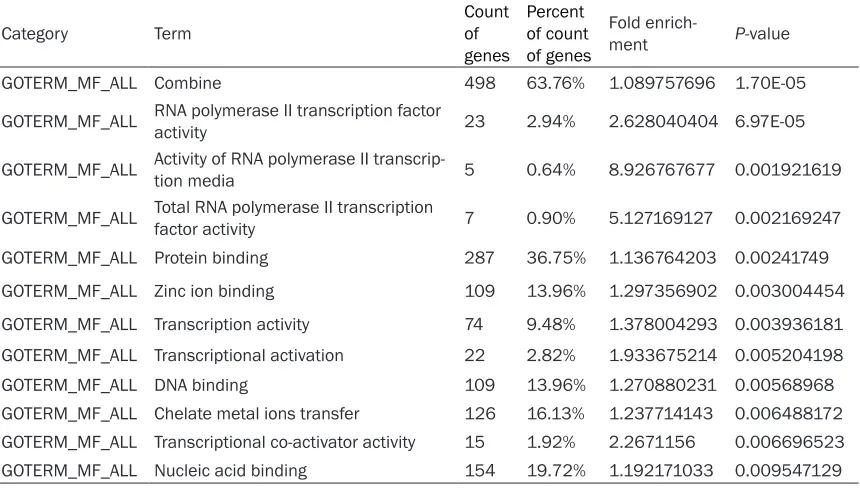
values of 22 functional description nodes were less than 0.01, which were mainly related to the regulation of cellular or cellular metabolic process, nucleic acid metabolic process and transcription, chromatin modification, intracel-lular signaling cascade, post-translational pro-tein modification, G-propro-tein signaling and so on (Table 1). In cellular component, 3 of functional description nodes were found less than 0.01, which were mainly located in the nucleus, the cell and local (Table 2). In molecular function , the P values of 12 functional description nodes were less than 0.01, such as DNA, RNA or pro-tein binding, zincion binding, transcription regu-lation and activation activity, etc. (Table 3).
Screening of the signaling pathways related to pancreatic cancer
KEGG Pathway analysis:The two highest
[image:3.612.91.289.70.330.2]scores of indicators of Enrichment and Count are hsa04150: Mammalian target of rapamycin (mTOR) signaling pathway, and hsa05211:
Table 1. GO (Gene Ontology) analysis of pancreatic cancer with different biological processes related to gene
Category Term Count of
genes
Percent of count of genes
Fold
enrichment P-value
GOTERM_BP_ALL Programmed cell 478 61.20% 1.072124483 2.30E-05 GOTERM_BP_ALL Programmed cell regulation 190 24.33% 1.249948604 2.05E-04 GOTERM_BP_ALL Biological regulation 219 28.04% 1.216286577 2.43E-04
GOTERM_BP_ALL Metabolic processes of biological macromolecules 220 28.17% 1.193807126 7.38E-04
GOTERM_BP_ALL Biological process regulation 198 25.35% 1.210338306 8.48E-04 GOTERM_BP_ALL Chromatin modification 16 2.05% 2.315694256 0.004008869 GOTERM_BP_ALL Intracellular signaling cascade 71 9.09% 1.380651434 0.004459034
GOTERM_BP_ALL DNA dependent transcriptional regulation 107 13.70% 1.279904306 0.004671531
GOTERM_BP_ALL Gene regulation 119 15.24% 1.25428195 0.0050482
GOTERM_BP_ALL DNAregulationdependent transcriptional 108 13.83% 1.263296414 0.00640369
GOTERM_BP_ALL Protein modification process 82 10.50% 1.32231405 0.006550157 GOTERM_BP_ALL Cellular metabolic process 313 40.08% 1.101155466 0.006596904
GOTERM_BP_ALL Post-transcriptional protein modification 71 9.09% 1.357066061 0.0066885
GOTERM_BP_ALL RNA synthesis 108 13.83% 1.26177437 0.006918278 GOTERM_BP_ALL G-protein signaling 11 1.41% 2.735042735 0.006968409 GOTERM_BP_ALL Transcriptional regulation 112 14.34% 1.254111554 0.006969324 GOTERM_BP_ALL Cellular metabolic process regulation 122 15.62% 1.23532576 0.007422386 GOTERM_BP_ALL Basic metabolic process 313 40.08% 1.098233572 0.007941417 GOTERM_BP_ALL Transcription 115 14.72% 1.241356047 0.008236733 GOTERM_BP_ALL Polymer metabolism 276 35.34% 1.112678895 0.008454144
GOTERM_BP_ALL Modification of biological macromolecules 84 10.76% 1.301884051 0.008663423
GOTERM_BP_ALL Regulate nucleic acid bases, nucleotides, nucleic acid metabolism 113 14.47% 1.2367467 0.009761074
Table 2. GO (Gene Ontology) analysis of pancreatic cancer associated with the cellular location of differentially expressed genes
Category Term Count of genes Percent of count of genes Fold enrichment P-value
GOTERM_CC_ALL Nucleus 196 25.10% 1.196364067 0.001908819
GOTERM_CC_ALL Cells 399 51.09% 1.077291739 0.005076384
[image:4.612.89.522.612.699.2]Renal cell carcinoma pathway (Table 4). In mTOR Pathway, the 9 key genes of ULK2, PIK3R3, PDPK1, EIF4EBP1, PGF, VEGFB, ULK3, RICTOR and PIK3R5 had significant difference (P < 0.05, Table 5). In renal carcinoma pathway, the 9 key genes of TGFB3, EPAS1, PIK3R3, EGLN1, PGF, ETS1, VEGFB, CREBBP and PIK3R5 had significant difference (P < 0.05,
Table 6).
Discussion
The DNA microarray gene expression profiles have been successful in the large-scale analy-sis of gene expression differences, particularly in cancer investigations. Recently, numerous studies have reported gene expression profiles of various cancers [9-14], including pancreatic cancer [15-18]. For example, Nakamura et al
[19] examined gene-expression profiles of 18
pancreatic cancers. The analysis identified 260 commonly up-regulated genes and 346 down-regulated genes in pancreatic cancer. In pres-ent study, through comparison of gene expres-sion profile of pancreatic cancer and paracancerous tissue, differentially expressed genes were screened and analyzed further by GO term analysis. Our results showed that there were 1276 differentially expressed genes associated with pancreatic cancer. 691 genes were up regulated and 585 were down regulat-ed in pancreatic cancer group. Furthermore, our study found that pancreatic cancer was related to an activation of the mTOR signaling pathway and renal cell carcinoma pathway. By GO analysis, genes of significant difference involved in biological process mainly related to regulation of cellular or cellular metabolic pro-cess, nucleic acid metabolic process and
tran-Table 3. GO (Gene Ontology) analysis of pancreatic cancer associated with molecular function of differentially expressed genes
Category Term Count of
genes
Percent of count of genes
Fold
enrich-ment P-value
GOTERM_MF_ALL Combine 498 63.76% 1.089757696 1.70E-05
GOTERM_MF_ALL RNA polymerase II transcription factor activity 23 2.94% 2.628040404 6.97E-05
GOTERM_MF_ALL Activity of RNA polymerase II transcrip-tion media 5 0.64% 8.926767677 0.001921619
GOTERM_MF_ALL Total RNA polymerase II transcription factor activity 7 0.90% 5.127169127 0.002169247
GOTERM_MF_ALL Protein binding 287 36.75% 1.136764203 0.00241749
GOTERM_MF_ALL Zinc ion binding 109 13.96% 1.297356902 0.003004454
GOTERM_MF_ALL Transcription activity 74 9.48% 1.378004293 0.003936181
GOTERM_MF_ALL Transcriptional activation 22 2.82% 1.933675214 0.005204198
GOTERM_MF_ALL DNA binding 109 13.96% 1.270880231 0.00568968
GOTERM_MF_ALL Chelate metal ions transfer 126 16.13% 1.237714143 0.006488172 GOTERM_MF_ALL Transcriptional co-activator activity 15 1.92% 2.2671156 0.006696523
[image:5.612.92.522.102.346.2]GOTERM_MF_ALL Nucleic acid binding 154 19.72% 1.192171033 0.009547129
Table 4. Pancreatic cancer KEGG pathway analysis result KEGG pathway ID
and name Count of genes Percent of count of genes NCBI entrez gene ID of genes Foldment enrich- P-value hsa04150:mTOR
signaling pathway 9 1.15% 9706, 8503, 5170, 1978, 5228, 7423, 25989, 253260, 23533, 4.5622519 6.23E-04 hsa05211:Renal
[image:5.612.92.524.391.464.2]Table 6.Key gene list of renal cell carcinoma pathway related to pancreatic cancer
Entrez
gene ID Genesymbol Cancer/paracancerous tissues ratio (2) P-value Description
7043 TGFB3 -1.05930128 0.00163691 Homo sapiens transforming growth fact β3 (TGFB3), mRNA [NM_003239]
2034 EPAS1 0.92358726 0.034126314 Human [NM_001430]Endothelial PAS Protein1(EPAS1), mRNA
8503 PIK3R3 -1.88099339 0.033034565 Homo sapiens Phosphatidylinositol 3-kinase regu-latory subunit gamma (p55, gamma) (PIK3R3) , mRNA [NM_003629]
54583 EGLN1 0.217749398 0.01410161 Homo sapiens EGLN1, mRNA [NM_022051]
5228 PGF -0.29988699 0.012464356 Homo sapiens placental growth factorascular endothelial growth factor-related protein(PGF), mRNA [NM_002632]
[image:6.612.90.524.428.708.2]2113 ETS1 0.670387076 0.049698783 Homo sapiens v-ets erythroblastosis virus E26 oncogene homolog 1 (ETS1), mRNA [NM_005238]
7423 VEGFB -0.31859571 0.044830214 Homo sapiens Vascular endothelial growth factor B (VEGFB), mRNA [NM_003377]
1387 CREBBP 0.410273283 0.004572845 Homo sapiens CREB binding protein (Rubinstein–Taybi syndrome) (CREBBP), mRNA [NM_004380]
23533 PIK3R5 -0.73838379 0.030512859 Homo sapiens Phosphoinositide 3-kinase regulatory subunit 5, p101 (PIK3R5), mRNA [NM_014308]
Table 5.Key gene list of mTOR signaling pathway related to pancreatic cancer Entrez
gene ID Genesymbol Cancer/paracancerous tissue ratio (2) P-value Description
9706 ULK2 -0.25170218 0.048056072 Homo sapiens unc-51-like kinase 2 (C. elegans) (ULK2), mRNA [NM_014683]
8503 PIK3R3 -1.88099339 0.033034565 Homo sapiens Phosphatidylinositol 3-kinase regulatory subunit gamma 3 (p55, gamma) (PIK3R3) , mRNA [NM_003629]
5170 PDPK1 0.39752027 0.033717114 Homo sapiens 3-phosphoinositide dependent protein kinase-1(PDPK1), mRNA [NM_002613]
1978 EIF4EBP1 -0.28287385 0.040891956 Homo sapiens eukaryotic translation initiation factor 4E bindingprotein 1 (EIF4EBP1), mRNA [NM_004095]
5228 PGF -0.29988699 0.012464356 Homo sapiens placental growth factorascular endothelial growth factor-related protein (PGF), mRNA [NM_002632]
7423 VEGFB -0.31859571 0.044830214 Homo sapiens Vascular endothelial growth fac-tor B (VEGFB), mRNA [NM_003377]
25989 ULK3 0.254669903 0.001672132 Human mRNA; cDNA DKFZp434C131 (from clone DKFZp434C131). [AL117482]
253260 RICTOR 0.649677387 0.009706675 Homo sapiens rapamycin-insensitive compan-ion of mTOR (RICTOR), mRNA [NM_152756]
scription, chromatin modification, intracellular signaling cascade, post-translational protein modification, G-protein signaling and so on. Genes of significant difference involved in cel-lular location mainly located at nucleus and intracellular part. Genes of significant differ-ence involved in molecular function mainly related to DNA, nucleic acid or protein binding, zinc ion binding, transcription regulator and activator activity, etc.
Cellular programming is essential to normal cell growth, proliferation and apoptosis, and this “procedure” is not a single process, but rather a component process. We found 478 signifi-cant abnormal genes involved in cellular pro-gramming associated with pancreatic cancer. Regulating the cellular programming supposed to inhibit tumor growth. Recently, Mouratidis et al [20]studied cell killing effect on pancreatic cancer of two new PDE4 is (Phosphodiesterase-4 inhibitors) inhibitor compounds CC-8075 and CC-8062 .The results showed significant anti-proliferation effect, which may be effected by intervening the activation of p38MAPK signal transduction pathway. In addition, cytokines such as TNF trigger intracellular signal trans-duction cascade and induce apoptosis, medi-ated through the activation of appropriate cell membrane receptors [21]. If the intracellular signal transduction cascade is affected, it will undoubtedly lead to abnormal apoptosis. It is well known that pancreatic cancer is closely associated with apoptosis [22, 23]. In this study, we found 71 significantly abnormal genes of pancreatic cancer patients were involved in intracellular signal transduction cascade.
Moreover, Bioinformatics analysis of KEGG pathway in this study showed that mTOR signal-ing pathway and renal cell carcinoma pathway had a most biological significance associated with pancreatic cancer. It has been shown that mTOR pathway functions in the downstream of the phosphatidylinositol 3-kinase (PI3K) /AKT pathway, which is often deregulated in human cancer and the deregulation of mTOR pathway will cause loss of growth control in tumor. In addition, several studies have reported that mTOR pathway whose activity increases in some tumors can be used as the target for can-cer treatment [24, 25]. In mTOR pathway, there were nine key genes differentially expressed
which included ULK2, PIK3R3, PDPK1, EIF4EBP1, PGF, VEGFB, ULK3, RICTOR and PIK3R5 (P < 0.05). Some of these genes have been reported to associate with pancreatic cancer or other cancers. For example, Soroceanu et al [26] reported that Insulin-Like Growth Factor II (IGF2)-PIK3R3 signaling axis involved in the promotion of occurrence of human neural glioblastoma. In addition, Zhang
et al [27]found that knockdown of PIK3R3
sig-nificantly increased the apoptosis in cultured ovarian cancer cell lines, and the result sug-gested that PIK3R3 may be a potential thera-peutic target of epithelial ovarian cancer. One Study has found that PDPK1 gene show aber-rant expression in gastric cancer, colon cancer and lung cancer [28]. Chakraborty et al [29] reported to confirme that mTOR signaling pathway and PDPK1 up-regulation play an important role in oral squamous cell carcinoma and several key members of this pathway can provide useful therapeutic targets. Our study has reported PDPK1 was significantly up-regu-lated in pancreatic cancer which was consis-tent with the previous research. Similarly, EIF4EBP1 [30], PGF [31], VEGFB [32] and RICTOR have associated with a variety of tumors, the former has tumor suppressor func-tion and can be used as an indicator of prog-nostic evaluation of gastrointestinal tumors. However, the relationships of PIK3R5, ULK2 and ULK3 with tumor have not been reported previously.
PAS domain protein 1 (EPAS1) was significantly increased in colorectal cancer tissue, super-vised class prediction using EPAS1, UBE2D3 and KIAA0101 correctly (77%) assigned pre-surgery samples to the CRC group and assigned post-surgery samples from the same patients to the healthy group. In our result, EPAS1 and EGLN1 were found to be over-expressed in pan-creatic cancer, which were consistent with some previous studies [37, 38]. Moreover, ETS1 was found over-expressed in pancreatic cancer and to be involved in the chemoresistance to gemcitabine in pancreatic cancer [39]. Dysregulation of CREBBP also contributes to various diseases including cancer [40], but there is no report about CREBBP activity in Pancreatic cancer. We found ETS1 (P < 0.05) and CREBBP (P < 0.01) were significantly increased in pancreatic cancer on whole genome detection. Their relationship with the biological behavior of pancreatic cancer is being further explored.
Taken together, our present study confirmed that the occurrence of pancreatic cancer was involved in multiple-gene interaction. Moreover, our study found that mTOR pathway and renal cell carcinoma pathway were closely related to pancreatic cancer. The research of these signal systems and the interactions of genes in pan-creatic cancer research will contribute to the early diagnosis and targeted therapy cancer. In addition, the action of these differentially expressed genes in the progress of pancreatic cancer pathogenesis and development needs further excavation and research.
Acknowledgement
This work was supported partly by Key Discipline Construction Project of Pudong Health Bureau of Shanghai, China (Grant No: PWZxkq2010-05) to HJ, and partly by China National 863 Project Foundation for Cancer Genomics (Pancreas Genomics) (Grant No: 1006AA02A302) to HG.
Address correspondence to: Hua Jiang, MD, PhD, Department of Gastroenterology, Shanghai East Hospital Affiliated to Tongji University, Shanghai 200120, China E-mail: huajiang666@yahoo.com. Hengjun Gao, MD, PhD, Department of Gastroenterology, Institute of Digestive Diseases,
Tongji Hospital Affiliated to Tongji University, Shanghai 200065, China. National Engineering Center for Biochip at Shanghai, 201203, China Tel: 86-021-51320287; Fax: 86-021-51320287; E-mail: hengjun_gao@shbiochip.com.
References
[1] Jemal A, Siegel R, Xu JQ and Ward E. Cancer Statistics, 2010. Ca-a Cancer Journal for Clinicians 2010; 60: 277-300.
[2] Li DH and Abbruzzese JL. New Strategies in Pancreatic Cancer: Emerging Epidemiologic and Therapeutic Concepts. Clinical Cancer Research 2010; 16: 4313-4318.
[3] Remmers N, Bailey J, Mohr A and Holling-sworth MA. Molecular pathology of early pan-creatic cancer. Cancer Biomarkers 2011; 9: 421-440.
[4] Lauth M and Toftgard R. Hedgehog Signaling and Pancreatic Tumor Development. Advances in Cancer Research 2011; 110: 1-17.
[5] Morris JP, Wang SC and Hebrok M. KRAS, Hedgehog, Wnt and the twisted developmental biology of pancreatic ductal adenocarcinoma. Nature Reviews Cancer 2010; 10: 683-695. [6] Wang Z, Banerjee S, Ahmad A, Li Y, Azmi AS,
Gunn JR, Kong D, Bao B, Ali S, Gao J, Moham-mad RM, Miele L, Korc M and Sarkar FH. Acti -vated K-ras and INK4a/Arf deficiency cooper -ate during the development of pancreatic cancer by activation of Notch and NF-kappaB signaling pathways. Plos One 2011; 6: e20537. [7] Liotta LA and Kohn EC. The microenvironment
of the tumour-host interface. Nature 2001; 411: 375-379.
[8] Wong HH and Lemoine NR. Pancreatic cancer: molecular pathogenesis and new therapeutic targets (vol 6, pg 412, 2009). Nature Reviews Gastroenterology & Hepatology 2009; 6: 440-440.
[9] Huang MZ, Li YQ, Wu GH and Zhang FC. Whole Genome Expression Profiling Reveals a Signifi -cant Role for the Cell Junction and Apoptosis Pathways in Breast Cancer Stem Cells. Molec-ular Biotechnology 2010; 45: 39-48.
[10] Nancarrow DJ, Clouston AD, Smithers BM, Got-ley DC, Drew PA, Watson DI, Tyagi S, Hayward NK and Whiteman DC. Whole genome expres-sion array profiling highlights differences in mucosal defense genes in Barrett’s esopha-gus and esophageal adenocarcinoma. Plos One 2011; 6: e22513.
expression profiles predictive of outcome and age in infant acute lymphoblastic leukemia: a Children’s Oncology Group study. Blood 2012; 119: 1872-1881.
[12] Salazar R, Roepman P, Capella G, Moreno V, Simon I, Dreezen C, Lopez-Doriga A, Santos C, Marijnen C, Westerga J, Bruin S, Kerr D, Kup-pen P, van de Velde C, Morreau H, Van Velthuy-sen L, Glas AM, Van’t Veer LJ and Tollenaar R. Gene Expression Signature to Improve Progno-sis Prediction of Stage II and III Colorectal Can-cer. Journal of Clinical Oncology 2011; 29: 17-24.
[13] Jennen DGJ, Magkoufopoulou C, Ketelslegers HB, van Herwijnen MHM, Kleinjans JCS and van Delft JHM. Comparison of HepG2 and HepaRG by Whole-Genome Gene Expression Analysis for the Purpose of Chemical Hazard Identification. Toxicological Sciences 2010; 115: 66-79.
[14] Zhang YZ, Zhang LH, Gao Y, Li CH, Jia SQ, Liu N, Cheng F, Niu DY, Cho WCS, Ji JF and Zeng CQ. Discovery and validation of prognostic markers in gastric cancer by genome-wide expression profiling. World Journal of Gastroenterology 2011; 17: 1710-1717.
[15] Morse DL, Balagurunathan Y, Hostetter G, Tris-sal M, Tafreshi NK, Burke N, Lloyd M, Enke -mann S, Coppola D, Hruby VJ, Gillies RJ and Han HY. Identification of novel pancreatic ade -nocarcinoma cell-surface targets by gene ex-pression profiling and tissue microarray. Bio-chemical Pharmacology 2010; 80: 748-754. [16] Hirono S, Yamaue H, Hoshikawa Y, Ina S, Tani
M, Kawai M, Ushijima M, Matsuura M, Saiki Y, Saiura A, Yamamoto J, Miki Y and Noda T. Mo -lecular markers associated with lymph node metastasis in pancreatic ductal adenocarcino-ma by genome-wide expression profiling. Can -cer Science 2010; 101: 259-266.
[17] Karanjawala ZE, Illei PB, Ashfaq R, Infante JR, Murphy K, Pandey A, Schulick R, Winter J, Sharma R, Maitra A, Goggins M and Hruban RH. New markers of pancreatic cancer identi -fied through differential gene expression anal -yses: Claudin 18 and annexin A8. American Journal of Surgical Pathology 2008; 32: 188-196.
[18] Huang H, Dong X, Kang MX, Xu B, Chen Y, Zhang B, Chen J, Xie QP and Wu YL. Novel blood biomarkers of pancreatic cancer-associated diabetes mellitus identified by peripheral blood-based gene expression profiles. The American journal of gastroenterol -ogy 2010; 105: 1661-1669.
[19] Nakamura T, Furukawa Y, Nakagawa H, Tsuno -da T, Ohigashi H, Murata K, Ishikawa O, Ohgaki K, Kashimura N, Miyamoto M, Hirano S, Kondo
S, Katoh H, Nakamura Y and Katagiri T. Ge -nome-wide cDNA microarray analysis of gene expression profiles in pancreatic cancers using populations of tumor cells and normal ductal epithelial cells selected for purity by laser mi-crodissection. Oncogene 2004; 23: 2385-2400.
[20] Mouratidis PXE, Colston KW, Bartlett JB, Muller GW, Man HW, Stirling D and Dalgleish AG. Anti-proliferative Effects of CC-8062 and CC-8075 in Pancreatic Cancer Cells. Pancreas 2009; 38: 78-84.
[21] Fotin-Mleczek M, Welte S, Mader O, Duchardt F, Fischer R, Hufnagel H, Scheurich P and Brock R. Cationic cell-penetrating peptides interfere with TNF signalling by induction of TNF receptor internalization. Journal of Cell Science 2005; 118: 3339-3351.
[22] Angst E, Dawson DW, Stroka D, Gloor B, Park J, Candinas D, Reber HA, Hines OJ and Eibl G. N-myc downstream regulated gene-1 expression correlates with reduced pancreatic cancer growth and increased apoptosis in vitro and in vivo. Surgery 2011; 149: 614-624.
[23] Thoennissen NH, Iwanski GB, Doan NB, Oka -moto R, Lin P, Abbassi S, Song JH, Yin D, Toh M, Xie WD, Said JW and Koeffler HP. Cucurbitacin B Induces Apoptosis by Inhibition of the JAK/ STAT Pathway and Potentiates Antiproliferative Effects of Gemcitabine on Pancreatic Cancer Cells (vol 69, pg 5876, 2009). Cancer Re-search 2009; 69: 8527-8527.
[24] Akcakanat A, Sahin A, Shaye AN, Velasco MA and Meric-Bernstam F. Comparison of Akt/ mTOR signaling in primary breast tumors and matched distant metastases. Cancer 2008; 112: 2352-2358.
[25] Chan S. Targeting the mammalian target of ra-pamycin (mTOR): a new approach to treating cancer. British Journal of Cancer 2004; 91: 1420-1424.
[26] Soroceanu L, Kharbanda S, Chen RH, Soriano RH, Aldape K, Misra A, Zha JP, Forrest WF, Ni-gro JM, Modrusan Z, Feuerstein BG and Phil-lips HS. Identification of IGF2 signaling through phosphoinositide-3-kinase regulatory subunit 3 as a growth-promoting axis in glioblastoma. Proceedings of the National Academy of Sci-ences of the United States of America 2007; 104: 3466-3471.
[28] Lee JW, Soung YH, Kim SY, Nam SW, Park WS, Lee JY, Yoo NJ and Lee SH. Mutational analysis of PDPK1 kinase domain in gastric, colorectal and lung carcinomas. Acta Oncologica 2006; 45: 340-341.
[29] Chakraborty S, Mohiyuddin SMA, Gopinath KS and Kumar A. Involvement of TSC genes and differential expression of other members of the mTOR signaling pathway in oral squamous cell carcinoma. Bmc Cancer 2008; 8:
[30] Martin ME, Perez MI, Redondo C, Alvarez MI, Salinas M and Fando JL. 4E binding protein 1 expression is inversely correlated to the progression of gastrointestinal cancers. International Journal of Biochemistry & Cell Biology 2000; 32: 633-642.
[31] Jinawath N, Chamgramol Y, Furukawa Y, Obama K, Tsunoda T, Sripa B, Pairojkul C and Nakamura Y. Comparison of gene expression profiles between Opisthorchis viverrini and non-Opisthorchis viverrini associated human intrahepatic cholangiocarcinoma. Hepatology 2006; 44: 1025-1038.
[32] Abdelrahim M, Baker CH, Abbruzzese JL, Sheikh-Hamad D, Liu SX, Cho SD, Yoon K and Safe S. Regulation of vascular endothelial growth factor receptor-1 expression by specificity proteins 1, 3, and 4 in pancreatic cancer cells. Cancer Research 2007; 67: 3286-3294.
[33] Vagenas K, Spyropoulos C, Gavala V and Tsamandas AC. TGFbeta1, TGFbeta2, and TGFbeta3 protein expression in gastric carcinomas: correlation with prognostics factors and patient survival. The Journal of sur-gical research 2007; 139: 182-188.
[34] Peng L, Mayhew CN, Schnekenburger M, Knud -sen ES and Puga A. Repression of Ah receptor and induction of transforming growth factor-beta genes in DEN-induced mouse liver tu-mors. Toxicology 2008; 246: 242-247.
[35] Fukumura Y, Kumasaka T, Mitani K, Karita K and Suda K. Expression of transforming growth factor beta1, beta2, and beta3 in chronic, can-cer-associated, obstructive pancreatitis. Ar-chives of pathology & laboratory medicine 2006; 130: 356-361.
[36] Collado M, Garcia V, Garcia JM, Alonso I, Lombardia L, Diaz-Uriarte R, Fernandez LA,
Za-ballos A, Bonilla F and Serrano M. Genomic profilingof circulating plasma RNA for the anal -ysis of cancer. Clinical chemistry 2007; 53: 1860-1863.
[37] Zhu DM, Li DC, Zhang ZX and Zhang XY. Effect of endothelial PAS domain protein 1 and hy-poxia inducible factor 1alpha on vascular en-dothelial growth factor expression in human pancreatic carcinoma. Chinese medical jour-nal 2008; 121: 2258-2264.
[38] Lee KA, Lynd JD, O‘Reilly S, Kiupel M, McCor-mick JJ and LaPres JJ. The biphasic role of the hypoxia-inducible factor prolyl-4-hydroxylase, PHD2, in modulating tumor-forming potential. Molecular cancer research : MCR 2008; 6: 829-842.
[39] Khanna A, Mahalingam K, Chakrabarti D and Periyasamy G. Ets-1 expression and gem-citabine chemoresistance in pancreatic can-cer cells. Cellular & molecular biology letters 2011; 16: 101-113.