INTRODUCTION
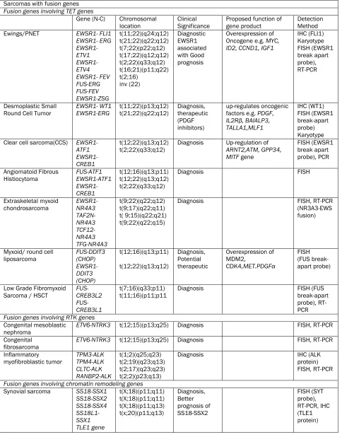
Sarcomas are relatively rare malignant tumors arising in connective tissues, including fat, mus-cle, blood vessels, deep skin tissues, nerves, bones and cartilage, comprising less than 10% of all malignancies. Sarcomas are widely be-lieved to develop as a result of genetic muta-tions in mesenchymal progenitor/ stem cells, but the precise cellular origin of most of these tumors remains unknown. Based upon molecu-lar cytogenetic analysis, sarcomas can be di-vided into two main categories (Table 1): (i) sar-comas with specific genetic alterations, which can further be subclassified based on a) recip-rocal translocations resulting in oncogenic
fu-sion transcripts e.g. EWSR1-FLI1 in Ewing sar-coma, FUS-DDT3 in myxoid liposarsar-coma, and SS18-SSX in synovial sarcoma, and b) specific oncogenic mutations e.g. KIT and PDGFRA mu-tations in gastrointestinal stromal tumors and (ii) sarcomas displaying multiple complex karyo-typic abnormalities with no specific pattern, in-cluding malignant fibrous histiocytoma (MFH / undifferentiated sarcoma), leiomyosarcoma, and pleomorphic liposarcoma. Fusion genes are most readily detected by fluorescence in situ hybridization and reverse transcription-PCR technologies. These specific translocations or characteristic genetic alterations aid in the diag-nosis of sarcomas type and, in some cases, impart prognostic or predictive information influ-encing clinical management. The genetic abnor-Int J Clin Exp Pathol 2010;3(4):416-429
www.ijcep.com/IJCEP1003008
Review Article
Molecular classification of soft tissue sarcomas and its
clinical applications
Shilpa Jain1, Ruliang Xu1, Victor G. Prieto2, Peng Lee1,3,4,5
Department of 1Pathology, 3Urology and 4Cancer Institute, New York University School of Medicine, 5New York Harbor
Healthcare System, New York, NY and 2Department of Pathology, UT, M. D. Anderson Cancer Center, Houston, TX
Received March 21, 2010, accepted April 15, 2010, available online April 23, 2010
Abstract: Sarcomas are a heterogeneous group of tumors that are traditionally classified according to the morphology and type of tissue that they resemble, such as rhabdomyosarcoma, which resembles skeletal muscle. However, the cell of origin is unclear in numerous sarcomas. Molecular genetics analyses have not only assisted in understanding the molecular mechanism in sarcoma pathogenesis but also demonstrated new relationships within different types of sarcomas leading to a more proper classification of sarcomas. Molecular classification based on the genetic altera-tion divides sarcomas into two main categories: (i) sarcomas with specific genetic alteraaltera-tions; which can further be subclassified based on a) reciprocal translocations resulting in oncogenic fusion transcripts (e.g. EWSR1-FLI1 in Ew-ing sarcoma) and b) specific oncogenic mutations (e.g. KIT and PDGFRA mutations in gastrointestinal stromal tumors) and (ii) sarcomas displaying multiple, complex karyotypic abnormalities with no specific pattern, including leiomyo-sarcoma, and pleomorphic liposarcoma. These specific genetic alterations are an important adjunct to standard mor-phological and immunohistochemical diagnoses, and in some cases have a prognostic value, e. g., Ewing family tu-mors, synovial sarcoma, and alveolar rhabdomyosarcoma. In addition, these studies may also serve as markers to detect minimal residual disease and can aid in staging or monitor the efficacy of therapy. Furthermore, sarcoma-specific fusion genes and other emerging molecular events may also represent potential targets for novel therapeutic approaches such as Gleevec for dermatofibrosarcoma protuberans. Therefore, increased understanding of the mo-lecular biology of sarcomas is leading towards development of newer and more effective treatment regimens. The review focuses on recent advances in molecular genetic alterations having an impact on diagnostics, prognostication and clinical management of selected sarcomas.
417 Int J Clin Exp Pathol 2010;3(4):416-429 malities associated with sarcomas also
repre-sent potential targets for novel therapeutic ap-proaches, which are desperately required to improve outcome in certain categories of sarco-mas. Here we review sarcomas that have asso-ciated molecular genetics and its clinical appli-cation to diagnosis, prognosis or clinical man-agement.
SARCOMAS WITH SPECIFIC GENETIC ALTERATIONS
Sarcomas with reciprocal translocation encoding specific fusion genes
The unique genetic events associated with sar-comas may give rise to at least three types of oncogenic mediators: aberrant transcription factors; constitutively active receptor tyrosine kinases (RTKs) and constitutively active growth factors. This leads to further subdivision of this category.
Fusion genes involving TET genes
Nearly half of the fusion proteins believed to participate in sarcoma initiation and develop-ment contain a portion of TET gene family prod-ucts, named after the initials of TLS/FUS, EWSR1, and TAFII68. TET-family proteins con-tain a characteristic 87-aminoacid-RNA recogni-tion motif that is thought to be implicated in protein-RNA binding and to participate in tran-scription and RNA metabolism. [1]
ET family members may be interchangeable in the development of a subgroup of sarcomas. Thus, both EWSR1-DDIT3 and FUS-DDIT3 are found in myxoid liposarcoma with an indistin-guishable phenotype; EWSR1-ERG as well as some FUS-ERG fusions are associated with Ew-ing Sarcoma Family Tumors, and both EWSR1 and TAFII68 fused to NR4A3 are found in myxoid chondrosarcoma [1].
Ewing sarcoma/Primitive peripheral neuroectodermal tumor (PNET)
Ewing sarcoma is a small blue cell tumor that primarily affects long bones or the vertebral area in young adults and children. Histologically, it is characterized by sheets of small round cells that may form Homer-Wright rosettes. Along with PNET, Ewing sarcomas are referred to as Ewing Sarcomas Family Tumors (ESFT), in which
lesions with evidence of neuroendocrine differ-entiation are sometimes subclassified as primi-tive neuroectodermal tumor (PNET) [2]. How-ever, the distinction between Ewing sarcoma and PNET is no longer considered to be critical for clinical management as their prognosis and therapy is similar.
ESFTs are the prototype of tumors with fusion genes involving the TET gene family. They char-acteristically harbor a recurring t(11;22) (q24;q12) that juxtaposes the FLI1 and EWSR1 genes encoding a chimeric RNA and protein. About 10% of Ewing family tumors have an al-ternative translocation involving the EWSR1 gene, implying that disruption of EWSR1 is the critical molecular event underlying tumorigene-sis. Except for FUS–ERG, all fusion transcripts identified so far in ESFTs consist of EWSR1 and an ET-family transcription factor, including the FLI1, ERG, ETV1, ETV4, and FEV genes[3]. Ap-proximately 85–90% of EFSTs are associated with the EWSR1–FLI1 fusion gene, 9–14% with EWR1–ERG, and the remaining 1–5% with other rare variants[3].
418 Int J Clin Exp Pathol 2010;3(4):416-429
Desmoplastic small round cell tumor (DSRCT)
DSRCT exhibits nests and cords of primitive small cells enclosed by an abundant colla-genous (desmoplastic) stroma arising most commonly in the abdomen of adolescents and young adults. DSRCT often resembles other small round cell tumors but it characteristically displays a multilineage differentiation immuno-phenotype with expression of epithelial, mesen-chymal (fibrous and/or myogenic), neural, and WT1 markers [8-10]. These tumors usually har-bor a recurring t(11;22) involving the EWSR1 gene on chromosome 22 and the WT1 (Wilms tumor 1) gene on the short arm of chromosome 11 (11p13). In contrast, the t(11;22) described above in association with Ewing sarcoma in-volves the FLI1 gene in the long arm of chromo-some 11 (11q24). The appearance of the de-rivative chromosome 11, as analyzed by karyo-type, can therefore distinguish these alternative gene partners. Another method to distinguish the two alternative gene partners for EWSR1 is cDNA amplification across the breakpoint of each fusion transcript. The 5' portion of the EWSR1 gene apparently controls the expression of the fused EWSR1-WT1 chimeric protein, whereas the 3' end of WT1 functions as a tran-scription factor that up-regulates oncogenic fac-tors, such as platelet-derived growth factor (PDGF). The fibrosis/desmoplasia that is typical of this subtype of sarcoma is probably a conse-quence of PDGF-related recruitment of fibro-blasts, and thus PDGF inhibitors are being ex-plored as novel treatment strategies.
Clear cell sarcoma (CCS)
CCS, also known as melanoma of soft parts, is a rare but distinct neoplasm, which has a predi-lection for occurring in the tendons and aponeu-roses of the distal extremities in adolescents and young adults.
CCS is characterized by a nested or fascicular growth of pale-staining or clear cells displaying phenotypic features shared with malignant melanoma, including the presence of melanin, ultrastructural evidence of melanosomes, and immunohistochemical expression of S-100 pro-tein and melanoma-associated markers such as gp100 (with HMB-45), Melan-A, and mi-crophthalmia transcription factor (MITF). The specific recurrent chromosomal translocation t (12;22)(q13;q12) or a resultant fusion of the
EWSR1 gene on 22q12 and the ATF1 gene on 12q13 are detected in most cases with CCS. ATF1 is a member of the CREB transcription factor family with bZip, via which ATF-1 binds to CRE following activation by cAMP-dependent protein kinase A (PKA). Upon fusion with EWSR1, the PKA site of ATF-1 was replaced by the N-terminus of EWSR1, resulting in a cAMP-independent and constitutively active transacti-vator that is driven by the EWSR1 promoter. EWSR1–ATF1 binds to the promoter region and induces the expression of the MITF gene, a mas-ter regulator of melanocyte differentiation. Upregulation of MITF expression is found in CCS, endowing tumor cells with melanocytic features. Recently, another variant fusion gene, EWSR1-CREB1, probably resulting from a yet elucidated chromosomal translocation t(2;22) (q34;q12), has been found in a subset of CCS that arise exclusively in the gastrointestinal tract [11]. The gene product afforded by this translo-cation, the EWSR-ATF1 fusion transcript, can be detected by RT-PCR, and thus allowing diagno-sis of clear cell sarcoma by molecular means.
Angiomatoid fibrous histiocytoma (AFH)
Angiomatoid fibrous histiocytoma (AFH) is a dis-tinct soft tissue tumor of intermediate malig-nancy with a partial myoid phenotype. Despite not having obvious melanocytic differentiation, it shares a similar fusion gene, EWSR1–ATF with CCS.
Extraskeletal Myxoid Chondrosarcoma
419 Int J Clin Exp Pathol 2010;3(4):416-429 implicated in various pathological conditions of
extracellular matrix degradation as well as in cancer [14].
Myxoid/Round Cell Liposarcoma
Myxoid/round cell liposarcoma comprises 50% of all liposarcoma arising usually in the thigh. Myxoid liposarcoma is composed of univacuolar and multivacuolar lipoblasts embedded in a richly myxoid ground substance. Characteristi-cally, there is an acute angle (plexiform) branch-ing capillary vasculature. Round cell, also called hypercellular liposarcoma is characterized by a relative increase in the cellularity of the tumor so that individual tumor cells lie in direct conti-nuity with each other without intervening matrix. Myxoid/round cell liposarcoma is treated by wide surgical excision with or without radiation therapy. Approximately 30% of patients develop distant metastases. Most myxoid/round cell liposarcomas demonstrate a specific chromoso-mal translocation t(12;16)(q13;p11) that re-sults in the rearrangement of the CHOP and FUS genes. A minority demonstrates variants of this translocation that typically also involve the 12q13 breakpoint.
Low-grade fibromyxoid sarcoma (LGFMS)
LGFMS, also known as Evans tumor, usually occurs in deep soft tissue, and is characterized by a very bland histological appearance. The translocation t(7;16)(q33;p11) so far seems to be specific for LGFMS resulting in the FUS-CREB3L2 fusion gene. Interestingly, most of the break points are located within exon 6, intron 6, or exon 7 of the FUS gene and within exon 5 of the CREB3L2 gene; those of other translocation-associated sarcomas are usually positioned within introns. As FUS and CREB3L2 gene seg-ments having break points within exons may be directly fused without interposed introns in many LGFMSs, DNA-based PCR for detecting the FUS-CREB3L2 fusion gene also seems to be applicable even when using formalin-fixed, par-affin-embedded (FFPE) tumor tissues, despite the limited length of extracted nucleic acids (e.g. DNA). FISH is another tool for detecting tumor-associated fusion genes using FFPE tu-mor specimens. FISH provides several advan-tages in comparison to RT-PCR-based or DNA-based PCR method: the possibility of contami-nation can be ruled out when using FISH on a histologic section, and one examination can
detect any variants of FUS rearrangement in-cluding FUS-CREB2L2 with variable breakpoints and FUS-CREB3L1.[17]
Fusion genes involvingthe genes encoding receptor tyrosine kinases (RTKs) genes
Congenital Mesoblastic Nephromas (CMN)
CMN are uncommon renal tumors diagnosed generally within the first 3 months of life. CMN are characterized by a variably cellular prolifera-tion of bland spindle cells arranged in interlac-ing bundles, and their clinical behavior is gener-ally benign. Recently, CMN were shown to con-tain a novel t(12;15)(p13;q25) translocation, resulting in ETV6-NTRK3 gene fusion. ETV6 is a member of the ETs family of transcription fac-tors containing a basic helix-loop-helix (bHLH) dimerization domain, which was originally found at the breakpoint in translocations in leukemia and myeloproliferative syndromes. NTRK3 is the cell surface receptor for neurotropin 3 ex-pressed primarily in the central nervous system. The ETV6–NTRK3 fusion protein forms a homodimer or heterodimer with wild-type NTRK3, which displays receptor tyrosine kinase (RTK) activity and undergoes autophosphoryla-tion at tyrosine domain [18].
Cytogenetic t(12;15) translocations may not identified in several cases (CMN4, CFS1, and CFS5), although RT-PCR and FISH analyses re-veal ETV6-region rearrangements in those same cases. The detection of ETV6-NTRK3 fusion transcripts in most CMN using RT-PCR methods with archival, FFPE tissues therefore is a useful diagnostic adjunct.
Congenital Fibrosarcoma (CFS)
[image:5.612.67.551.93.711.2]
420 Int J Clin Exp Pathol 2010;3(4):416-429 Table 1. Sarcomas with specific alterations
Sarcomas with fusion genes Fusion genes involving TET genes
Gene (N-C) Chromosomal
location
Clinical Significance
Proposed function of gene product
Detection Method Ewings/PNET EWSR1- FLI1
EWSR1- ERG EWSR1- ETV1 EWSR1- ETV4 EWSR1- FEV FUS-ERG FUS-FEV EWSR1-ZSG t(11;22)(q24;q12) t(21;22)(q22;q12) t(7;22)(p22;q12) t(17;22)(q12;q12) t(2;22)(q33;q12) t(16;21)(p11;q22) t(2;16) inv (22) Diagnostic EWSR1 associated with Good prognosis Overexpression of Oncogene e.g. MYC, ID2, CCND1, IGF1
IHC (FLI1) Karyotype FISH (EWSR1 break apart probe), RT-PCR Desmoplastic Small Round Cell Tumor
EWSR1- WT1 EWSR1-ERG t(11;22)(p13;q12) t(21;22)(q22;q12) Diagnosis, therapeutic (PDGF inhibitors) up-regulates oncogenic factors e.g. PDGF, IL2Rβ, BAIALP3, TALLA1,MLF1 IHC (WT1) FISH (EWSR1 break-apart probe) Karyotype Clear cell sarcoma(CCS) EWSR1-
ATF1 EWSR1-CREB1
t(12;22)(q13;q12)
t(2;22)(q33;q12) Diagnosis Up-regulation of ARNT2,ATM, GPP34, MITF gene
FISH (EWSR1 break apart probe), PCR
Angiomatoid Fibrous
Histiocytoma FUS-ATF1 EWSR1-ATF1 EWSR1-CREB1
t(12;16)(q13;p11) t(12;22)(q13;q12) t(2;22)(q33;q12)
Diagnosis FISH
Extraskeletal myxoid
chondrosarcoma EWSR1-NR4A3 TAF2N- NR4A3 TCF12- NR4A3 TFG-NR4A3 t(9;22)(q22;q12) t(9;17)(q22;q11) t( 9;15)(q22;q21) t(9;22)(q22;q15)
Diagnosis FISH, RT-PCR
(NR3A3-EWS fusion)
Myxoid/ round cell
liposarcoma FUS-DDIT3 (CHOP) EWSR1-DDIT3 (CHOP) t(12;16)(q13;p11) t(12;22)(q13;q12) Diagnosis, Potential therapeutic Overexpression of MDM2, CDK4,MET.PDGFα FISH (FUS break-apart probe)
Low Grade Fibromyxoid
Sarcoma / HSCT FUS-CREB3L2 FUS-CREB3L1
t(7;16)(q33;p11)
t(11;16)(p11;p11 Diagnosis FISH (FUS break-apart probe), RT-PCR Fusion genes involving RTK genes
Congenital mesoblastic
nephroma ETV6-NTRK3 t(12;15)(p13;q25) Diagnosis FISH, RT-PCR Congenital
fibrosarcoma
ETV6-NTRK3 t(12;15)(p13;q25) Diagnosis FISH, RT-PCR
Inflammatory
myofibroblastic tumor TPM3-ALK TPM4-ALK CLTC-ALK RANBP2-ALK t(1;2)(q25;q23) t(2;19)(q23;q13) t(2;17)(q23;q23) t(2;2)(p23;q13)
Diagnosis IHC (ALK
protein) FISH, RT-PCR
Fusion genes involving chromatin remodeling genes Synovial sarcoma SS18-SSX1
421 Int J Clin Exp Pathol 2010;3(4):416-429 (Continued Table 1)
Endometrial stromal
sarcoma JAZF1-SUZ12 JAZF1-PHF1 EPC1-PHF1
t(7;17)(p15;q21) t(6;7)(p21;p15) t(6;10)(p21;p11)
Diagnosis RT-PCR
Fusion genes involving growth factors genes Dermatofibrosarcoma
protuberans COL1A1-PDGFB t(17;22)(q22;q13) Diagnosis, Therapeutic (Gleevec responsive)
Up-regulate the
expression of PDGFR FISH, RT-PCR
Giant Cell
Fibroblastoma COL1A1-PDGFB t(17;22)(q22;q13) Diagnosis FISH, RT-PCR
Other type of fusion genes Alveolar
Rhabdomyosarcoma PAX3-FOXO1A PAX7-FOXO1A PAX3-MLLT7 PAX3-NCOAI
t(2;13)(q35;q14) t(1;13)(q36;q14) T(2;X)(p35;q13) T(2;2)(q35;q23)
Diagnostic Better
Prognosis with PAX7-FOXO1A
FISH (FOXO1A Break-apart probe), Karyotype, RT-PCR Alveolar soft part
sarcoma ASPSL-TFE3 t(X;17)(p11;q25) Diagnosis IHC ( TFE3), RT-PCR Aneurysmal bone cyst
CDH11-USP6 THRAP3-USP6 CNBP-USP6 OMD-USP6 COL1A1-USP6
t(16;17) t(1;17) t(3;17) t(9;17) t(17;17)
Diagnosis FISH, RT-PCR
Tenosynovial giant cell
tumor CSF1-COL6A3 t(1;2) Diagnosis Hemangiopericytoma t(12;19) Diagnosis Pericytoma ACTB-GLI1 t(7;12) Diagnosis Sarcomas with specific oncogenic mutation
Gastrointestinal Stromal Tumors
KIT or PGDFRA
Occult 4q12 Diagnosis, C-Kit Gleevac responsive
Activation Tyrosine kinase Receptor
IHC ( C-Kit), PCR
Rhabdoid tumor SMARCB1 del 22q11.22 Diagnosis LOH IHC (loss of INI1) Atypical lipomatous
tumor/ Well-differentiated liposarcoma
giant marker and inMicatio3
Cyclin dependent
kinaser
FISH ( MDM2, CDK4 amplification)
Fibromatosis APC inactivation
Trisomies 8 and 20
Deletion of 5q
Diagnosis IHC( β
catenin)
422 Int J Clin Exp Pathol 2010;3(4):416-429
Inflammatory Myofibroblastic Tumor (IMT)
IMTs are mesenchymal solid tumors that occur preferentially in children and young adults. They present as myofibroblastic cell proliferations accompanied by plasmocytes and lymphocytes. Recent cytogenetic studies showed abnormali-ties of chromosomal band 2p23 resulting in a rearrangement of the ALK gene, thus suggesting a neoplastic rather than a reactive inflammatory process. ALK is a tyrosine kinase oncogene ini-tially found to be rearranged in anaplastic large-cell lymphomas (ALCL). NPM-ALK fusions, found in 80% of ALCL, have not been identified in IMT and TMP3-ALK fusions are rare in ALCL [20]. Interestingly, the breakpoints within ALK, and also within some of the ALK fusion gene part-ners, such as TPM3 or CLTC, are similar in IMT and ALCL. The consistent involvement of ALK, together with the diversity of partner genes, underlines both the central role of ALK constitu-tive activation in IMT development, as well as the importance of homodimerization mecha-nisms of the chimeric fusion proteins in this activation. Immunohistochemical analyses per-formed on PPFE sections have shown positive ALK expression with cytoplasmic localization in half of the IMT cases containing the molecular ALK rearrangement. Therefore, detection of a rearrangement of ALK by FISH is a useful, com-plementary tool for IMT diagnosis.
Fusion genes involving chromatin-remodeling genes
Synovial Sarcoma (SS)
SS, often arises deep in the soft tissue near a joint in the extremity of a young adult patient. Most synovial sarcomas are characterized by t (X;18)(p11.2;q11.2), resulting in a fusion be-tween the SS18(SYT) gene on chromosome 18 and one of the SSX genes on the X chromo-some, creating SS18-SSX1, SS18-SSX2 or SS18 -SSX4 chimeric genes [21-27]. There are two common histologic and genetic variants of syno-vial sarcoma: (i) a monophasic variant com-prised of vimentin-expressing spindle cells, usu-ally carrying the SS18-SSX2 translocation and (ii) a biphasic variant comprised of a mixture of vimentin-expressing spindle cells and keratin-expressing glandular epithelial cells harboring the SS18-SSX1 or SS18-SSX2 translocation [28]. The biphasic variant may resemble adeno-carcinoma but more typically resembles
carcino-sarcoma and, at least in early stage patients, carries a worse prognosis.
Karyotype is helpful when it is positive, but negative results could either reflect failure of the tumor cells to divide sufficiently in culture or other mechanisms of false negative results as alluded to above. The two translocations are indistinguishable using traditional cytogenetics. FISH using an SS18 break-apart probe is helpful for demonstrating t(X;18), but it cannot distin-guish which partner gene is involved for prog-nostic purposes. RNA from either frozen or par-affin-embedded tissue is suitable for RT-PCR to detect and distinguish the two common translo-cation variants [29]. From a mechanistic stand-point, the translocation creates a chimeric gene encoding a fusion protein that redirects the transcription factor function of SS18. Relevant downstream targets include cyclin D1 (CCND1) that enhances cell cycle progression. TLE1 en-codes a transcriptional co-repressor that is over-expressed in synovial sarcomas. Recently, gene and tissue microarray studies have identified TLE1 as an excellent bio-marker for distinguish-ing the synovial sarcoma from other soft tissue malignancies on immunohistochemistry [30].
Endometrial Stromal Sarcoma (ESS)
423 Int J Clin Exp Pathol 2010;3(4):416-429
Fusion gene involving growth factors
Dermatofibrosarcoma Protuberans (DFSP)
DFSP is a rare skin tumor of low-grade malig-nant behavior that shows frequent local recur-rence. Either the reciprocal chromosomal trans-location t(17;22) (q11;q13.1) or a supernumer-ary ring chromosome derived from t(17;22) may be found in this type of tumor, resulting in the fusion of the COL1A1 gene on chromosome 17 with the PDGFB gene on chromosome 22 [31]. Since the point of fusion is highly specific for PDGFB but spread over almost the entire locus for COL1A1, the role of the COL1A1 gene may be simply to up-regulate the expression of PDGFR, which acts as an auto- or paracrine growth factor. Imatinib mesylate (gleevec®) is an inhibitor of tyrosine kinases, among them PDGFR, showing a dramatic response in adult in treatment. The use of other PDGFR inhibitors such as sunitinib and sorafenib is being tested in patients with metastatic DFSP [32].
Giant Cell Fibroblastoma (GCF)
GCF represents the juvenile form of dermatofi-brosarcoma protuberans (DFSP). Although his-tologically different these two diseases share a number of similarities: clinical localization and course, CD34 positivity, and most importantly, genetic background. The same fusion gene COL1A1 with PDGFB is also found in giant-cell fibroblastoma [31].
Other types of fusion genes
Alveolar Rhabdomyosarcoma (ARMS)
Rhabdomyosarcoma (RMS) is the most common soft tissue sarcoma in children and young adults, often presenting as a muscle mass in the extremities, paranasal sinus, or retroperito-neum. The main histological subtypes are alveo-lar (ARMS) (20%), embryonal (ERMS) (60%), and pleomorphic (PRMS); for these subtypes sur-vival rates vary from <25% to >95%, respec-tively [33]. In alveolar rhabdomyosarcoma, ex-pression profiling has identified further subsets of patients having good, intermediate, and poor outcomes [34]. Histologically, there is striated muscle differentiation with concomitant expres-sion of vimentin, muscle-specific actin, desmin, myogenin, and MyoD1. PAX3 and PAX7 are tran-scription factors that initiate myogenesis in
muscle stem cells, and the aberrant fusion of their DNA binding domain with the transactiva-tion domain of FOXO1A creates a potent tran-scription factor that stimulates myogenesis and resists apoptosis. Prognosis of ARMS is poor compared with embryonal rhabdomyosarcoma, justifying the effort to distinguish these two tu-mor types.
In alveolar rhabdomyosarcomas, 70% of ARMS harbor the translocation t(2;13)(q35:q14), which fuses the 5' end of PAX3 with the 3' end of the FOXO1A gene. An additional 10% of ARMS are associated with fusion of PAX7 to the FOXO1A gene. The remaining 20% of ARMS do not have these fusion genes detectable by rou-tine RT-PCR and comprise cases with a very low expression of a fusion gene, a rare variant fu-sion, or are true fusion negative cases. FISH using a break-apart probe identifies rearrange-ment of the FOXO1A gene [35, 36]. Karyotype analysis typically reveals either t(2;13) (q35;q14) PAX3-FOXO1A or t(1;13)(p36;q14) PAX7-FOXO1A. The latter is less common but is associated with a better prognosis. ARMS tend to have a poor prognosis overall, especially when presenting with disseminated disease, so genetic testing may be not relevant in stage IV patients. Testing is most useful when the his-tologic features are not classic (e.g. mixed al-veolar and embryonal patterns).
Alveolar Soft-Part Sarcoma (ASPS)
424 Int J Clin Exp Pathol 2010;3(4):416-429 sarcomas.
Sarcomas with specific oncogenic mutations
Gastrointestinal Stromal Tumors (GIST)
GIST is the most common mesenchymal tumor in the gastrointestinal tract. Activating muta-tions of the c-kit gene, which encodes a tyrosine kinas receptor for stem cell / platelet-derived growth factor receptor, were found in 75–80% of cases, and those of PDGFRA were found among the rest (5–10%). GISTs without detect-able c-kit/PDGFRA mutation are extremely rare. The c-kit protein comprises a long extracellular domain, a transmembrane segment, and an intracellular part. In all cases, mutated recep-tors transmit growth signals in a ligand-independent manner, inducing dysregulated cell proliferation. Mutations generally occur in the DNA encoding the intracellular part exon 11, but can also occur in exons 9 and rarely 13 and 1. Mutations in c-kit and PDGFRA are mutually exclusive. Detection of either overexpression or mutation of KIT/ PDGFRA has become a stan-dard diagnostic tool for GISTs. Targeting the activating tyrosine kinase with medications such as imatinib mesylate (Glivec/Gleevec®), has been used successfully to treat patients with metastatic GISTs showing the above described mutations. D816V point mutations in c-kit exon 17 are responsible for resistance to targeted therapy drugs like imatinib mesylate [37].
Malignant Rhabdoid Tumors (MRT)
MRT are highly aggressive pediatric tumors that develop mainly in the kidney but also at virtually any extra-renal anatomic site, particularly affect-ing infants. SMARCB1/hSN5/INI1 is a compo-nent of the SWI/SNF complex and represses the expression of cyclin D1, which promotes cell cycle progression acting as a tumor suppressor. Loss-of-function mutations of the INI1 gene ac-companied by a loss of the wild-type allele are frequently found in this type of tumor. Loss of INI-1 staining by immunohistochemistry corre-lates well with mutations or deletion of the INI-1/hSNF5/SMARCB1/BAF47 locus and is a well-established diagnostic marker for MRT [38]. Recently, frequent loss of INI1 protein expres-sion was reported in epithelioid sarcomas, which are highly malignant adult sarcomas of unknown origin, although the frequency of ge-netic deletions was low [39].
SARCOMAS WITH VARIABLE COMPLEX GENETIC ALTERATIONS LACKING A SPECIFIC PATTERN
Tumors in this group account for about 50% of soft tissue sarcomas and characterized by pleo-morphic/spindle cell morphology, including Pleomorphic Malignant Fibrous Histiocytoma / undifferentiated (PMFH), pleomorphic liposar-coma (PLPS), leiomyosarliposar-coma (LMS), pleomor-phic rhabdomyosarcoma (PRMS) and malignant peripheral nerve sheath tumor (MPNST). Chro-mosomal breakpoints in these tumors are widely scattered, with no predilection of any of the recurrent breakpoints and losses to any of the morphological subtypes. However, high grade dedifferentiated liposarcoma in addition have distinctive genomic abnormalities (giant chromosome, ring chromosomes, amplification of 12q13-q21), similar to well-differentiated liposarcoma.
Pleomorphic Malignant Fibrous Histiocytoma/ Undiffrentiated sarcoma (PMFH)
PMFH has been considered to be the most fre-quent soft tissue sarcomas in adults. The cellu-lar origin of PMFH has long been unknown, al-though different theories claimed that the tumor originates from histiocytes, fibroblasts, or primi-tive mesenchymal cells.
425 Int J Clin Exp Pathol 2010;3(4):416-429 as a marker of cancer progression and a
poten-tial target for cancer therapy. It plays a key role in cell morphology, adhesion, migration, and organization. Ezrin is over-expressed in many neoplasms including PMFH, and this over-expression was found to correlate with in-creased metastatic potential and reduced sur-vival [40].
Pleomorphic Liposarcoma (PLPS)
PLPS is the least common variant of liposar-coma, accounting for less than 5%. It usually occurs in elderly people (median age 55–65 years) predominantly in deep soft tissues (75% of cases) of the lower extremities, especially the thigh. PLSP characteristically shows high chro-mosome counts and complex structural ar-rangements. Deletion of 13q14.2-q14.3, target-ing the RB1 pathway, is observed in about 60% of PLPS [41]. Despite their morphologic similar-ity, PLPS and high-grade dedifferentiated LPS show distinct chromosomal imbalances. Gains of 5p13-p15, 1p21, 1q21-q22, and 7q22 are more frequently observed in PLPS whereas high -level amplifications within chromosomal sub-region 12q13-q21 are observed in dedifferenti-ated LPS. MAD2, a gene involved in the RB1 signaling pathway over-expressed by PLPS (and dedifferentiated LPS), might constitute a thera-peutic target [42].
Leiomyosarcoma (LMS)
LMS accounts for about 8–10% of adult soft tissue sarcomas. These malignant neoplasms show varying degrees of smooth muscle differ-entiation and can develop anywhere in the body. LMS usually show complex karyotypic al-terations that differ generally from one tumor to another. Many LMSs show chromosomal imbal-ances or aberrations in the form of gains (chromosomes 1, 5, 6, 8, 15, 16, 17, 19, 20, 22, X), losses (chromosomes 1p, 2, 3, 4, 6q, 8, 9, 10p, 11p, 12q, 11q, 13, 16, 17p, 18 19, 22q), and amplifications (chromosomes 1, 5, 8, 12, 13, 17, 19, 20). Some gains and losses of chromosomal material are more frequently ob-served and tend to correlate with poor outcome, large tumor size, and metastatic dissemination (e.g. loss of 1p12-pter, 2p, 13q14-q21 (targeting the Rb pathway), 10q (targeting PTEN) , and 16q; gains of 17p, 8q, and 5p14 pter). Activation of the PI3K–AKT pathway through different mechanisms (e.g., activation
of IGFR, inactivation of PTEN (a negative regula-tor of the PI3K–AKT) also plays a crucial role in the development and maintenance of LMS. This activation leads to the concomitant activation of downstream effectors such as mTOR and its targets (ß-catenin, pS6, p4E-BP1, etc.), as well as to MDM2 stabilization. Recent clinical trials showed that analogs of rapamycin, such as the mTOR inhibitor everolimus (RAD001), have some efficacy in patients with LMS [43]. It has also been realized that the more differentiated retroperitoneal LMSs tend to behave more ag-gressively and that this was mainly dependent upon amplification/overexpression of myocar-din. Myocardin is a transcriptional cofactor of SRF that regulates smooth muscle differentia-tion. Inactivation of the myocardin pathway results in a significant reduction of smooth mus-cle differentiation, cell proliferation, and cell migration and was associated with less differen-tiated histology. These data suggest that myo-cardin might constitute a promising therapeutic target.
Pleomorphic Rhabdomyosarcoma (PRMS)
PRMS usually occurs in the extremities, espe-cially the thigh, often in middle-aged men. It is an aggressive tumor, prone to recurrence and metastasis to the lungs. It shows nonspecific complex karyotypes. Numerical and unbalanced structural abnormalities are common with gains (chromosomes 1, 5, 8, 14, 18, 20, and 22) and loss (chromosomes 2, 5, 6, 10, 11, 13, 14, 15, 16, 17, 18, 19 and Y) of chromosomes, of which losses of chromosomes 2, 13, 14, 15, 16, and 19 are the most frequent. PRMS does not contain the t(2;13) or t(1;13) characteristic of alveolar rhabdomyosarcoma.
Malignant peripheral Nerve Sheath Tumor (MPNST)
426 Int J Clin Exp Pathol 2010;3(4):416-429 17, 19p and 22q in NF1-associated
neurofibro-mas, suggesting inactivation of tumor suppres-sor genes in the development, however, in MPNST gains are more frequent than losses, probably relating to proto-oncogene activation during MPNST progression. Losses of chromo-somes (1p12-13, 1p21, 1p36, 3p21-pter, 9p13 -21, 9p22-24, 10, 10p11-15, 11p, 11q21-25, 13q14, 15p, 16/16q24, 17/17p, 17q11-12, 17q21-25, 22, 22p, 22q13, and 22q11-12) were the most frequent abnormalities observed. Gains mainly involved the regions 7p21-q36, 7p22, 7q, 8, 8q11-23, 1q25-44, and 5q13-35. Gain of 7p15-p21 was also found associated with poor prognosis and shortened survival [44]. cDNA microarray analysis has found that six genes (keratin 18, survivin, tenascin C, adeno-sine deaminase, collagen type VIa3, and colla-gen type VIIa1) were significantly upregulated in MPNST, whereas one gene, insulin-like growth factor binding protein 6, was downregulated in MPNST. MPNST is a highly aggressive sarcoma for which no effective treatment currently exists. PDGF-BB was identified as the most effective factor that induces MPNST cell invasion and proliferation. Expression of PDGF-BB receptor (PDGFR-β) mRNA was detected more frequently and its protein was expressed at higher levels in MPNST tissues than benign peripheral nerve sheath tumors (schwannomas and neurofibro-mas). The effectiveness of imatinib mesylate in vitro suggests that targeting PDGFR-β may re-sult in the establishment of novel treatments for MPNST [45]. Survivin and tenascin C expression was validated on RT-PCR. Immunohistochemis-try confirmed upregulation of survivin in MPNST at the protein level.
In summary, genetic analysis of soft tissue sar-comas has proven to be invaluable for diagnos-tic and clinical management. In the future, clini-cal decisions will increasingly be based on a combination of histologic criteria and the mo-lecular identification of genetic abnormalities that are indicative of biologic properties. Fur-thermore, elucidation of the functions of sar-coma-specific fusion proteins will continue to improve understanding of the oncogenic proc-ess and will lead to the identification of new therapeutic targets for the treatment of sarco-mas.
Please address correspondence to: Victor Prieto, MD, PhD, Department of Pathology, UT, MD Anderson Cancer Center, 1515 Holcombe Blvd, Houston, TX 77030, Phone: (713) 792-3187, E-mail:
[email protected]; or Peng Lee, MD, PhD, Departmentof Pathology and Urology, New York Uni-versity School of Medicine, 423 e.23RD Street, New
York, NY 10010, Phone: (212)9513-418, E-mail: [email protected]
References
[1] Riggi N, Cironi L, Suva ML and Stamenkovic I. Sarcomas: genetics, signalling, and cellular ori-gins. Part 1: The fellowship of TET. J Pathol 2007; 213: 4-20.
[2] Olsen SH, Thomas DG and Lucas DR. Cluster analysis of immunohistochemical profiles in synovial sarcoma, malignant peripheral nerve sheath tumor, and Ewing sarcoma. Mod Pathol 2006; 19: 659-668.
[3] de Alava E and Gerald WL. Molecular biology of the Ewing's sarcoma/primitive neuroectodermal tumor family. J Clin Oncol 2000; 18: 204-213. [4] Kilpatrick SE, Bergman S, Pettenati MJ and
Gul-ley ML. The usefulness of cytogenetic analysis in fine needle aspirates for the histologic subtyping of sarcomas. Mod Pathol 2006; 19: 815-819. [5] Lin PP, Brody RI, Hamelin AC, Bradner JE, Healey
JH and Ladanyi M. Differential transactivation by alternative EWS-FLI1 fusion proteins correlates with clinical heterogeneity in Ewing's sarcoma. Cancer Res 1999; 59: 1428-1432.
[6] Vermeulen J, Ballet S, Oberlin O, Peter M, Pier-ron G, Longavenne E, Laurence V, Kanold J, Chastagner P, Lejars O, Blay JY, Marec-Berard P, Michon J, Delattre O and Schleiermacher G. Inci-dence and prognostic value of tumour cells de-tected by RT-PCR in peripheral blood stem cell collections from patients with Ewing tumour. Br J Cancer 2006; 95: 1326-1333.
[7] Hameed M. Small round cell tumors of bone. Arch Pathol Lab Med 2007; 131: 192-204. [8] Ladanyi M and Gerald W. Fusion of the EWS and
WT1 genes in the desmoplastic small round cell tumor. Cancer Res 1994; 54: 2837-2840. [9] Murphy AJ, Bishop K, Pereira C, Chilton-MacNeill
S, Ho M, Zielenska M and Thorner PS. A new molecular variant of desmoplastic small round cell tumor: significance of WT1 immunostaining in this entity. Hum Pathol 2008; 39: 1763-1770. [10] Nishio J, Iwasaki H, Ishiguro M, Fukuda T,
Chu-man H, Kaneko Y and Kikuchi M. Intra-abdominal small round cell tumour with EWS-WT1 fusion transcript in an elderly patient. Histo-pathology 2003; 42: 410-412.
[11] Hisaoka M, Ishida T, Kuo TT, Matsuyama A, Ima-mura T, Nishida K, Kuroda H, Inayama Y, Oshiro H, Kobayashi H, Nakajima T, Fukuda T, Ae K and Hashimoto H. Clear cell sarcoma of soft tissue: a clinicopathologic, immunohistochemical, and molecular analysis of 33 cases. Am J Surg Pathol 2008; 32: 452-460.
427 Int J Clin Exp Pathol 2010;3(4):416-429 (9;22)(q22;q12) is a primary cytogenetic
abnor-mality in extraskeletal myxoid chondrosarcoma. Int J Cancer 1995; 62: 398-402.
[13] Clark J, Benjamin H, Gill S, Sidhar S, Goodwin G, Crew J, Gusterson BA, Shipley J and Cooper CS. Fusion of the EWS gene to CHN, a member of the steroid/thyroid receptor gene superfamily, in a human myxoid chondrosarcoma. Oncogene 1996; 12: 229-235.
[14] Sjogren H, Meis-Kindblom JM, Orndal C, Bergh P, Ptaszynski K, Aman P, Kindblom LG and Sten-man G. Studies on the molecular pathogenesis of extraskeletal myxoid chondrosarcoma-cytogenetic, molecular genetic, and cDNA mi-croarray analyses. Am J Pathol 2003; 162: 781-792.
[15] Hisaoka M, Ishida T, Imamura T and Hashimoto H. TFG is a novel fusion partner of NOR1 in ex-traskeletal myxoid chondrosarcoma. Genes Chromosomes Cancer 2004; 40: 325-328. [16] Antonescu CR, Argani P, Erlandson RA, Healey
JH, Ladanyi M and Huvos AG. Skeletal and ex-traskeletal myxoid chondrosarcoma: a compara-tive clinicopathologic, ultrastructural, and mo-lecular study. Cancer 1998; 83: 1504-1521. [17] Matsuyama A, Hisaoka M, Shimajiri S and
Hashi-moto H. DNA-based polymerase chain reaction for detecting FUS-CREB3L2 in low-grade fibro-myxoid sarcoma using formalin-fixed, paraffin-embedded tissue specimens. Diagn Mol Pathol 2008; 17: 237-240.
[18] Rubin BP, Chen CJ, Morgan TW, Xiao S, Grier HE, Kozakewich HP, Perez-Atayde AR and Fletcher JA. Congenital mesoblastic nephroma t(12;15) is associated with ETV6-NTRK3 gene fusion: cyto-genetic and molecular relationship to congenital (infantile) fibrosarcoma. Am J Pathol 1998; 153: 1451-1458.
[19] Knezevich SR, McFadden DE, Tao W, Lim JF and Sorensen PH. A novel ETV6-NTRK3 gene fusion in congenital fibrosarcoma. Nat Genet 1998; 18: 184-187.
[20] Iwasaki H, Nabeshima K, Nishio J, Jimi S, Aoki M, Koga K, Hamasaki M, Hayashi H and Mogi A. Pathology of soft-tissue tumors: daily diagnosis, molecular cytogenetics and experimental ap-proach. Pathol Int 2009; 59: 501-521.
[21] Oda Y, Sakamoto A, Saito T, Kinukawa N, Iwa-moto Y and Tsuneyoshi M. Expression of hepato-cyte growth factor (HGF)/scatter factor and its receptor c-MET correlates with poor prognosis in synovial sarcoma. Hum Pathol 2000; 31: 185-192.
[22] Kanemitsu S, Hisaoka M, Shimajiri S, Ma-tsuyama A and Hashimoto H. Molecular detec-tion of SS18-SSX fusion gene transcripts by cRNA in situ hybridization in synovial sarcoma using formalin-fixed, paraffin-embedded tumor tissue specimens. Diagn Mol Pathol 2007; 16: 9-17.
[23] Skytting B, Nilsson G, Brodin B, Xie Y, Lundeberg J, Uhlen M and Larsson O. A novel fusion gene,
SYT-SSX4, in synovial sarcoma. J Natl Cancer Inst 1999; 91: 974-975.
[24] Nishio J, Iwasaki H, Althof PA, Naumann S, Ishi-guro M, Haraoka S, Iwashita A, Iwasaki A, Kaku Y, Kaneko Y, Kikuchi M and Bridge JA. Identifica-tion of a ring chromosome with spectral karyo-typing in a pleural synovial sarcoma. Cancer Genet Cytogenet 2005; 160: 174-178.
[25] Nishio J, Iwasaki H, Ishiguro M, Ohjimi Y, Fujita C, Isayama T, Naito M, Oda Y, Kaneko Y and Kiku-chi M. Establishment of a new human synovial sarcoma cell line, FU-SY-1, that expresses c-Met receptor and its ligand hepatocyte growth factor. Int J Oncol 2002; 21: 17-23.
[26] Nishio J, Iwasaki H, Ishiguro M, Ohjimi Y, Isayama T, Naito M, Kaneko Y, Kamada N and Kikuchi M. Synovial sarcoma with a secondary chromosome change der(22)t(17;22)(q12;q12). Cancer Genet Cytogenet 2002; 137: 23-28. [27] Nishio J, Iwasaki H, Ishiguro M, Ohjimi Y,
Isayama T, Naito M and Kikuchi M. Identification of syt-ssx fusion transcripts in both epithelial and spindle cell components of biphasic synovial sarcoma in small tissue samples isolated by membrane-based laser microdissection. Vir-chows Arch 2001; 439: 152-157.
[28] Ladanyi M, Antonescu CR, Leung DH, Woodruff JM, Kawai A, Healey JH, Brennan MF, Bridge JA, Neff JR, Barr FG, Goldsmith JD, Brooks JS, Gold-blum JR, Ali SZ, Shipley J, Cooper CS, Fisher C, Skytting B and Larsson O. Impact of SYT-SSX fusion type on the clinical behavior of synovial sarcoma: a multi-institutional retrospective study of 243 patients. Cancer Res 2002; 62: 135-140. [29] Hill DA, Riedley SE, Patel AR, Shurtleff SA, Hyer J, Cain AM, Billups CA, Downing JR and Pappo AS. Real-time polymerase chain reaction as an aid for the detection of SYT-SSX1 and SYT-SSX2 transcripts in fresh and archival pediatric syno-vial sarcoma specimens: report of 25 cases from St. Jude Children's Research Hospital. Pediatr Dev Pathol 2003; 6: 24-34.
[30] Jagdis A, Rubin BP, Tubbs RR, Pacheco M and Nielsen TO. Prospective Evaluation of TLE1 as a Diagnostic Immunohistochemical Marker in Synovial Sarcoma. Am J Surg Pathol 2009; 33 (12):1743-51.
[31] Simon MP, Pedeutour F, Sirvent N, Grosgeorge J, Minoletti F, Coindre JM, Terrier-Lacombe MJ, Mandahl N, Craver RD, Blin N, Sozzi G, Turc-Carel C, O'Brien KP, Kedra D, Fransson I, Guil-baud C and Dumanski JP. Deregulation of the platelet-derived growth factor B-chain gene via fusion with collagen gene COL1A1 in dermatofi-brosarcoma protuberans and giant-cell fibroblas-toma. Nat Genet 1997; 15: 95-98.
[32] McArthur GA. Dermatofibrosarcoma protuber-ans: a surgical disease with a molecular savior. Curr Opin Oncol 2006; 18: 341-346.
428 Int J Clin Exp Pathol 2010;3(4):416-429 Baker KS, Paidas CN and Crist WM. Prognostic
factors and clinical outcomes in children and adolescents with metastatic rhabdomyosarcoma --a report from the Intergroup Rhabdomyosar-coma Study IV. J Clin Oncol 2003; 21: 78-84. [34] Davicioni E, Finckenstein FG, Shahbazian V,
Buckley JD, Triche TJ and Anderson MJ. Identifi-cation of a PAX-FKHR gene expression signature that defines molecular classes and determines the prognosis of alveolar rhabdomyosarcomas. Cancer Res 2006; 66: 6936-6946.
[35] Mehra S, de la Roza G, Tull J, Shrimpton A, Valente A and Zhang S. Detection of FOXO1 (FKHR) gene break-apart by fluorescence in situ hybridization in formalin-fixed, paraffin-embedded alveolar rhabdomyosarcomas and its clinicopathologic correlation. Diagn Mol Pathol 2008; 17: 14-20.
[36] Matsumura T, Yamaguchi T, Seki K, Shimoda T, Wada T, Yamashita T and Hasegawa T. Advan-tage of FISH analysis using FKHR probes for an adjunct to diagnosis of rhabdomyosarcomas. Virchows Arch 2008; 452: 251-258.
[37] Miettinen M and Lasota J. Gastrointestinal stro-mal tumors: review on morphology, molecular pathology, prognosis, and differential diagnosis. Arch Pathol Lab Med 2006; 130: 1466-1478. [38] Morgenstern DA, Gibson S, Brown T, Sebire NJ
and Anderson J. Clinical and pathological fea-tures of paediatric malignant rhabdoid tumours. Pediatr Blood Cancer 2010; 54: 29-34.
[39] Flucke U, Slootweg PJ, Mentzel T, Pauwels P and Hulsebos TJ. Re: Infrequent SMARCB1/INI1 gene alteration in epithelioid sarcoma: a useful tool in distinguishing epithelioid sarcoma from malig-nant rhabdoid tumor: Direct evidence of muta-tional inactivation of SMARCB1/INI1 in epi-thelioid sarcoma. Hum Pathol 2009; 40: 1361-1362; author reply 1362-1364.
[40] Guillou L and Aurias A. Soft tissue sarcomas with complex genomic profiles. Virchows Arch 2009; [41] Taylor BS, Barretina J, Socci ND, Decarolis P,
Ladanyi M, Meyerson M, Singer S and Sander C. Functional copy-number alterations in cancer. PLoS One 2008; 3: e3179.
[42] Singer S, Socci ND, Ambrosini G, Sambol E, De-carolis P, Wu Y, O'Connor R, Maki R, Viale A, Sander C, Schwartz GK and Antonescu CR. Gene expression profiling of liposarcoma identifies distinct biological types/subtypes and potential therapeutic targets in well-differentiated and dedifferentiated liposarcoma. Cancer Res 2007; 67: 6626-6636.
[43] Mita MM and Tolcher AW. The role of mTOR in-hibitors for treatment of sarcomas. Curr Oncol Rep 2007; 9: 316-322.
[44] Bridge RS, Jr., Bridge JA, Neff JR, Naumann S, Althof P and Bruch LA. Recurrent chromosomal imbalances and structurally abnormal break-points within complex karyotypes of malignant peripheral nerve sheath tumour and malignant triton tumour: a cytogenetic and molecular cyto-genetic study. J Clin Pathol 2004; 57: 1172-1178.