Original Article
Effects of Baimai ointment medicated plasma on
osteogenic differentiation of mesenchymal
stem cells in vitro
Yan Xue1,2*, Daofang Ding3*, Dapeng Han2, Weitao Zhai2, Yang Sun2, Ding Jiang1,2, Xuezong Wang1,2, Jian
Pang1,2, Yuxin Zheng1,2, Yuelong Cao1,2
1Shi’s Center of Orthopedics and Traumatology, Shuguang Hospital Affiliated to Shanghai University of Traditional
Chinese Medicine, Shanghai 201203, China; 2Institute of Arthritis Research, Shanghai Academy of Chinese
Medi-cal Sciences, Guanghua Integrative Medicine Hospital, Shanghai University of T.C.M, Shanghai 200052, China;
3School of Rehabilitation, Shanghai University of Traditional Chinese Medicine, Shanghai 201203, China. *Equal
contributors.
Received November 28, 2018; Accepted April 8, 2019; Epub June 15, 2019; Published June 30, 2019
Abstract: The aim of the current study was to observe the effects of Baimai ointment (MPBM) medicated plasma for stimulation of osteogenesis in mesenchymal stem cells (MSCs) in vitro. This study tested both rat plasma and medicated plasma (BM to stimulate MSCs). CCK-8 assays were employed to detect proliferation levels of MSCs. This study also applied alkaline phosphatase (ALP) staining and Alizarin Red S staining to detect reactive osteogenic dif-ferentiation of MSCs. Moreover, mRNA levels of ALP, OCN, Runx2, Col1a1, Col1a2, OPN, and OPG were quantified using RT-qPCR. Protein levels of Akt and Wnt/β-catenin signaling pathways were measured using Western blotting. Compared with the control group, there were significant increases in proliferation of MSCs, as well as increased mRNA expression of ALP, OCN, Runx2, Col1A1, ColA2, OPN, and OPG, in the MPBM group (P < 0.05). Accordingly, phosphorylated Akt (Ser473, Thr308) and β-catenin signals were strongly activated in the MPBM group (P < 0.05). Results indicate that MPBM enhanced proliferation and osteoblastic differentiation of MSCs via Akt and Wnt/β-catenin signaling pathways, suggesting BM as a promising therapeutic drug for improved fracture healing.
Keywords: Baimai ointment (BM), rat, mesenchymal stem cell (MSC), osteogenic differentiation
Introduction
Bone fractures are typically caused by falls, trauma, and other secondary diseases, includ-ing diminished bone density and osteoporosis. Fractures are often accompanied by various degrees of injury to surrounding soft tissues. This results in compromised mobility, negative-ly impacting patient quality of life [1, 2]. Th- erefore, clinical management of fractures focuses on obtaining bone healing in the short-est possible time frame. The aim is to obtain the best possible functional recovery with the least number of complications. Some problems with bone healing can occur after bone trauma. Approximately 5% to 10% of bone healing cases are complicated by delayed union or non-union [3, 4].
MSCs, also known as multipotent
mesenchy-mal stem cells. These cells mainly reside inside
the bone marrow. They have been identified in a
wide range of other tissues in the human body, including the brain, thymus, lungs, liver, spleen, and kidneys, among others [5-7]. MSCs are able to self-replicate and differentiate into a variety of cell types, including chondrocytes, osteocytes, and adipocytes [8, 9]. Osteogenic differentiation of MSCs is essential for mainte-nance of bone quality and quantity. Increased osteogenic potential of MSCs has been associ-ated with increased bone formation and mechanical strength, translating to accelerated bone healing [10]. Cell therapy for delayed union and non-union after bone fractures using MSCs has been proposed [11, 12].
to relax tendons and activate collaterals. It is usually applied for treatment of traumatic inju-ries to meridians and tendons, tendon rigidity, clonus of the hands and feet, paralysis, hemi-plegia, and limps. A previous clinical study pro-vided evidence that topical application of BM can promote bone healing and nerve function recovery [13].
Some studies have demonstrated that comple-mentary and alternative medicine can
acceler-ate bone healing under specific conditions [14]. It would be beneficial to understand the active
ingredients that promote bone healing [15]. Based on clinical observations, it was hypothe-sized that BM may promote bone fracture heal-ing through stimulation of MSC osteogenic dif-ferentiation in vitro. The current study, therefore, investigated the effects of Baimai ointment (MPBM) medicated-plasma on osteogenic dif-ferentiation of MSCs in vitro.
Materials and methods
Cell culture
Rat primary MSCs were purchased from Cyagen (Cyagen Biosciences Inc. in Guangzhou, China).
MSCs were cultured in Dulbecco’s Modified
Eagle’s Medium (DMEM, Biowest, France), sup-plemented with 10% fetal bovine serum (FBS, Biowest, France) and 1%
penicillin-streptomy-cin (T150731G001, Cyagen) in humidified 5%
and monitored by the China Food and Drug Administration (CFDA).
Preparation of rat medicated plasma
Rat medicated plasma was prepared according to published protocol [16, 17]. Twenty male Sprague-Dawley rats (Sippr BK Laboratory Animals Ltd, Shanghai, China), weighing approx-imately 200 grams, were randomly divided into control (n = 10) and MPBM groups (n = 10). They were housed in a 20°C-25°C air-condi-tioned room with a humidity level of 45% to 65%. They were housed under a 12-hour light-dark cycle and fed a standard diet with free access to tap water. Before preparation of the plasma, the rats fasted for 16 hours. The MPBM group was exposed to BM (0.09 g/kg body weight) by application to the dorsal skin of the mice, twice a day for three consecutive days. For the control group, only the vehicle sol-vent of BM was applied. One hour after the last administration, blood from the rats was collect-ed and centrifugcollect-ed (3,000 rpm/min for 20 min-utes), obtaining the plasma. Finally, the plasma
was sterilized by filtration and stored at -20°C
prior to use. Treatment of MSCs
[image:2.612.92.318.94.257.2]MSCs were cultured with either 5% rat plasma in DMEM (control group) or 5% MPBM in DMEM (MPBM group), respectively. The groups were
Table 1. Ingredients of Qingpeng ointment (Cheezheng®*)
Ingredients Chinese name Dosage
Curcumaelongae Rhizoma Jiang Huang 36.3 g Myristicae Semen Rou Dou Kou 12.1 g Nardostachyos Radix Et Rhizoma Gan Song 19.4 g
Actinolite Yang Qi Shi 12.1 g
licorice Gan Cao 17.0 g
Moschus She Xiang 0.17 g
Zingiberis Rhizoma Gan Jiang 24.2 g Carumcarvi Zang Hui Xiang 31.5 g Acorus calamus Zang Chang Pu 17.0 g Zanthoxyli Pericarpium Hua Jiao 12.1 g
Alkali flowers Jian Hua 18.2 g
*Standard number: Guo Jia Yao Pin Biao Zhun
YBZ14322006-2012Z; License number: Guo Yao Zhun Zi Z20043178. Baimai ointment quality was examined according to Pharmacopoeia of The people’s republic of China (Chinese Pharmacopoeia), edition 2010, Part I, appendix I R.
CO2 at 37°C. Media were replaced every ot- her day.
Preparation of BM
BM was obtained from Cheezheng Tibet Me- dicine using an herbal medicine license (Number Z20043178 Cheezheng Tibet Me- dicine Co. Ltd., Gansu, China). The ointment contained eleven traditional medicinal com-ponents (Table 1). Preparation of BM includ-ed the following steps. Except for moschus, the other 10 herbs were mixed and crushed
into fine dust powder. This powder was added to a mixture of liquid paraffin, glycer -ol, polysorbate 80, methyl 4-hydroxybenzo-ate, and water. The mixture was stirred con-tinuously at 80°C, then cooled. When the temperature dropped to 38°C, moschus was added. The mixture was stirred well
cultured for 24, 48, or 72 hours, then harvest-ed for further analysis.
Cell counting kit (CCK-8) assay
Cell proliferation was measured using CCK8 assays (Dojindo, Japan). MSCs (3,000 cells/ well) were incubated in 96-well plates. They were treated, as previously described, for 24, 48, and 72 hours. Following treatment, 10 µL of
shed with PBS three times. Alkaline phospha-tase (ALP) staining was performed using an ALP stain kit (Takara Bio Inc., USA), following
manu-facturer specifications. For detection of miner -alized nodules, the cells were washed three
times with PBS and fixed in 95% ethanol.
Alizarin red S (Cat. No A5533; Sigma-Aldrich, Switzerland) was left on the plates overnight. The cells were then washed with water and examined under an upright microscope (Ol- ympus BX53, Japan).
Western blotting
To examine protein expression, the cells were collected in lysis buffer (Beyotime, P0013B) containing PMSF. The lysates were centrifuged at 12,000×g at 4°C for 10 minutes. Protein concentrations were measured using a BCA Protein Assay Kit (Cat. No 23227, Pi- erce, USA). Proteins were separated using 15% SDS-PAGE and electroblotted to polyvinylidene
fluoride (PVDF) membranes, using standard
[image:3.612.91.340.83.348.2]protocol. These membranes were incubated with primary antibodies and probed with corre-sponding secondary antibodies. Enhanced ch-
Table 2. Primer sequences for qPCR Gene
name GeneBank accession no. mRNA sequences (5’-3’) ALP-F NM_013059.1 TTCAACGGCACAGTCAAGG
ALP-R CTCAGCACCAGCATCACC
OCN-F M25490.1 GCAGGGATAACGGACTGAAG
OCN-R GAGTAAAGTGGTCATAGTTCAGCTTG
Runx2-F NM_001278483.1 ATCTTCAAGGCGCTGCAA
Runx2-R CGGTGGACCCTGAGATTG
OSX-F AY177399.1 AGCCATCCTGTTCACCAGAG
OSX-R CATTCCCAGGGTGTCACAT
Col1a1-F NM_053304.1 CATGTTCAGCTTTGTGGACCT
Col1a1-R GCAGCTGACTTCAGGGATGT
Col1a2-F NM_053356.1 CCTGGCTCTCGAGGTGAAC
Col1a2-R CAATGCCCAGAGGACCAG
OPN-F M14656.1 CCTCTGCATGAAGACGACATAA
OPN-R GGTCAGGTTTAGAGCCACGA
OPG-F U94330.1 AAGGAGCACAAACATGGCTG
OPG-R TCTTAGGGTCTCGGAGGGAA
GAPDH-F NM_031054.2 GTCGCCCATCATCAAGTTCC
GAPDH-R GCATGGTCTCGATGGTGTTC
ALP = Alkaline phosphatase; OCN = Osteocalcin; RUNX2 = runt-related transcription factor 2; OSX = Osterix; Col1a1 = type I collagen, alpha 1 chain; Col1a2 = type I collagen, alpha 2 chain; OPN = osteopontin; OPG
= osteoprotegerin; GAPDH = glyceraldehyde-3-phosphate dehydroge-nase.
CCK-8 solution was added to each well, incubating for 0.5 hours at 37°C. Optical densities of each well were determined using a microplate reader (SYNERGY4, USA) at a wavelength of 450 nm. At least three independent experiments were performed.
Real-time quantitative PCR (RT-qPCR)
Total RNA was collected from cultured cells using TRIzol Reagent (Cat. No 15596-026, Invitrogen). First strand cDNA was synthesized using the RT Reagent Kit (Takara code DRR037A). Moreover, qPCR was performed with SYBR Premix Ex TaqTM (Cat. no.
RR420R). The delta-delta Ct method was employed to analyze results.
Specific primers for ALP, OCN, Runx2,
OSX, Col1a1, Col1a2, OPN, and OPG are listed in (Table 2). These experi-ments were independently repeated three times.
Alkaline phosphatase (ALP) and Aliza-rin Red S (ARS) staining
The cells were fixed in 4% paraformal -dehyde at room temperature, then wa-
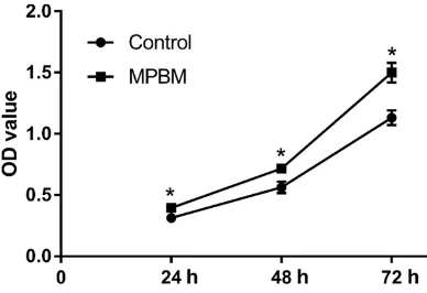
Figure 1. Comparison of proliferation abilities of MSCs between the groups. *P < 0.05, compared with
[image:3.612.91.285.419.552.2]Effects of BM on MSCs ALP staining and Alizarin Red S staining
ALP activity is a marker of ear-ly-stage osteogenesis. MSCs were observed via ALP histo-chemical staining after osteo-genic differentiation for 7 days (Figure 2A). Compared to the control group, higher ALP activity was observed in the MPBM group.
[image:4.612.90.376.73.298.2]Mineralization of inducted MSCs was evaluated using Alizarin Red S staining on day 3 and day 7 of osteogenic dif-ferentiation (Figure 2B). The cells were enclosed by a min-eralized matrix that was rich in calcium deposits, as evi-denced by Alizarin Red S-po- sitive regions. Compared to
Figure 2. A. ALP histochemical staining of induced MSCs at day 7. Original magnification: 100×. B. Microscopic illustrations of induced MSCs stained by Alizarin Red S staining at day 21. Original magnification: 100×.
emiluminescence (Pierce Biotechnology, Ro- ckford, USA) was used for protein visualization. The following antibodies were utilized: p-Akt Ser473 (1:1,000 dilution, CST, USA), p-Akt Thr308 (1:1,000 dilution, CST, USA), Akt
(1:1,000 dilution, CST, USA), β-catenin (1:1,000
dilution, CST, USA), and GAPDH (1:2,000 dilu-tion, CST, USA).
Statistical analysis
Data are expressed as mean ± standard devia-tion (SD). Addidevia-tionally, Student’s t-test was used
to determine statistically significant differences
between both groups. P-values ≤ 0.05 indicate statistical significance. Statistical analysis was
conducted using SPSS 16.0 (SPSS Inc, Chicago, IL, USA) software.
Results
Effects of BM on MSCs proliferation
Proliferation of MSCs was measured via CCK-8 assays. CCK-8 results suggested a faster growth rate of MSCs in the MPBM group. OD490 values of the MPBM group were much higher than those of the control group at 24, 48, and 72 hours. Compared to the control group, the proliferation ability of treated MSCs was highest at 72 hours (Figure 1).
the control group, the MPBM group showed more Alizarin Red S-positive regions.
Effects of BM on MSC differentiation
Expression of osteogenic genes, including ALP, OCN, Runx2, OSX, Col1a1, Col1a2, OPN, and OPG, was determined via RT-qPCR after 7 days of induction. RT-qPCR analysis revealed that mRNA levels of genes associated with osteo-genic differentiation, except for OSX, were
sig-nificantly higher in the MPBM group than in the
control group (Figure 3).
Effects of BM on MSCs Akt and β-catenin signaling activation
To determine the effects of MPBM on Akt and
β-catenin signaling, Western blot analysis was conducted on the MSCs. There were significant
differences in p-Akt (Ser473), p-Akt (Thr308),
and β-catenin protein expression between the two groups. MPBM significantly increased p-Akt (Ser473), p-Akt (Thr308), and β-catenin protein
expression, compared to the control group (Figure 4).
Discussion
MSCs in vitro. Present findings
suggest that BM could assist in bone fracture healing th- rough the promotion of prolif-eration of MSCs. For example, BM was shown to stimulate BMSCs and promote osteo-genic differentiation, as as- sessed by ALP and Alizarin Red S staining. ALP, a func-tional enzyme in osteoblasts, plays a major role in bone for-mation and bone mineraliza-tion. Therefore, the activity of
ALP is often used to confirm
the presence of osteoblasts and the degree of osteogenic differentiation [20, 21]. Al- izarin Red S staining is gener-ally employed to determine the degree of extracellular bone matrix nodules [22].
MPBM not only significantly
upregulated expression of ALP, but also exhibited superi-or ability in stimulating MSCs to perform calcium deposi-tion, compared with normal rat plasma.
The biological behavior of MSCs is closely connected with cell signaling pathways.
Akt and Wnt/β-Catenin signal -ing pathways play important roles in proliferation and os- teogenic differentiation of MSCs [23, 24]. Akt is a down-stream serine-threonine ki- nase that transmits survival signals from growth factors
[25]. β-Catenin, the core mol
-ecule of canonical
[image:5.612.91.372.63.575.2]Wnt/β-Catenin signaling, also plays an important role in MSC pro-liferation and differentiation,
Figure 3. Osteogenic gene expression of induced MSCs at day 7. *P < 0.05
versus the control group.
remodeling and bone fracture healing [18]. The
efficiency of MSCs, in terms of proliferation and
differentiation, is crucial in the treatment of patients with bone non-unions or bone defects. Thus, exploring safe and effective treatments that regulate proliferation and osteogenic
dif-ferentiation of MSCs is of vital significance [19].
The present study, using CCK-8 assays, showed
especially in osteoblast genesis and bone for-mation [26]. Compared to the untreated control group, the current study found that not only had
β-Catenin increased expression, but expres -sion levels of P-Akt (Th308 and Thr473) were also elevated following MPBM treatment. During the osteogenic differentiation process, MPBM promoted expression of osteogenic
well as PI3K/Akt related genes, Col1a2, and OPN [29, 30]. They all play essential roles in the commitment of MSCs to osteoblastic lineage.
The activity of OCN typically confirms the pres -ence of osteoblasts, as well as the degree of osteogenic differentiation. Runx2 plays a vital
role in the early stages of bone calcification.
Col1 is the most abundant protein in the bone matrix. It is directly involved in mineralization and maturation of osteoblasts [31]. Expression of OPN has been linked to the mineralization front of the osteoid [32]. OPG can inhibit the differentiation and maturation of osteoclasts [33]. Current results indicate that MPBM could upregulate expression of these osteogenesis related genes, promoting the osteogenic differ-entiation of MSCs by modulation of Akt and
Wnt/β-catenin signaling pathways.
To the best of our knowledge, this is the first
study to systematically explore the effects of MPBM on osteogenic differentiation of MSCs in vitro. Current observations signified that a com
-bination of Akt and β-catenin activation, in
response to MPBM, could promote MSC gr- owth and increase expression of osteogenic biomarkers. However, there were some limita-tions to the current study. As a result of meth-odological restrictions in herbal compounds and allowances for a more pharmacologically relevant assessment of mechanisms, this study applied medicated plasma instead of using the components of BM in the culture
directly. As a result, the current study could not target which active ingredients of BM exert osteogenic effects.
Conclusion
In summary, the current study validated the suggestion that MPBM promotes proliferation and osteoblastic differentiation of MSCs via Akt
and β-catenin signaling pathways. Current find -ings suggest that BM could be a promising alternative medicine for improved bone frac-ture healing.
Acknowledgements
This study was supported by the National Natural Science Foundation of China (81- 503598; 81373665), Summit Plateau Team Project in Traumatology of Shanghai University of TCM, Shanghai Municipal Commission of Health and Family Planning (201740224), the Graduate Innovation Training Project of Shang- hai University of TCM in 2018 (Y201820), and the Key Clinical Discipline Construction Project “Orthopedics and Traumatology of Traditional Chinese Medicine” (2017Z02024).
Disclosure of conflict of interest
None.
[image:6.612.95.522.71.271.2]Address correspondence to: Dr. Yuelong Cao, Shi’s Center of Orthopedics and Traumatology, Shuguang
Hospital Affiliated to Shanghai University of Tr-aditional Chinese Medicine, No. 528 Zhang Heng Road, Pudongxin Qu, Shanghai 201203, China. Tel: +86-21-20256519; Fax: +86-2120256588; E-mail:
ningtcm@126.com
References
[1] O’Keefe RJ and Mao J. Bone tissue engineer-ing and regeneration: from discovery to the clinic--an overview. Tissue Eng Part B Rev 2011; 17: 389-92.
[2] Bigham-Sadegh A and Oryan A. Basic concepts regarding fracture healing and the current op-tions and future direcop-tions in managing bone fractures. Int Wound J 2015; 12: 238-47. [3] Fong K, Truong V, Foote CJ, Petrisor B, Williams
D, Ristevski B, Sprague S and Bhandari M. Pre-dictors of nonunion and reoperation in pa-tients with fractures of the tibia: an observa-tional study. BMC Musculoskelet Disord 2013; 14: 103.
[4] Gómez-Barrena E, Rosset P, Lozano D, Stanovi-ci J, Ermthaller C, Gerbhard F. Bone fracture healing: cell therapy in delayed unions and nonunions. Bone 2015; 70: 93-101.
[5] Wang X, Wang Y, Gou W, Lu Q, Peng J and Lu S. Role of mesenchymal stem cells in bone re-generation and fracture repair: a review. Int Orthop 2013; 37: 2491-8.
[6] da Silva Meirelles L, Chagastelles PC and Nar-di NB. Mesenchymal stem cells reside in virtu-ally all post-natal organs and tissues. J Cell Sci 2006; 119: 2204-13.
[7] Kovach TK, Dighe AS, Lobo PI and Cui Q. Inter-actions between MSCs and immune cells: im-plications for bone healing. J Immunol Res 2015; 2015: 752510.
[8] Song F, Jiang D, Wang T, Wang Y, Lou Y, Zhang Y, Ma H and Kang Y. Mechanical stress regu-lates osteogenesis and adipogenesis of rat mesenchymal stem cells through PI3K/Akt/ GSK-3beta/beta-catenin signaling pathway. Biomed Res Int 2017; 2017: 6027402.
[9] Dreger T, Watson JT, Akers W, Molligan J, Achil-efu S, Schon LC and Zhang Z. Intravenous ap-plication of CD271-selected mesenchymal stem cells during fracture healing. J Orthop Trauma 2014; 28 Suppl 1: S15-19.
[10] Long H, Sun B, Cheng L, Zhao S, Zhu Y, Zhao R and Zhu J. miR-139-5p represses BMSC osteo-genesis via targeting Wnt/beta-catenin signal-ing pathway. DNA Cell Biol 2017; 36: 715-724. [11] Gharaibeh B, Lavasani M, Cummins JH and
Huard J. Terminal differentiation is not a major determinant for the success of stem cell thera-py-cross-talk between muscle-derived stem cells and host cells. Stem Cell Res Ther 2011; 2: 31.
[12] Xu L, Huang S, Hou Y, Liu Y, Ni M, Meng F, Wang K, Rui Y, Jiang X and Li G. Sox11-modified mes -enchymal stem cells (MSCs) accelerate bone fracture healing: Sox11 regulates differentia-tion and migradifferentia-tion of MSCs. FASEB J 2015; 29: 1143-1152.
[13] Zhang XP, Xu GR, Xu SQ, Lu ZM and Huang L. [Case-control study on Tibetan Baimai oint-ment (see symbol in text) for the treatoint-ment of wrist-dysfunction after distal radius fracture]. Zhongguo Gu Shang 2014; 27: 920-4.
[14] Hsueh TP and Chiu HE. Traditional Chinese medicine speeds-up humerus fracture healing: two case reports. Complement Ther Med 2012; 20: 431-433.
[15] Hejazi ZA, Namjooyan F and Khanifar M. Com-plementary and alternative medicine for osteo-porosis. Iran J Med Sci 2016; 41: S27. [16] He Q, Liu Q, Chen Y, Meng J and Zou L.
Long-Zhi Decoction medicated serum promotes an-giogenesis in human umbilical vein endothelial cells based on autophagy. Evid Based Comple-ment Alternat Med 2018; 2018: 6857398. [17] Erdogan H, Fadillioglu E, Kotuk M, Iraz M,
Tas-demir S, Oztas Y and Yildirim Z. Effects of Gink-go biloba on plasma oxidant injury induced by bleomycin in rats. Toxicol Ind Health 2006; 22: 47-52.
[18] Dong P, Gu X, Zhu G, Li M, Ma B and Zi Y. Mela-tonin induces osteoblastic differentiation of mesenchymal stem cells and promotes frac-ture healing in a rat model of femoral fracfrac-ture via neuropeptide Y/neuropeptide Y receptor Y1 signaling. Pharmacology 2018; 102: 272-280.
[19] Lu W, Xiu X, Zhao Y and Gui M. Improved prolif-eration and differentiation of bone marrow mesenchymal stem cells into vascular endo-thelial cells with sphingosine 1-phosphate. Transplant Proc 2015; 47: 2035-2040.
[20] Jiang T, Zhou B, Huang L, Wu H, Huang J, Liang T, Liu H, Zheng L and Zhao J. Andrographolide exerts pro-osteogenic effect by activation of Wnt/beta-catenin signaling pathway in vitro. Cell Physiol Biochem 2015; 36: 2327-39. [21] He F, Liu M, Chen Z, Liu G, Wang Z, Liu R, Luo
J, Tang J, Wang X, Liu X, Zhou H, Chen X, Liu Z and Zhang W. Corrigendum to assessment of human tribbles homolog 3 genetic variation (rs2295490) effects on type 2 diabetes pa-tients with glucose control and blood pressure lowering treatment” [EBioMedicine 13 (2016) 181-189]. EBioMedicine 2017; 17: 239. [22] Li-Yu J, Clayburne GM, Sieck MS, Walker SE,
Athreya BH, DeHoratius RJ, Schumacher HR Jr. Calcium apatite crystals in synovial fluid rice bodies. Ann Rheum Dis 2002; 61: 387-90. [23] Dong K, Hao P, Xu S, Liu S, Zhou W, Yue X,
alle-viates high-glucose suppressed osteogenic dif-ferentiation of MC3T3-E1 cells via antioxidant effect and PI3K/Akt signaling pathway. Cell Physiol Biochem 2017; 42: 1897-1906. [24] Tao K, Xiao D, Weng J, Xiong A, Kang B and
Zeng H. Berberine promotes bone marrow-de-rived mesenchymal stem cells osteogenic dif-ferentiation via canonical Wnt/beta-catenin signaling pathway. Toxicol Lett 2016; 240: 68-80.
[25] Leavens KF, Easton RM, Shulman GI, Previs SF and Birnbaum MJ. Akt2 is required for hepatic lipid accumulation in models of insulin resis-tance. Cell Metab 2009; 10: 405-418.
[26] Zhou L, Zhang T, Sun S, Yu Y and Wang M. Cryptochrome 1 promotes osteogenic differen-tiation of human osteoblastic cells via Wnt/ beta-Catenin signaling. Life Sci 2018; 212: 129-137.
[27] Song HB, Jiang Y, Liu JX, Wang GQ, Zhang DP, Jiang YC, Ren SJ, Liu HP and Jiang XY. Stimula-tion of osteogenic differentiaStimula-tion in bone mar-row stromal cells via Wnt/beta-catenin path-way by Qili Jiegu-containing serum. Biomed Pharmacother 2018; 103: 1664-1668. [28] Pacheco-Costa R, Kadakia JR, Atkinson EG,
Wallace JM, Plotkin LI and Reginato RD. Con-nexin37 deficiency alters organic bone matrix, cortical bone geometry, and increases Wnt/ beta-catenin signaling. Bone 2017; 97: 105-113.
[29] Tao X, Qi Y, Xu L, Yin L, Han X, Xu Y, Wang C, Sun H and Peng J. Dioscin reduces ovariecto-my-induced bone loss by enhancing osteoblas-togenesis and inhibiting osteoclasosteoblas-togenesis. Pharmacol Res 2016; 108: 90-101.
[30] Yu X, Zheng Y, Zhu X, Gao X, Wang C, Sheng Y, Cheng W, Qin L, Ren N, Jia H and Dong Q. Os-teopontin promotes hepatocellular carcinoma progression via the PI3K/AKT/Twist signaling pathway. Oncol Lett 2018; 16: 5299-5308. [31] Wu Z, Weng S, Yan D, Xie Z, Zhou Q, Li H, Bai B,
Boodhun V, Shen Z, Tang J, Zhou L, Tao Z and Yang L. Administration of cinnamaldehyde pro-motes osteogenesis in ovariectomized rats and differentiation of osteoblast in vitro. J Pharmacol Sci 2018; 138: 63-70.
[32] Holm E, Gleberzon JS, Liao Y, Sorensen ES, Beier F, Hunter GK and Goldberg HA. Osteo-pontin mediates mineralization and not osteo-genic cell development in vitro. Biochem J 2014; 464: 355-364.
[33] Fu Y, Gu J, Wang Y, Yuan Y, Liu X, Bian J and Liu ZP. Involvement of the Ca2+ signaling pathway