ORIGINAL RESEARCH
EXTRACRANIAL VASCULAR
Vitamin D and Vulnerable Carotid Plaque
XJ.S. McNally, X T.M. Burton, XB.W. Aldred, XS.-E. Kim, X M.S. McLaughlin, XL.B. Eisenmenger, XG.J. Stoddard, XJ.J. Majersik, D.V. Miller, XG.S. Treiman, and XD.L. Parker
ABSTRACT
BACKGROUND AND PURPOSE:MR imaging– detected carotid intraplaque hemorrhage indicates vulnerable plaque with high stroke risk. Angiotensin II stimulates intraplaque hemorrhage in animal models, and the angiotensin system is highly regulated by vitamin D. Our purpose was to determine whether low vitamin D levels predict carotid intraplaque hemorrhage in humans.
MATERIALS AND METHODS: In this cross-sectional study, 65 patients with carotid disease underwent carotid MR imaging and blood draw. Systemic clinical confounders and local lumen imaging markers were recorded. To determine the association of low vitamin D levels with MR imaging detected intraplaque hemorrhage, we performed multivariable Poisson regression by using generalized estimating equations to account for up to 2 carotid arteries per patient and backward elimination of confounders. MR imaging detected intraplaque hemorrhage volume was also correlated with vitamin D levels and maximum plaque thickness. Thirty-five patients underwent carotid endarterectomy, and histology-detected intraplaque hemorrhage was correlated with vitamin D levels and total plaque area.
RESULTS:Low vitamin D levels (⬍30 ng/mL, prevalence ratio⫽2.05,P⫽.03) were a significant predictor of MR imaging detected intraplaque hemorrhage, along with plaque thickness (prevalence ratio⫽1.40,P⬍.001). MR imaging detected intraplaque hemorrhage volume linearly correlated with plaque thickness (partialr⫽0.45,P⬍.001) and low vitamin D levels (partialr⫽0.26,P⫽.003). Additionally, histology-detected intraplaque hemorrhage area linearly correlated with plaque area (partialr⫽0.46,P⬍.001) and low vitamin D levels (partialr⫽0.22,P⫽.03). The association of intraplaque hemorrhage volume with low vitamin D levels was also higher with ischemic stroke.
CONCLUSIONS: Low vitamin D levels and plaque thickness predict carotid intraplaque hemorrhage and outperform lumen markers of vulnerable plaque. This research demonstrates a significant link between low vitamin D levels and carotid intraplaque hemorrhage.
ABBREVIATIONS:AT1R⫽angiotensin II type 1 receptor; IPH⫽intraplaque hemorrhage; NADPH⫽nicotinamide adenine dinucleotide phosphate; PR⫽prevalence ratio
L
arge-artery atherosclerosis, including carotid disease, is a sig-nificant cause of ischemic stroke and an important therapeutic target.1Reports estimate that carotid disease accounts for 10%– 15% of ischemic strokes.2,3Additionally, recurrent stroke is more highly associated with carotid atherosclerosis than other causes.4,5While only 10%–15% of patients with stroke have large-artery atherosclerosis, approximately one-third of early recurrence oc-curs in this group.6,7
These past estimates are based on whether moderate or severe stenosis is present, defined for the carotid arteries as NASCET stenosis of⬎50%.8More recent studies have questioned stenosis, finding that other markers predict unstable plaque better, includ-ing intraplaque hemorrhage (IPH). Carotid IPH is a better esti-mate of recurrent stroke risk, with a⬃5-fold higher risk of recur-rent stroke in all stenosis categories in multiple recent studies and meta-analyses.9-11Carotid IPH can be accurately detected with heavily T1-weighted sequences, including MPRAGE. Both Received November 6, 2015; accepted after revision April 26, 2016.
From the Department of Radiology and Imaging Sciences, Utah Center for Ad-vanced Imaging Research (J.S.M., B.W.A., S.-E.K., M.S.M., L.B.E., D.L.P.); Department of Neurology (T.M.B., J.J.M.); Department of Orthopedics, Study Design and Biostatis-tics Center (G.J.S); Department of Pathology (D.V.M.); and Department of Surgery at the University of Utah and VA Salt Lake City Health Care System, Salt Lake City, Utah (G.S.T.).
This work was supported by a Radiological Society of North America Research Scholar Grant; a University of Utah Intramural Seed Grant; and a grant for the Study Design and Biostatistics Center, with funding, in part, from the National Center for Research Resources and the National Center for Advancing Transla-tional Sciences, NaTransla-tional Institutes of Health, grant 8UL1TR000105 (formerly UL1RR025764).
Paper previously presented at: Annual Meeting of the American Society of Neuro-radiology and the Foundation of the ASNR Symposium, April 25–30, 2015; Chicago, Illinois.
Please address correspondence to J. Scott McNally, MD, PhD, University of Utah, Department of Radiology and Imaging Sciences, 30 North 1900 East #1A071, Salt Lake City, UT 84132-2140; e-mail: scott.mcnally@hsc.utah.edu
Indicates open access to non-subscribers at www.ajnr.org
Indicates article with supplemental on-line table.
MPRAGE and TOF have low false-negative rates (3% versus 4%), but the MPRAGE sequence has a lower false-positive rate (20%) compared with TOF (44%).12
Despite MR imaging detection of IPH, no treatment has been shown to reverse these lesions. A potential treatment target is the angiotensin system, a major determinant of carotid plaque insta-bility and stroke risk.13Angiotensin II stimulates adventitial neo-vascularity and is implicated in animal models of IPH.14 Angio-tensin II increases plaque microvessel angiogenesis through the endothelial angiotensin II type 1 receptor (AT1R).15AT1R acti-vation leads to reactive oxygen species formation through the nic-otinamide adenine dinucleotide phosphate (NADPH) oxidase and plaque inflammation.15Extrapolating from this, inhibition of the angiotensin system may prevent or decrease IPH.
The angiotensin system is highly regulated by an endogenous inhibitory axis, including vitamin D and the vitamin D receptor.16 The vitamin D receptor is downregulated in atherosclerotic plaque in animal models.17Vitamin D deficiency has been asso-ciated with increased intima-media and plaque thickness in sub-clinical carotid atherosclerosis.18Serum vitamin D levels are neg-atively correlated with carotid intima-media thickness (r ⫽
⫺0.51).19Prior studies have demonstrated a high prevalence of vitamin D insufficiency (⬍30 ng/mL, 63.6%) and increased stroke risk in the Mountain West population of the United States.20 This mirrors the prevalence of vitamin D deficiency (⬍20 ng/mL) in the United States as a whole (41.6%).21
Our purpose was to determine whether low vitamin D levels predict carotid IPH in patients with carotid disease. Our hypoth-esis was that low vitamin D levels are associated with carotid IPH when controlling for systemic and local plaque confounders. If confirmed, this pathway may represent an important treatment target in patients with carotid IPH.
MATERIALS AND METHODS
Clinical Study DesignThis was a cross-sectional study of patients with carotid disease, defined asⱖ2-mm-thick carotid plaque. Patients were consecu-tively recruited from neurovascular outpatient and inpatient ser-vices. Exclusions included patients younger than 18 years of age, prisoners, pregnancy, or those with contraindications to MR im-aging (eg, pacemakers). There were no exclusion/inclusion crite-ria for carotid stenosis other than complete occlusion. Sixty-five patients were recruited and completed carotid MR imaging and blood draw.
Ethics. Institutional review board approval was obtained along with informed consent from all subjects. In subjects with im-paired decisional capacity, legal authorized representative con-sent and patient ascon-sent was obtained.
Serologic Analysis of Vitamin D and Angiotensin II All patients underwent blood draw following IV placement for MR imaging. Samples were taken directly to the Associated Re-gional and University Pathologists for serum vitamin D analysis by quantitative high-performance liquid chromatography tan-dem mass spectrometry (25-hydroxyvitamin D2and D3; http://
ltd.aruplab.com/Tests/Pub/2002348) and plasma angiotensin II analysis by quantitative immunoassay (http://ltd.aruplab.com/
Tests/Pub/0098771). Vitamin D levels were considered low/insuf-ficient at⬍30 ng/mL. Angiotensin II levels were considered high at⬎18 ng/L.
Research MR Imaging Protocol
Images were obtained on 3T MR imaging scanners (Trio, Verio, Skyra and Prisma scanners; Siemens, Erlangen, Ger-many) with custom-made carotid coils.22Each MR imaging included brain DWI and carotid MPRAGE sequences acquired with custom neck coils described below.
Subject-Specific Radiofrequency Coils
A modular system of subject-specific radiofrequency coil arrays was used to maximize signal to noise.23The head coil provides head immobilization, essential to carotid imaging.24 Either 7-channel or 9-channel coils were used, connected to preamplifi-ers through low-resistance connectors, allowing them to be inter-changed to fit necks of different shapes and sizes. These coils can image simultaneously with clinical head coils without extra posi-tioning hardware.
Carotid MPRAGE and IPH Determination
MPRAGE parameters were the following: 3D, TR/TE/TI⫽6.39/ 2.37/370 ms, flip angle⫽15°, FOV⫽130 ⫻130⫻48 mm3,
matrix⫽256⫻256⫻48, voxel⫽0.5⫻0.5⫻1.0 mm3, fat
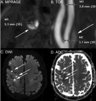
saturation, acquisition time⫽ ⬃5 minutes as described previ-ously.25Images were obtained from 20 mm below to 20 mm above the carotid bifurcation at a 1.0-mm section thickness. Ca-rotid IPH was defined by MPRAGE-positive plaque withⱖ2-fold signal compared with the sternocleidomastoid muscle (Fig 1).26 We have previously shown that MPRAGE-positive plaque corre-sponds to carotid IPH on histology.26IPH volume was deter-mined quantitatively with volumes traced from the PACS.
Carotid Lumen Measurements
All non-IPH carotid lumen measurements were determined by consensus of 2 reviewers, blinded to brain MR imaging and clin-ical covariates. The entire carotid plaque was considered 1 unit, from 20 mm above to 20 mm below the bifurcation. Maximum plaque thickness was measured in the transverse plane on MPRAGE images, perpendicular to the center axis of the lumen (Fig 1). Lumen markers (stenosis, ulceration, and intraluminal thrombus) were determined from noncontrast TOF with param-eters as follows: 3D, TR/TE⫽25/4.02 ms, flip angle⫽20°, FOV⫽ 160⫻160⫻90 mm, matrix⫽256⫻256, section thickness⫽ 0.64 mm, 144 sections, acquisition time⫽ ⬃3.5 minutes. In all cases, duplex sonography was performed before study entry and was used as a complementary measure of stenosis. In cases of
de-fined by visible bulb stenosis, a distal ICA diameter ofⱕ3 mm, and a distal ICA/distal external carotid artery ratio of
ⱕ1.25.29,30Ulceration was determined by using a size threshold of 2 mm.31Intraluminal thrombus was determined by a filling defect and confirmed in all cases with contrast CTA or MRA.32
Ischemic Stroke Determination
Ischemic stroke was determined by using the American Heart Association definition of CNS infarction: brain or retinal cell death attributable to ischemia based on the following: 1) imaging evidence of cerebral or retinal ischemia in the carotid distribution, or 2) clinical symptoms persisting forⱖ24 hours, with other eti-ologies excluded.33We reviewed neurovascular clinic or inpatient charts to determine the presence of recent stroke occurring at the time of recruitment. Asymptomatic (“silent”) recent infarcts were included as strokes and determined on brain DWI performed in all patients to supplement clinical determination of infarct by showing acuity and distribution (Fig 1). DWI-positive carotid territory infarcts were detected using DTI trace images, which are superior to conventional DWI in detecting recent infarcts.34,35 The DTI parameters were 2D, 128⫻128 matrix, 3-mm section thickness,b⫽2000, 20 directions. Brain DWI was interpreted by a Certificate of Added qualification– certified neuroradiologist.
Clinical Characteristics
Clinical characteristics were determined by chart review, including cerebrovascu-lar risk factors of age, male sex, diabetes, hypertension, hyperlipidemia, renal in-sufficiency, body mass index, and smok-ing status. We recorded the followsmok-ing cerebrovascular medications: antiplate-lets, anticoagulants, statins, and antihy-pertensives, including antiangiotensin medications: angiotensin-converting en-zyme inhibitors and angiotensin receptor blockers.
Histologic Processing
In 35 patients who underwent carotid endarterectomy, each specimen was fixed in 10% neutral buffered formalin for histology. The ratio of the fixative to the specimen was at least 10:1. Speci-mens were decalcified in 1% Enhanced Decalcification Formulation (SL85–32; Statlab, Lewisville, Texas). Tissue cas-settes were processed on an automated Vacuum Infiltrating Processor (Sakura, Alphen aan den Rijn, the Netherlands), embedded in paraffin wax, sectioned at 3- to 4-mm intervals, and stained with hematoxylin-eosin.
Histology Interpretation of IPH A pathologist outlined recent IPH by using hematoxylin-eosin, blinded to MPRAGE results. “Recent” IPH was de-fined by intact red blood cells or degen-erated red blood cells on hematoxylin-eosin. Each carotid area positive for IPH was then compared with vitamin D levels.
Statistical Analysis
Statistical modeling was performed by using generalized estimat-ing equations to account for data clusterestimat-ing, with up to 2 carotid arteries per patient. Carotid arteries were treated as separate units grouped within subjects because IPH may be associated with local carotid plaque markers (eg, plaque thickness) and systemic clini-cal factors (eg, age). Because⬎1 marker for IPH was being stud-ied, potential confounding was investigated on the outcome vari-able, IPH. Therefore only 1 data table was required withPvalues from univariable generalized estimating equation Poisson regres-sion models. Poisson regresregres-sion directly estimates the prevalence ratio (PR), which is more intuitive to interpret than an odds ratio from a logistic regression approach (Zou and Donner36). Next, all potential confounders withP ⬍.20 from a univariable model were placed in an initial multivariable generalized estimating equa-tion Poisson regression model for IPH, then eliminated in a back-ward fashion until all remaining variables met the thresholdP⬍.10. A liberal significance criterion,P⬍.10, was used to protect against residual confounding (Maldonado and Greenland37).
[image:3.594.55.373.49.383.2]For hypothesis testing of markers predictive of IPH, we used the traditionalP⬍.05. In binary outcome models, 5 outcome events for every predictor variable are sufficient to avoid overfit-ting.38With 48 carotid plaques positive for IPH and 80 negative for IPH, 48/5 or 9.6 or up to 9 predictor variables could be in-cluded in the model without overfitting, exceeding the number of variables in our final model. Additionally, we identified the fol-lowing potential confounders with a scientific basis for confound-ing the association between vitamin D and IPH: angiotensin, an-giotensin-converting enzyme inhibitor use, and angiotensin receptor blocker use. Because of the high potential for these to confound the vitamin D and IPH association, we used a combi-nation of the 10% change in estimate and backward elimicombi-nation and kept these potential confounders in a secondary analysis even though they were eliminated in the primary analysis. We also calculated IPH volume as a continuous variable and used a linear mixed-effects regression model to correlate carotid IPH with maximum plaque thickness and vitamin D levels, again account-ing for up to 2 carotid arteries per patient. For the histology-outlined IPH area correlation with vitamin D levels, a random intercept linear regression model was used with an autoregressive correlation structure repeated-measures analysis of the 35 sub-jects that had underwent carotid endarterectomy. In this analysis, the “time” repetition variable was each histology slide, and the “group” variable was each carotid plaque. All statistical analyses were performed with STATA 13.1 (StataCorp, College Station, Texas).
RESULTS
Clinical Characteristics
Sixty-five patients were recruited, with a mean age 72.3⫾8.3 years, 90.8% men and 3.1% minorities. Patients had multiple cerebrovascular risk factors (69.2% current or prior smokers, 87.7% hypertensive, 84.6% hyperlipidemic, 44.6% diabetic) and were aggressively managed before enrollment (84.6% on antihy-pertensives, 89.2% on statins, 81.5% on antiplatelets) (On-line Table). These characteristics reflect the high prevalence of carotid disease in our population.
Imaging and Clinical Characteristics by Vessel
Imaging and clinical characteristics were also listed by vessel and broken down by IPH status (positive versus negative) in the
On-line Table. Each patient contributed 2 carotid plaques, with the exception of 2 carotid occlusions that were excluded, leaving 128 carotid arteries for the final sample. Stenosis was worse in carotid ar-teries with IPH-positive versus negative plaque (NASCET stenosis of 53.9% ver-sus 35.7% and millimeter stenosis of 2.25 versus 3.17 mm,P⫽.003). Maxi-mum plaque thickness was also higher (5.53 versus 4.08 mm,P⬍ .001), and there was a higher prevalence of ulcer-ation (64.6% versus 37.5%,P⫽.02) and intraluminal thrombus, though rare (6.3% versus 0.0%,P⫽.05). Some fac-tors were potential confounders be-tween IPH-positive and negative status (P⬍.20), requiring mul-tivariable regression to determine essential IPH predictors.
Multivariable Generalized Estimating Equation Poisson Regression Analysis for Carotid IPH Prediction
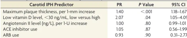
Multivariable generalized estimating equation Poisson regression analysis was performed to determine predictors of carotid IPH from the On-line Table. The initial model included 9 predictors withP⬍.20: NASCET stenosis, millimeter stenosis, maximum plaque thickness, ulceration, intraluminal thrombus, high angio-tensin II (⬎18 ng/L), vitamin D level ng/mL (continuous vari-able), low vitamin D level (⬍30 ng/mL), and male sex.Table 1 shows the final model after sequential backward elimination of confounders withP⬎.10. The final model included maximum plaque thickness (PR⫽1.40; 95% CI, 1.18 –1.67;P⬍.001) and low vitamin D levels (PR⫽2.05; 95% CI, 1.06 –3.96;P⫽.03) as significant predictors of carotid IPH.Table 2illustrates the lack of confounding by angiotensin II, angiotensin-converting enzyme inhibitor, and angiotensin receptor blockers use.
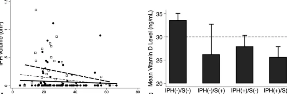
Linear Mixed-Effects Regression Model of IPH Volume with Maximum Plaque Thickness and Low Vitamin D Levels Carotid IPH volume significantly correlated with maximum plaque thickness (r⫽0.45,P⬍.001) and low vitamin D levels (r⫽0.26,P⫽ .003) as depicted in the linear regression plot (Fig 2).
Carotid IPH Histology and Vitamin D Levels
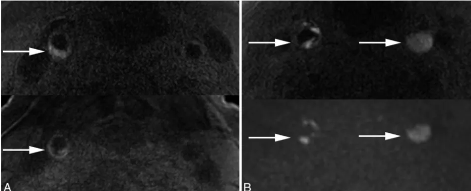
Histology-defined IPH area (Fig 3A) negatively correlated with vitamin D levels (Fig 3B). In addition, IPH area correlated with plaque area (partialr⫽0.46,P⬍.001) and low-versus-normal vitamin D levels (partialr⫽0.22,P⫽.03) (Fig 3C).
Carotid IPH, Vitamin D Levels, and Stroke
IPH volume more negatively correlated with vitamin D levels with ischemic stroke (hollow squares, thick dashed line, partialr⫽
⫺0.21,P⫽.27) compared to without it (solid circles, thick solid line, partialr⫽ ⫺0.06,P⫽.53) (Fig 4A). Vitamin D levels were also lower in patients with versus without stroke in the setting of IPH-negative plaque (mean, 33.5⫾13.2 ng/mL in 73 carotids versus 26.2⫾17.4 ng/mL in 7 carotids,P⫽.17) and less so with IPH-positive plaque (27.8⫾12.6 ng/mL in 25 carotids versus 25.6⫾11.1 ng/mL in 23 carotids,P⫽.51) (Fig 4B).
Table 1: Final MRI-IPH prediction modela
Carotid IPH Predictor PR PValue 95% CI
Maximum plaque thickness, per 1-mm increase 1.40 ⬍.001 1.18–1.67 Low vitamin D level of⬍30 ng/mL, low versus high 2.05 .03 1.06–3.96 a
[image:4.594.53.377.130.196.2]The final prediction model for the presence of MRI-detected carotid IPH depended on 2 factors: maximum plaque thickness and low vitamin D levels.
Table 2: IPH prediction model with angiotensin system confoundersa
Carotid IPH Predictor PR PValue 95% CI
Maximum plaque thickness, per 1-mm increase 1.40 ⬍.001 1.18–1.67 Low vitamin D level,⬍30 ng/mL, low versus high 2.07 .04 1.05–4.09 Angiotensin II level (ng/L), per 1-U increase 1.00 .80 0.99–1.01
ACE inhibitor use 1.05 .87 0.56–1.99
ARB use 0.93 .90 0.31–2.77
Note:—ARB indicates angiotensin receptor blockers.
a
Carotid IPH Volume Follow-Up
Two patients were re-evaluated after 1 year (Fig 5). Both were treated with medical therapy (statins, antiplatelets, and
anti-hypertensives) and both patients’ primary care physicians were alerted to low vitamin D levels (⬍30 ng/mL). Patient 1 was supplemented, vitamin D increased (baseline/1 year: 18.1/26.8 FIG 2. Linear regression of MR imaging– detected IPH volume, plaque thickness, and vitamin D status. Pooled data demonstrate the IPH volume negative correlation with vitamin D level (A) and correlation with maximal plaque thickness (partialr⫽0.45,P⬍.001,thin dashed line) and low-versus-normal vitamin D levels (partialr⫽0.26,P⫽.003; low vitamin D level:empty squaresandthick dashed line; normal vitamin D levels: solid circlesandthick solid line,B).
FIG 3.Linear regression of IPH area on histology, plaque area, and vitamin D status. Representative MPRAGE-positive plaque (upper image) and IPH area outlined on the corresponding hematoxylin-eosin stain (lower image) (A). Pooled data demonstrate the IPH area negatively correlating with vitamin D levels (B) and positively correlating with plaque area (partialr⫽0.46,P⬍.001,thin dashed line) and low-versus-normal vitamin D levels (partialr⫽0.22,P⫽.03, low vitamin D level:empty squaresandthick dashed line; normal vitamin D level:solid circlesandthick solid line) (C).
FIG 4. Association of IPH volume, vitamin D levels, and ischemic stroke. IPH volume was negatively correlated with vitamin D levels as inFig 2A
[image:5.594.49.530.46.207.2] [image:5.594.58.531.268.391.2] [image:5.594.58.530.467.621.2]ng/mL), carotid IPH volume decreased by 24.1% (baseline/1 year: 0.151/0.115 cm3), and he remained asymptomatic.
Pa-tient 2 was not supplemented, vitamin D levels decreased (baseline/1 year: 20.9/10.7 ng/mL), carotid IPH volume mini-mally decreased by 4.3% (baseline/1 year: 1.041/0.996 cm3),
and he had sequential bilateral strokes.
DISCUSSION
This work shows that both the presence and volume of carotid IPH are strongly associated with plaque thickness and low vitamin D levels, and not necessarily with percentage stenosis or other measures of carotid architecture. This finding supports the idea that low vitamin D levels may play a role in atherosclerosis and stroke risk. Considering its multiple health benefits, vitamin D testing may be worthwhile in patients with vulnerable carotid plaque.
Low vitamin D levels have been linked to cardiovascular dis-ease in many prior studies.39Two cohort studies evaluating par-ticipants in the Framingham Heart Study (Offspring Cohort) and the Copenhagen City Heart Study found increased risk of isch-emic stroke with lower vitamin D levels,40,41and a low vitamin D level is an independent predictor of ischemic stroke volume.42 Most important, vitamin D supplementation decreases muscle atrophy, falls, hip fractures, and cognitive impairment and leads to functional improvement in patients with stroke.43,44 Vita-min D also has many direct beneficial effects on the vascula-ture. Within 1 minute of treatment, vitamin D increases endo-thelial cell nitric oxide production.45 Active vitamin D also improves endothelial function by decreasing AT1R and NADPH oxidase expression and increasing superoxide dismu-tase expression.46
An intriguing possibility is that low vitamin D levels may allow disinhibition of the local angiotensin system, leading to microvessel leakage of blood products. Alternatively, vitamin
D may have an effect on the lipid-rich necrotic core where microhemorrhages occur. Most interesting, while statins have been shown to decrease the carotid lipid-rich necrotic core47 and decrease plaque inflammation,48 statins do not ensure plaque stabilization. In trials in patients with IPH, including those with symptomatic low-grade (⬍50%) carotid stenosis, there is an extremely high stroke recurrence rate (46.0% per patient-year), despite aggressive therapy with statins, aspirin, and antihypertensives.49Most interesting, supplementing with vitamin D allows most statin-intolerant patients to tolerate statins and reach current low-density lipoprotein targets.50 This finding suggests an interaction between statins and vita-min D, and optimizing both may be important in treating pa-tients with carotid IPH.
One limitation of our study is its cross-sectional nature, mak-ing it difficult to determine causation between low vitamin D levels and carotid IPH. Another limitation is that unknown con-founders may exist that we did not have data to control for in the regression analysis. These would include variables related to both predictor (vitamin D) and outcome (IPH). While we evaluated multiple factors that may influence both vitamin D levels and carotid IPH, including age, sex, body mass index, angiotensin system markers, and carotid markers including plaque thickness, we did not find a significant association between IPH and any of the factors listed except for plaque thickness. Still, it is possible that low vitamin D levels are linked to some other undiscovered confounder or sedentary lifestyle, which even surveys may fail to appropriately quantify.51 Finally, our study recruited patients from the neurovascular clinic and inpatient settings who were predominantly overweight (average body mass index⫽28.6), white (97%), and men (91%) and should be applied primarily to that population. Still, low vitamin D levels are highly prevalent in African American and Hispanic populations, and given the higher FIG 5. Vitamin D supplementation and follow-up.A, Patient 1: Baseline (upper arrow) versus 1-year follow-up (lower arrow) with vitamin D supplementation and medical therapy, including statins and antiplatelet and antihypertensive medications, demonstrates decreased IPH volume in a patient with no interval stroke (vitamin D baseline/1 year: 18.1/26.8 ng/mL; carotid IPH baseline/1 year: 0.151/0.115 cm3, or 24.1%
[image:6.594.54.533.48.242.2]rates of stroke and heart attack in these groups, further study in such minorities would be worthwhile.
Despite these limitations, in patients with carotid IPH, vita-min D screening and supplementation may be warranted. A trial of vitamin D supplementation in patients with carotid disease with low vitamin D levels could clarify whether vitamin D reduces or prevents IPH. Randomized controlled clinical trials may be warranted to determine the effect of vitamin D on preventing or reversing carotid IPH and its influence on future stroke risk. The relationship between vitamin D levels and IPH could be further investigated in animal models, including theapolipoprotein E knockout mouse model of atherogenesis coupled with vitamin D deficiency or vitamin D receptor knockout models.
CONCLUSIONS
Vitamin D insufficiency was associated with both the presence and volume of carotid IPH in patients with carotid atherosclero-sis. These results link low vitamin D levels with plaque vulnera-bility. Future clinical trials are needed to determine whether vita-min D supplementation can decrease IPH and subsequent stroke risk. Animal studies may also allow further insight into the role of vitamin D and receptor status in pathways leading to IPH.
Disclosures: Statistical analysis for this study was supported by the Study Design and Biostatistics Center grant* with funding in part from the National Center for Re-search Resources and the National Center for Advancing Translational Sciences, National Institutes of Health, grant 8UL1TR000105. J. Scott McNally—RELATED: Grants:Radiological Society of North America Research Scholar Grant* and Univer-sity of Utah Intramural Seed Grant. Tina M. Burton—OTHER RELATIONSHIPS: Vas-cular Neurology Fellow at the National Institutes of Health/National Institute of Neurological Disorders and Stroke, from July 1, 2015, to present. Work related to this submission was performed prior to July 1, 2015. Jennifer J. Majersik—UNRELATED: Grants/Grants Pending:National Institutes of Health/National Institute of Neuro-logical Disorders and Stroke. Dylan V. Miller—UNRELATED: Royalties:Elsevier Pub-lishing;Travel/Accommodations/Meeting Expenses Unrelated to Activities Listed: College of American Pathologists, United States and Canadian Academy of Pathol-ogy. *Money paid to the institution.
REFERENCES
1. Go AS, Mozaffarian D, Roger VL, et al; American Heart Association Statistics Committee and Stroke Statistics Subcommittee.Heart Dis-ease and Stroke Statistics—2013 Update: a report from the Ameri-can Heart Association. Circulation 2013;127:e6 – e245 CrossRef Medline
2. Petty GW, Brown RD Jr, Whisnant JP, et al.Ischemic stroke subtypes: a population-based study of incidence and risk factors.
Stroke1999;30:2513–16CrossRef Medline
3. Flaherty ML, Kissela B, Khoury JC, et al.Carotid artery stenosis as a cause of stroke.Neuroepidemiology2013;40:36 – 41CrossRef Medline
4. Lee BI, Nam HS, Heo JH, et al; Yonsei Stroke Team.Yonsei Stroke Registry: analysis of 1,000 patients with acute cerebral infarctions.
Cerebrovasc Dis2001;12:145–51CrossRef Medline
5. Tsantilas P, Ku¨hnl A, Kallmayer M, et al.Stroke risk in the early period after carotid related symptoms: a systematic review.J Car-diovasc Surg (Torino)2015;56:845–52Medline
6. Lovett JK, Coull AJ, Rothwell PM.Early risk of recurrence by sub-type of ischemic stroke in population-based incidence studies. Neu-rology2004;62:569 –73CrossRef Medline
7. Redfors P, Jood K, Holmegaard L, et al.Stroke subtype predicts out-come in young and middle-aged stroke sufferers.Acta Neurol Scand
2012;126:329 –35CrossRef Medline
8. Adams HP Jr, Bendixen BH, Kappelle LJ, et al.Classification of sub-type of acute ischemic stroke: definitions for use in a multicenter
clinical trial—TOAST. Trial of Org 10172 in Acute Stroke Treat-ment.Stroke1993;24:35– 41CrossRef Medline
9. Hosseini AA, Kandiyil N, Macsweeney ST, et al.Carotid plaque hem-orrhage on magnetic resonance imaging strongly predicts recur-rent ischemia and stroke.Ann Neurol2013;73:774 – 84CrossRef Medline
10. Gupta A, Baradaran H, Schweitzer AD, et al.Carotid plaque MRI and stroke risk: a systematic review and meta-analysis.Stroke2013;44: 3071–77CrossRef Medline
11. Saam T, Hetterich H, Hoffmann V, et al.Meta-analysis and system-atic review of the predictive value of carotid plaque hemorrhage on cerebrovascular events by magnetic resonance imaging.J Am Coll Cardiol2013;62:1081–91CrossRef Medline
12. Ota H, Yarnykh VL, Ferguson MS, et al.Carotid intraplaque hemor-rhage imaging at 3.0-T MR imaging: comparison of the diagnostic performance of three T1-weighted sequences.Radiology2010;254: 551– 63CrossRef Medline
13. Regoli D, Plante GE, Gobeil F, Jr.Impact of kinins in the treatment of cardiovascular diseases.Pharmacol Ther2012;135:94 –111CrossRef Medline
14. da Cunha V, Martin-McNulty B, Vincelette J, et al.Angiotensin II induces histomorphologic features of unstable plaque in a murine model of accelerated atherosclerosis.J Vasc Surg2006;44:364 –71
CrossRef Medline
15. Skultetyova D, Filipova S, Riecansky I, et al.The role of angiotensin type 1 receptor in inflammation and endothelial dysfunction. Re-cent Pat Cardiovasc Drug Discov2007;2:23–27Medline
16. Forman JP, Williams JS, Fisher ND.Plasma 25-hydroxyvitamin D and regulation of the renin-angiotensin system in humans. Hyper-tension2010;55:1283– 88CrossRef Medline
17. Gupta GK, Agrawal T, Del Core MG, et al.Decreased expression of vitamin D receptors in neointimal lesions following coronary ar-tery angioplasty in atherosclerotic swine.PLoS One2012;7:e42789
CrossRef Medline
18. Carrelli AL, Walker MD, Lowe H, et al.Vitamin D deficiency is as-sociated with subclinical carotid atherosclerosis: the Northern Manhattan study.Stroke2011;42:2240 – 45CrossRef Medline
19. Liu JX, Xiang J, Bu RF, et al.Serum 25-hydroxyvitamin D concen-tration is negatively related to carotid artery intima-media thick-ness in type 2 diabetic patients[In Chinese].Zhonghua Xin Xue Guan Bing Za Zhi2012;40:115–19Medline
20. Anderson JL, May HT, Horne BD, et al; Intermountain Heart Collab-orative (IHC) Study Group.Relation of vitamin D deficiency to car-diovascular risk factors, disease status, and incident events in a gen-eral healthcare population.Am J Cardiol2010;106:963– 68CrossRef Medline
21. Forrest KY, Stuhldreher WL.Prevalence and correlates of vitamin D deficiency in US adults.Nutr Res2011;31:48 –54CrossRef Medline
22. Hadley JR, Roberts JA, Goodrich KC, et al.Relative RF coil perfor-mance in carotid imaging. Magn Reson Imaging 2005;23:629 –39
CrossRef Medline
23. Tate Q, Kim SE, Treiman G, et al.Increased vessel depiction of the carotid bifurcation with a specialized 16-channel phased array coil at 3T.Magn Reson Med2013;69:1486 –93CrossRef Medline
24. Chapman BE, Minalga ES, Brown C, et al.Reducing morphological variability of the cervical carotid artery in serial magnetic reso-nance imaging using a head and neck immobilization device.J Magn Reson Imaging2008;28:258 – 62CrossRef Medline
25. Zhu DC, Ferguson MS, DeMarco JK.An optimized 3D inversion recovery prepared fast spoiled gradient recalled sequence for ca-rotid plaque hemorrhage imaging at 3.0 T.Magn Reson Imaging
2008;26:1360 – 66CrossRef Medline
26. McNally JS, Yoon HC, Kim SE, et al.Carotid MRI detection of intra-plaque hemorrhage at 3T and 1.5T.J Neuroimaging2015;25:390 –96
CrossRef Medline
28. Fox AJ. How to measure carotid stenosis.Radiology 1993;186: 316 –18CrossRef Medline
29. Fox AJ, Eliasziw M, Rothwell PM, et al.Identification, prognosis, and management of patients with carotid artery near occlusion.
AJNR Am J Neuroradiol2005;26:2086 –94Medline
30. Bartlett ES, Walters TD, Symons SP, et al.Quantification of carotid stenosis on CT angiography.AJNR Am J Neuroradiol2006;27:13–19
Medline
31. U-King-Im JM, Fox AJ, Aviv RI, et al.Characterization of carotid plaque hemorrhage: a CT angiography and MR intraplaque hemor-rhage study.Stroke2010;41:1623–29CrossRef Medline
32. Menon BK, Singh J, Al-Khataami A, et al; Calgary CTA Study Group. The donut sign on CT angiography: an indicator of reversible in-traluminal carotid thrombus? Neuroradiology 2010;52:1055–56
CrossRef Medline
33. Sacco RL, Kasner SE, Broderick JP, et al; American Heart Association Stroke Council, Council on Cardiovascular Surgery and Anesthesia, Council on Cardiovascular Radiology and Intervention, Council on Cardiovascular and Stroke Nursing, Council on Epidemiology and Prevention, Council on Peripheral Vascular Disease, Council on Nu-trition, Physical Activity and Metabolism.An updated definition of stroke for the 21st century: a statement for healthcare professionals from the American Heart Association/American Stroke Associa-tion.Stroke2013;44:2064 – 89CrossRef Medline
34. McNally JS, Kim SE, Yoon HC, et al.Carotid magnetization-pre-pared rapid acquisition with gradient-echo signal is associated with acute territorial cerebral ischemic events detected by diffusion-weighted MRI.Circ Cardiovasc Imaging 2012;5:376 – 82CrossRef Medline
35. Chou MC, Tzeng WS, Chung HW, et al.T2-enhanced tensor diffu-sion trace-weighted image in the detection of hyper-acute cerebral infarction: comparison with isotropic diffusion-weighted image.
Eur J Radiol2010;74:e89 –94CrossRef Medline
36. Zou GY, Donner A.Extension of the modified Poisson regression model to prospective studies with correlated binary data.Stat Meth-ods Med Res2013;22:661–70CrossRef Medline
37. Maldonado G, Greenland S.Simulation study of confounder-selec-tion strategies.Am J Epidemiol1993;138:923–36Medline
38. Vittinghoff E, McCulloch CE.Relaxing the rule of ten events per variable in logistic and Cox regression.Am J Epidemiol2007;165: 710 –18CrossRef Medline
39. Norman PE, Powell JT.Vitamin D and cardiovascular disease.Circ Res2014;114:379 –93CrossRef Medline
40. Wang TJ, Pencina MJ, Booth SL, et al.Vitamin D deficiency and risk of cardiovascular disease. Circulation 2008;117:503–11 CrossRef Medline
41. Brondum-Jacobsen P, Nordestgaard BG, Schnohr P, et al. 25-hy-droxyvitamin D and symptomatic ischemic stroke: an original study and meta-analysis. Ann Neurol 2013;73:38 – 47 CrossRef Medline
42. Turetsky A, Goddeau RP Jr, Henninger N.Low serum vitamin D is independently associated with larger lesion volumes after ischemic stroke.J Stroke Cerebrovasc Dis2015;24:1555– 63CrossRef Medline
43. Sato Y, Iwamoto J, Kanoko T, et al.Low-dose vitamin D prevents muscular atrophy and reduces falls and hip fractures in women af-ter stroke: a randomized controlled trial.Cerebrovasc Dis2005;20: 187–92CrossRef Medline
44. Yalbuzdag SA, Sarifakioglu B, Afsar SI, et al.Is 25(OH)D associated with cognitive impairment and functional improvement in stroke? A retrospective clinical study. J Stroke Cerebrovasc Dis2015;24: 1479 – 86CrossRef Medline
45. Molinari C, Uberti F, Grossini E, et al.1␣ ,25-dihydroxycholecalcif-erol induces nitric oxide production in cultured endothelial cells.
Cell Physiol Biochem2011;27:661– 68CrossRef Medline
46. Dong J, Wong SL, Lau CW, et al.Calcitriol protects renovascular function in hypertension by down-regulating angiotensin II type 1 receptors and reducing oxidative stress. Eur Heart J 2012;33: 2980 –90CrossRef Medline
47. Zhao XQ, Dong L, Hatsukami T, et al.MR imaging of carotid plaque composition during lipid-lowering therapy a prospective assess-ment of effect and time course.JACC Cardiovasc Imaging2011;4: 977– 86CrossRef Medline
48. Dong L, Kerwin WS, Chen H, et al.Carotid artery atherosclerosis: effect of intensive lipid therapy on the vasa vasorum– evaluation by using dynamic contrast-enhanced MR imaging.Radiology2011;260: 224 –31CrossRef Medline
49. Yoshida K, Sadamasa N, Narumi O, et al.Symptomatic low-grade carotid stenosis with intraplaque hemorrhage and expansive arte-rial remodeling is associated with a high relapse rate refractory to medical treatment. Neurosurgery 2012;70:1143–50; discussion 1150 –51CrossRef Medline
50. Khayznikov M, Kumar A, Wang P, et al.Statin intolerance and vita-min D supplementation.N Am J Med Sci2015;7:339 – 40Medline