http://www.scirp.org/journal/ce ISSN Online: 2151-4771
ISSN Print: 2151-4755
DOI: 10.4236/ce.2018.91007 Jan. 31, 2018 77 Creative Education
Direct Measurements of Root Canal Length in
Primary Anterior Teeth for Educational and
Clinical Purposes
Andréa Vaz Braga Pintor
1, Juliana Kluft Ponce de Almeida
2, Clara de Oliveira Antunes
2,
Aline Borburema Neves
1, Lizandra Ferrari Guimarães
1, Aline de Almeida Neves
1,
Roberta Barcelos
3, Laura Guimarães Primo
1*1Department of Pediatric Dentistry and Orthodontics, Universidade Federal do Rio de Janeiro, Rio de Janeiro, Brazil 2School of Dentistry, Universidade Federal do Rio de Janeiro, Rio de Janeiro, Brazil
3Department of Specific Formation, Universidade Federal Fluminense, Nova Friburgo, Brazil
Abstract
Background: The root canal length (RCL) estimates is an essential step for total endodontic therapy procedures. The aim of this study was to measure the RCL in a sample of extracted primary anterior teeth and compare the data obtained with the literature for educational and clinical purposes. Materials and methods: Seventy extracted primary anterior upper and lower teeth were selected according to the following inclusion criteria: absence of root resorp-tion and/or internal root canal anatomy complicaresorp-tions, and no previous root canal manipulation. The teeth were assigned to six groups: upper central in-cisors (UCI; n = 15), upper lateral inin-cisors (ULI; n = 13), upper canines (UC; n = 20), lower central incisors (LCI; n = 6), lower lateral incisors (LLI; n = 3), and lower canines (LC; n = 13). RCL was measured by the direct method after insertion of a #15 K-file with a silicone stopper in the root canal until the root apex and measuring with a millimetre rule. Results: Mean RCL values (mm) obtained were UCI = 14.33 ± 1.69; ULI = 14.00 ± 1.63; UC = 16.07 ± 2.43; LCI = 15.41 ± 3.20; LLI = 15.00 ± 1.73; and LC = 16.38 ± 1.51. Conclusions: The RCL direct measurement method and the results obtained were in accordance with the literature reports and can be useful for educational purposes aiming at knowledge of dental anatomy and the clinical determination of root canal treatment viability and treatment of primary teeth, as RCL is one of the para-meters for the indication/contra-indication of this therapy.
Keywords
Root Canal Therapy, Root Canal Preparation, Root Canal, Tooth, Deciduous, Dentition, Primary, Education, Clinical Competence
How to cite this paper: Pintor, A. V. B., de Almeida, J. K. P., de Oliveira Antunes, C., Neves, A. B., Guimarães, L. F., de Almeida Neves, A., Barcelos, R., & Primo, L. G. (2018). Direct Measurements of Root Canal Length in Primary Anterior Teeth for Edu-cational and Clinical Purposes. Creative Education, 9, 77-83.
https://doi.org/10.4236/ce.2018.91007
Received: January 3, 2018 Accepted: January 28, 2018 Published: January 31, 2018
Copyright © 2018 by authors and Scientific Research Publishing Inc. This work is licensed under the Creative Commons Attribution International License (CC BY 4.0).
http://creativecommons.org/licenses/by/4.0/
DOI: 10.4236/ce.2018.91007 78 Creative Education
1. Introduction
Paediatric dentistry encompasses all procedures aimed at the maintenance of healthy primary dentition until normal physiological exfoliation. Therefore, when non-resorbed primary teeth show irreversible pulp disease or signs of ne-crosis, they are indicated for total endodontic treatment (pulpectomy). The pro-cedure includes root canal debridement, shaping, disinfection, filling with a proper root canal filling material (AAPD, 2015), and a final crown restoration. Although different protocols for pulpectomy in primary teeth have been de-scribed in the literature (Mortazavi & Mesbahi, 2004;Trairatvorakul & Chunla-sikaiwan, 2008;Barcelos et al., 2012), they all agree that pulpectomy procedures are feasible only in teeth exhibiting minimal physiological or pathological root resorption, not exceeding 1/3 of the root length.
The root canal length (RCL) estimates is a fundamental step for establishing endodontic therapy procedures, as it guides the chemo-mechanical preparation and the final root canal sealing, avoiding damage to both the periapical tissues and the permanent successor tooth germ. Clinically, RCL estimates have been conventionally performed by pre-operative radiographic evaluation (Rodd et al., 2006;Neena et al., 2011;Nelson-Filho et al., 2011;Chougule, Padmanabhan, & Mandal, 2012;Saritha et al., 2012; Ahmad & Pani, 2015). However, anatomical variation in the apical foramen as well as superimposition of structures and pe-riapical lesions may hinder appropriate RCL determination (Haffner, Folcwacz-ny, Galler, & Hicckel, 2005).
In vitro measurements of RCL in a large sample (n = 70) of extracted primary teeth obtained by the standard direct tactile method might contribute to the knowledge of the anatomy of such teeth and to clinical endodontic procedures (Kottor, Albuquerque, Velmurugan, & Kuruvilla, 2013), since few studies had been published (Mello-Moura et al., 2010;Subramaniam, Konde, & Mandanna,
2005;Oznurhan et al., 2014;Wankhade, Kumar, Singh, & Chandra, 2013), most
with smaller samples (Mello-Moura et al., 2010;Subramaniam, Konde, &
Man-danna, 2005; Oznurhan et al., 2014). In addition, teaching and exploring the
dental anatomy in integration with clinical procedures emphasizes the clinical relevance of basic sciences (Hagen, Cooke, & Wright, 2017) while increases the clinical competence of dental students to perform endodontic procedures. Therefore, the aim of this study was to describe the RCL direct tactile method, to measure the RCL in a sample of extracted primary anterior teeth and compare the data obtained with the literature for educational and clinical purposes.
2. Materials and Methods
2.1. Specimen Selection
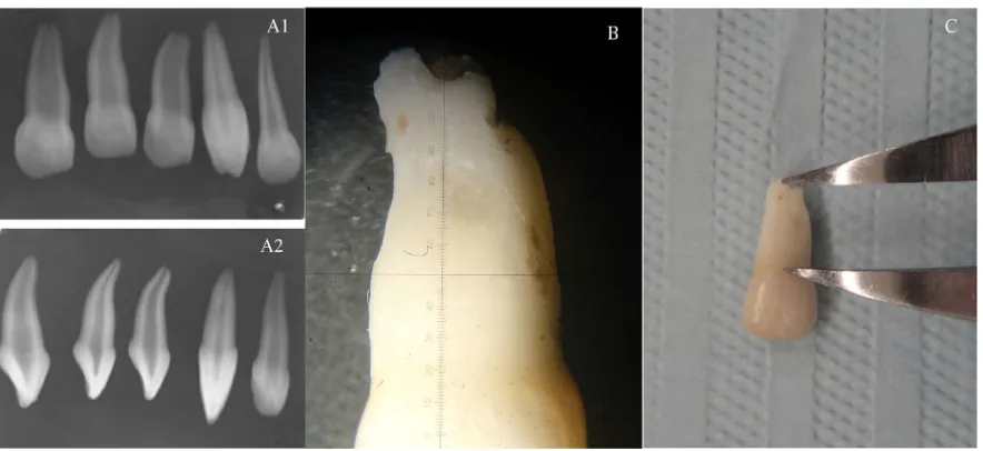
DOI: 10.4236/ce.2018.91007 79 Creative Education following eligibility criteria: visual absence of root resorption; absence of internal root canal anatomy alterations, as observed by periapical radiographic examina-tion; and no previous root canal manipulation (Figure 1). The sample was col-lected from 2007 to 2016 and kept in saline solution (pH 7.0) under refrigeration until measurements were performed. No gender or age classifications were made. Seventy upper and lower anterior primary teeth were selected and as-signed to six subgroups: 1) upper central incisors (UCI; n = 15), 2) upper lateral incisors (ULI; n = 13), 3) upper canines (UC; n = 20), 4) lower central incisors (LCI; n = 6), 5) lower lateral incisors (LLI; n = 3), and 6) lower canines (LC; n = 13).
2.2. Root Length Measurements
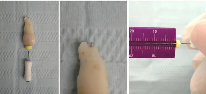
[image:3.595.99.541.466.669.2]Three experienced operators conducted the study. The teeth were examined at a 35x stereomicroscopy magnification (Olympus SZ-TR-BR-SIT, Micronal S. A., São Paulo, Brazil), and the root length was measured using a digital caliper (Beerendonk 042-750, Dentaurum, Germany) to confirm tooth integrity (Figure 1). Access cavities were prepared with a No. 2 round bur (Maillefer, São Paulo, Brazil) mounted on a high-speed handpiece and under water-cooling. RCL was measured by the direct method using a #15 K-file (Mani, Kyohara, Japan) pas-sively inserted in the root canal until the tip could be noticed at the root apex. A silicone stop was then adjusted at the incisal/cuspid reference face, and the reg-ister was performed with the aid of a millimetre rule (Prisma produtos Odon-tológicos, São Paulo, Brazil) with 0.5 mm accuracy (Mello-Moura et al., 2010; Krishnan & Sreedharan, 2012) (Figure 2). Undergraduate dental students at-tended the activity and were trained to perform the RCL measurements by the direct method taught.
Figure 1. Evaluated tooth sample showing no signs of internal root canal anatomy alterations and no previous root
DOI: 10.4236/ce.2018.91007 80 Creative Education
2.3. Statistical Analysis
Data were tabulated in Excel worksheets, and statistical analysis was performed with SPSS (Chi., IL, USA) software. Descriptive statistics were presented as mean ± standard deviation for each group of primary anterior teeth.
3. Results
The RCL values obtained in this study are reported in Table 1. The overall mean RCL of upper incisors was 14.99 ± 2.19 mm, whereas the overall mean of the whole evaluated sample was 15.28 ± 2.18 mm.
4. Discussion
Descriptive and comparative studies of human primary dentition have been performed to understand the morphology of primary dentition and assist un-dergraduate teaching in dentistry (Barker, Parsons, Williams, & Mills, 1975). Knowledge of dental anatomy is essential for learning clinical operative dentistry procedures (Kottor, Albuquerque, Velmurugan, & Kuruvilla, 2013). Therefore, teaching clinical procedures substantiated by data on human dental anatomy reinforces its clinical relevance and the need for training (Hagen, Cooke, & Wright, 2017).
[image:4.595.209.538.390.541.2]The terms root canal length (RCL) and working length (WL) have been used
[image:4.595.210.539.592.726.2]Figure 2. Root length measurement obtained by the direct method.
Table 1. Distribution of root canal length data (mm) for anterior primary teeth.
Arch Dental groups n Minimum Maximum Mean SD
Upper
Central incisors 15 11.0 16.5 14.33 1.69 Lateral incisors 13 11.0 16.5 14.00 1.63
Canines 20 11.0 19.0 16.07 2.43
Lower
Central incisors 6 11.0 19.0 15.41 3.20
Lateral incisors 3 14.0 17.0 15.00 1.73
DOI: 10.4236/ce.2018.91007 81 Creative Education indistinctively in primary teeth (Neena et al., 2011). However, the former refers to actual measurements made with the direct method or the estimated measure obtained from an initial radiograph (Mello-Moura et al., 2010), whereas the lat-ter refers to the length at which chemo-mechanical preparation and sealing should be performed, usually reducing 1 mm from the RCL (Nelson-Filho et al.,
2011). As an anatomical reference, RCL measurements obtained through the
di-rect method are generally considered as the gold standard (Chougule, Padma-nabhan, & Mandal, 2012; Mello-Moura et al., 2010; Subramaniam, Konde, & Mandanna, 2005; Oznurhan et al., 2014; Basso, Jeremias, Cordeiro, &
San-tos-Pinto, 2015). The present study described the RCL measurements obtained
by the direct method and discussed the results obtained within the searched lite-rature.
Considering the overall mean values for RCL of primary anterior teeth, the current results (15.28 ± 2.18 mm) were very similar to those reported by Subra-maniam et al. (2005) for 20 single-rooted teeth evaluated through the direct me-thod (15.91 ± 2.06 mm), and to those obtained by Wankhade et al. (2013) for 70 elements (16.44 ± 0.79 mm). In addition, the mean values for RCL of the central and lateral upper incisors obtained in the present study (14.99 ± 2.19 mm) were similar to WL data reported by Saritha et al. (2012) measured clinically using an electronic apex locator (15.55 ± 1.32 mm) and a digital radiograph method (15.93 ± 1.49 mm). The RCLs obtained in this Brazilian sample were similar to the values obtained in the pertinent literature for Indian populations.
5. Conclusion
The RCL direct measurement method and the results obtained in this Brazilian sample were in accordance with the literature reports and can be useful for edu-cational purposes aiming at knowledge of dental anatomy and the clinical de-termination of root canal treatment viability and treatment of primary teeth, as RCL is one of the parameters for the indication/contra-indication of this thera-py.
Acknowledgements
The work was supported by the Department of Orthodontics and Pediatric Den-tistry, School of DenDen-tistry, Universidade Federal do Rio de Janeiro, Brazil and Fundação Carlos Chagas Filho de Amparo à Pesquisa do Estado do Rio de Janeiro (FAPERJ).
Conflict of Interest
The authors declare that they have no conflict of interest.
References
AAPD (2015). Guideline on Pulp Therapy for Primary and Immature Permanent Teeth.
DOI: 10.4236/ce.2018.91007 82 Creative Education
Ahmad, I. A., & Pani, S. C. (2015). Accuracy of Electronic Apex Locators in Primary Teeth: A Meta-Analysis. International Endodontic Journal, 48, 298-307.
https://doi.org/10.1111/iej.12315
Barcelos, R., Tannure, P. N., Gleiser, R. et al. (2012). The Influence of Smear Layer Re-moval on Primary Tooth Pulpectomy Outcome: A 24-Month, Double-Blind, Rando-mized, and Controlled Clinical Trial Evaluation. International Journal of Paediatric Dentistry, 22, 369-381. https://doi.org/10.1111/j.1365-263X.2011.01210.x
Barker, B. C., Parsons, K. C., Williams, G. L., & Mills, P. R. (1975). Anatomy of Root Canals: IV Deciduous Teeth. Australian Dental Journal, 20, 101-106.
https://doi.org/10.1111/j.1834-7819.1975.tb04337.x
Basso, M. D., Jeremias, F., Cordeiro, R. C. L., & Santos-Pinto, L. (2015). Digital Radio-graphy for Determination of Primary Tooth Length: in Vivo and ex Vivo Studies.
Scientific World Journal, 2015, Article ID: 939045. https://doi.org/10.1155/2015/939045
Chougule, R. B., Padmanabhan, M. Y., & Mandal, M. S. (2012). A Comparative Evalua-tion of Root Canal Length Measurement Techniques in Primary Teeth. Pediatric Den-tistry, 34, 53-56.
Haffner, C., Folcwaczny, M., Galler, K., & Hicckel, R. (2005). Accuracy of Electronic Apex Locators in Comparison to Actual Length—An in Vivo Study. Journal of Dental Re-search, 33, 353-360. https://doi.org/10.1016/j.jdent.2004.11.017
Hagen, M., Cooke, B. K., & Wright, A. (2017). A Five-Year Review of Enhanced Learning through Integration: Anatomy and Clinical Practice. Creative Education, 8, 1774-1781.
https://doi.org/10.4236/ce.2017.811121
Kottor, J., Albuquerque, D., Velmurugan, N., & Kuruvilla, J. (2013). Root Anatomy and Root Canal Configuration of Human Mandibular Premolars: A Systematic Review.
Anatomy Research International, 2013, 14. https://doi.org/10.1155/2013/254250
Krishnan, I. S., & Sreedharan, S. (2012). A Comparative Evaluation of Electronic and Ra-diographic Determination of Root Canal Length in Primary Teeth: An in Vitro Study.
Contemporary Clinical Dentistry, 3, 416-420.
https://doi.org/10.4103/0976-237X.107430
Mello-Moura, A. C. V., Moura-Netto, C., Araki, A. T. et al. (2010). Ex Vivo Performance of Five Methods for Root Canal Length Determination in Primary Anterior Teeth. In-ternational Endodontic Journal, 43, 142-147.
https://doi.org/10.1111/j.1365-2591.2009.01667.x
Mortazavi, M., & Mesbahi, M. (2004). Comparison of Zinc Oxide and Eugenol, and Vita-pex for Root Canal Treatment of Necrotic Primary Teeth. International Journal of Pae-diatric Dentistry, 14, 417-424. https://doi.org/10.1111/j.1365-263X.2004.00544.x
Neena, I., Praveen, P., Rani, P. et al. (2011). Comparison of Digital Radiography and Apex Locator with the Conventional Method in Root Length Determination of Primary Teeth. Official Journal of the Indian Society of Pedodontics and Preventive Dentistry, 29, 300. https://doi.org/10.4103/0970-4388.86371
Nelson-Filho, P., Romualdo, P. C., Bonifácio, K. C. et al. (2011). Accuracy of the iPex Multi-Frequency Electronic Apex Locator in Primary Molars: An ex Vivo Study. In-ternational Endodontic Journal, 44, 303-306.
https://doi.org/10.1111/j.1365-2591.2010.01827.x
Oznurhan, F., Tüzüner, T., Baygin, O. et al. (2014). Accuracy of Three Different Apex Locators and Visual Exam in Primary Teeth with and without Root Resorption in Vi-tro. European Journal of Paediatric Dentistry, 15, 381-384.
DOI: 10.4236/ce.2018.91007 83 Creative Education https://doi.org/10.1111/j.1365-263X.2006.00774.x
Saritha, S., Uloopi, K. S., Vinay, C. et al. (2012). Clinical Evaluation of Root ZX II Elec-tronic Apex Locator in Primary Teeth. European Archives of Paediatric Dentistry, 13,
32-35. https://doi.org/10.1007/BF03262838
Subramaniam, P., Konde, S., & Mandanna, D. K. (2005). An in Vitro Comparison of Root Canal Measurement in Primary Teeth. Journal of Indian Society of Pedodontics and Preventive Dentistry, 23, 124-125. https://doi.org/10.4103/0970-4388.16883
Trairatvorakul, C., & Chunlasikaiwan, S. (2008). Success of Pulpectomy with Zinc Oxide-Eugenol vs Calcium Hydroxide/Iodoform Paste in Primary Molars: A Clinical Study. Pediatric Dentistry, 30, 303-308.