ORIGINAL RESEARCH
MR Imaging Findings in Patients with Secondary
Intracranial Hypertension
A.C. Rohr C. Riedel M.-C. Fruehauf A. van Baalen T. Bartsch J. Hedderich K. Alfke L. Doerner O. Jansen
BACKGROUND AND PURPOSE: IH can alter the configuration of anatomic structures of the central nervous system. We determined the sensitivity and specificity of MR imaging to detect these changes in patients with secondary IH.
MATERIALS AND METHODS: Patients (n ⫽ 36) with IH were prospectively investigated with MR imaging and were matched to 36 controls. MR images were evaluated for elongation and edema of the optic nerves, protrusion of the optic disc, flattening of the posterior sclera, height of the pituitary gland, and width of the optic nerve sheath. On MRV, we recorded venous sinus abnormalities and measured the luminal width of the superior ophthalmic veins. A grading score was introduced to define cranial venous outflow obstruction.
RESULTS: Cranial venous outflow obstruction and ONS hydrops were the most valid signs indicating IH with a sensitivity of 94% and 92% and a specificity of 100% and 89%, respectively. Sensitivities and specificities were 56% and 97% for reduced pituitary height, 64% and 78% for flattening of the posterior sclera, 31% and 97% for widening of the superior ophthalmic veins, 33% and 100% for optic disc protrusion, 14% and 100% for optic nerve edema, and 6% and 100% for elongation of the optic nerve. At least 2 MR imaging findings could be demonstrated in each patient but in none of the controls. The number of positive MR imaging findings correlated with CSF pressure (r⫽0.62,P⫽.01).
CONCLUSIONS:The combination of cranial and orbital MR imaging and MRV can be highly sensitive and specific in the diagnosis of patients with IH.
ABBREVIATIONS:CI⫽confidence interval; CVOO⫽cranial venous outflow obstruction; FLAIR⫽ fluid-attenuated inversion recovery; ICC ⫽ intraclass correlation coefficient; IH ⫽ intracranial hypertension; IIH⫽idiopathic intracranial hypertension; IJV⫽internal jugular vein; LP⫽lumbar puncture; LTS⫽left transverse sinus, MIP⫽maximum intensity projection, MRI⫽MR imaging; MRV⫽MR venography; ONS⫽optic nerve sheath; OR⫽odds ratio; PRES⫽posterior reversible encephalopathy syndrome; RTS⫽right transverse sinus; SIH⫽secondary intracranial hyperten-sion; SOV⫽superior ophthalmic vein; SSS⫽superior sagittal sinus; STIR⫽short tau inversion recovery; T1WI⫽T1-weighted imaging; T2WI⫽T2-weighted imaging; TS⫽transverse/sigmoid sinus; TSE⫽turbo spin-echo; VI⫽visual impairment
I
H can be caused by numerous conditions (SIH) but can also be observed without any evidence of an underlying condi-tion (pseudotumor cerebri, IIH1-4). The direct measurementof intracranial or lumbar CSF pressure is regarded as the cri-terion standard in the diagnostic work-up of IH, but due to its invasive nature, the dependence on patient compliance, and the susceptibility for false-positive or false-negative results, it has potential limitations.4 Furthermore, clinical signs and neurologic symptoms are variably expressed and might not be specific for IH.2,5-7Therefore, a complementary noninvasive
diagnosis of IH is preferable in clinical routine. Recently, ve-nous sinus stenoses, which might serve as diagnostic markers, have been reported in patients with IIH but are not
well-documented as occurring in patients with SIH.8-12 Other
MR imaging signs of IH have been partly investigated but not in a larger series of patients with SIH and lacking an inte-grative imaging protocol.13-22Reported findings are the fol-lowing: elongation of the optic nerve, protrusion of the optic nerve head, widening of the ONS, deformation of the pituitary gland, and dilation of the SOV.
The aim of our study was to determine the sensitivity and specificity of quantitative and qualitative MR imaging findings for IH in patients with SIH, by using a standardized MR imaging protocol, which combines orbital, cranial, and venous imaging.
Materials and Methods Patients
Thirty-six patients with SIH due to various causes were prospectively included in the study, among them 7 children. Clinical characteristics are given in Table 1. IH was proved by lumbar CSF pressure measure-ments in 22 patients, and in 3 patients, elevated pressure was reported intraoperatively with CSF gushing out at high pressure on opening the dura mater. In the remaining 11 patients without CSF pressure measurement, IH was diagnosed by positive funduscopy findings showing bilateral papilledema, clinical symptoms, and follow-up im-aging showing a reduction of MR imim-aging signs of increased intra-cranial pressure going along with clinical improvement. Patients with Received September 16, 2010; accepted after revision October 24.
From the Departments of Neuroradiology (A.C.R., C.R., K.A., O.J.), Neuropediatrics (A.v.B.), Neurology (T.B.), Medical Computer Science and Statistics (M.-C.F., J.H.), and Neurosurgery (L.D.), University Schleswig-Holstein Campus Kiel, Germany.
This work was supported by Archimedes Pharma (approximately 30,000 Euro).
Disclosure: Lutz Doerner is a member of the Speaker Bureau for Archimedes Pharma. He earned approximately 5000 Euro.
Paper previously presented in part at: Annual Meeting of the American Society of Neuroradiology and the Neuroradiology Education and Research Foundation Symposium, May 16 –21, 2009; Vancouver, British Columbia, Canada.
Please address correspondence to Axel C. Rohr, MD, Department of Neuroradiology, UKSH Campus Kiel, Arnold-Heller-Str 9, 24105 Kiel, Germany; e-mail: Axel.Rohr@gmx.de
DOI 10.3174/ajnr.A2463
BRAIN
ORIGINAL
visual disturbances (32/36) were grouped into 2 categories: 1) patients with little or transient visual disturbances and visual acuity of⬎70% (n⫽20), and 2) patients with severe visual loss (n⫽12). Four pa-tients had undergone surgery before inclusion in the study, including 3 children (7–14 years of age) with ventriculoperitoneal shunt oper-ations and 1 adult (51 years of age) with occlusion of the dominant IJV due to neck surgery. Patients with IIH were not included in this study.
Control Group
[image:2.594.50.533.60.600.2]Thirty-six individuals matched for age and sex with the SIH patient group served as controls. These individuals had cranial MR imaging performed for various reasons, mostly psychiatric disease, and did not have any clinical evidence of IH, such as headache or visual distur-bances. Written informed consent was obtained from both patients with SIH and controls, and institutional review board approval was
Table 1: Clinical characteristics in 36 patients with IH and 36 control subjects
Patient Groupa
Opening CSF Pressure on LP in cm H2O
(mean) (range)
Demographics Diagnosis, Clinical Signs, and Symptoms
Group Size, Sex Patient Age (mean)
(range) Diagnosis Papilledema Mild
VI Severe
VI Headache Nausea/
Vomiting Other Symptoms
All patients 39.8 n⫽36 40.9 yr Meningeal disease (n⫽10), 23/35 20/36 12/36 30/36 9/36 See below with IH (24–70) F (n⫽23)
M (n⫽13)
2–76 yr thrombosis or occlusion of venous sinus or the dominant IJV (n⫽10), intracranial tumor (n⫽
8), hydrocephalus (other than caused by tumor,n ⫽5), other (n⫽5), see below, multiple (n⫽3)
66% 56% 33% 83% 25%
Patient Subgroup I
29.5 (22–38)
n⫽12 F (n⫽7) M (n⫽5)
41.6 yr 32–66 yr
Occlusion of a venous sinus or IJV by meningioma or postoperatively (n⫽3), viral meningitis (n⫽3), bacterial meningitis (n⫽
1), venous sinus thrombosis (n⫽1), excessive arterial hypertension (n⫽1), head trauma with hygromas (n⫽1), hydrocephalus (n⫽1), minocycline medication (n ⫽1) 7/12 58% 7/12 58% 3/12 25% 12/12 100% 4/12 33%
Fever, meningism (n⫽
4), transient aphasia (n ⫽3), vertigo (n⫽2), transient apraxia (n⫽
1), cough (n⫽1), hypacusis (n⫽1), tinnitus (n⫽1), reduced consciousness (n⫽1), paraesthesia (n⫽1)
Patient Subgroup II
52 40–70
n⫽10 F (n⫽6) M (n⫽4)
34.5 yr 2–62 yr
Thrombosis of venous sinus or IJV (n⫽4), hydrocephalus (n⫽3), meningiosis (n⫽2), neurosarcoid meningitis (n ⫽1) 7/9 78% 4/10 40% 5/10 50% 8/10 80% 3/10 30% Vertigo, dysaesthesia (n⫽1), tinnitus (n⫽
1), double vision (n⫽
1), head enlargement (n⫽1), unspecific feeling of illness (n⫽
1) Patient
Subgroup III
Not testedb n⫽14
F (n⫽10) M (n⫽4)
44.8 yr 9–76 yr
Meningioma alone or combined with stenosis or occlusion of a venous sinus (n⫽4), venous sinus thrombosis alone or combined with brain abscess or dural metastasis (n⫽3), brain tumor with occlusive hydrocephalus (n⫽2), meningeosis (n⫽2), arterial hypertension, PRES (n⫽1), hydrocephalus (n⫽1)
9/14 64% 9/14 64% 4/14 29% 10/14 71% 2/14 14%
Double vision (n⫽4), paresis (n⫽4), reduced consciousness (n⫽3), nystagmus (n ⫽1). vertigo (n⫽1), fever (n⫽1)
Controls Not tested. n⫽36 39 yr Mostly psychiatric Not tested 0/36 0/36 0/36 0/36
F (n⫽23) 2–80 yr 0% 0% 0% 0%
M (n⫽13)
aPatients with IH (n⫽36) were subgrouped into those with CSF pressure⬍40 cm H
2O (n⫽12, group I), those with⬎40 cm H2O (n⫽10, group II), and those without direct pressure
measurements (n⫽14, group III).
obtained. No individual fulfilled the usual exclusion criteria for MR imaging.
MR Imaging
MR imaging was performed in all patients by using a standardized protocol with a 3T unit and a head-sense-coil, including the following sequences: 1) T2 TSE axial: TR/TE, 4021/100 ms; section thickness, 3 mm; 2) STIR coronal: TR/TE/TI, 3838/90/180 ms; section thickness, 3 mm, covering the orbit and pituitary gland; and 3) 3D phase-contrast MRV: TR/TE, 17/7.9 ms; velocity encoding, 15 cm/s. Additional con-trast-enhanced T1 sequences (n⫽29), contrast-enhanced time-of-flight angiography (n⫽3), time-resolved contrast-enhanced MR an-giography (n⫽1), digital subtraction angiography (n⫽4), and/or CT venography (n⫽1) were used to rule out venous sinus thrombo-sis in uncertain cases. Follow-up MR imaging was performed in 27 patients.
Analysis of MR Imaging Findings
Apart from the presence of space-occupying lesions with or without mass effect or obstruction of adjacent venous sinuses, special atten-tion was paid to the following MR imaging findings: 1) nonquantita-tive cross-sectional findings: ventriculomegaly, periventricular edema caused by transependymal CSF flow (Fig 1A), flattening of the posterior sclera (Fig 2A), optic papilla protrusion (Fig 1A), optic nerve edema (Fig 2B), and elongation of the optic nerve (Fig 1A); and
2) quantitative cross-sectional criteria: pituitary height (Fig 3C, -F) and ONS width as measured at 4 different locations (3, 6, 10, and 20 mm behind the globe; Figs 2 and 4) by using coronal STIR se-quences. With the source images of MRV, the maximum diameters of the SOV were measured (Fig 1C). Intracranial venous anatomy and pathology were judged by using MIPs, source images of MRV, and cross-sectional MR images. Gross venous anatomic variants, signal-intensity changes reflecting thrombosis within the venous sinuses (Fig 3A, -D), and venous sinus pathologies regarded as stenoses (but not typical for thrombosis) were further analyzed by using the pri-mary MRV sections.
A simple classification system was used for grading venous sinus pathologies. A reduction of the diameter of a dural sinus of⬍50% was regarded as being within the normal range of variation (grade 0 lesion), whereas a more severe narrowing or a complete signal-intensity loss was categorized as a significant stenosis or occlusion (grade 1 lesion, Figs 1B, 2C, 3B, and 4D). CVOO was assumed in cases in which a grade 1 lesion affected the following: both TSs or at least the dominant TS if the opposite TS was severely hypoplastic or absent (type I), the mid or dorsal part of the SSS (type II), or a combination of the previous 2 types (type III). MR images were blinded for the presence of IH and were evaluated by 3 readers (A.C.R., an experi-enced certified radiologist/neuroradiologist; C.R., a board-certified radiologist and Specialist Registrar in neuroradiology; and M.-C.F., a senior medical student who was trained to evaluate cranial
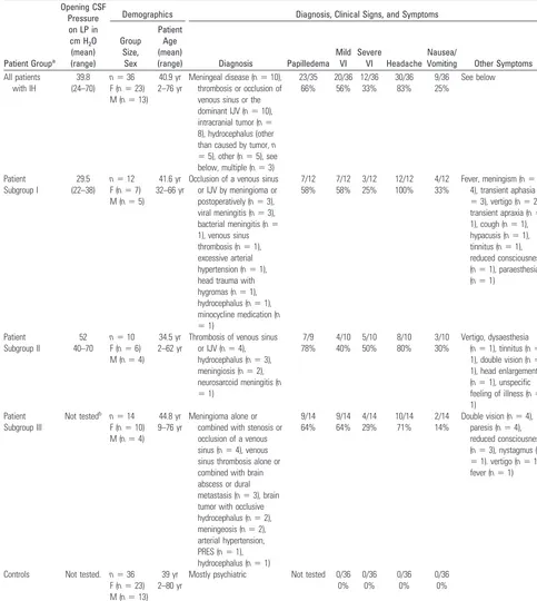
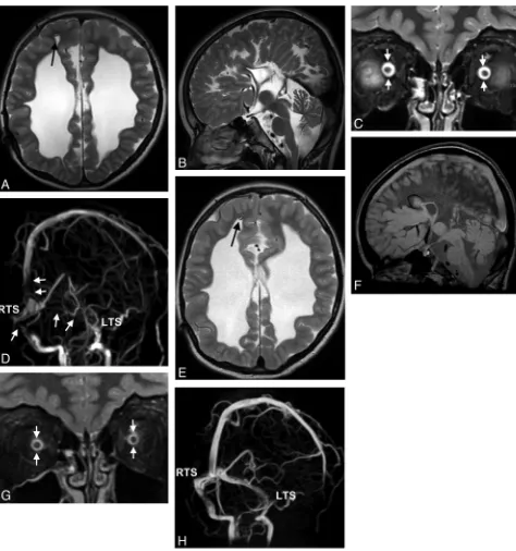
Fig 1.A 15-year-old boy with double vision and reduced acuity has bilateral papilledema.A, On axial T2WI, ventriculomegaly with periventricular CSF flow (black arrows) due to an obstructing tumor within the fourth ventricle is found. Optic nerve sheath hydrops, elongation of the optic nerve (white arrowsinA), and optic papilla protrusion are seen (black arrowhead).
[image:3.594.135.455.41.389.2]MR images with respect to features of IH). The results of the first reader were used for the primary analysis. The results of all 3 readers were used to calculate interobserver reliabilities.
Statistical Analysis
On the basis of cross-tabulations, the sensitivity, specificity, and OR of different MR imaging findings were calculated (95% CI). A Pear-son correlation coefficient test was used to find correlations between CSF pressure and quantitative MR imaging signs of IH. A Spearman correlation coefficient test was chosen to find correlations between CSF pressure values and the number of positive MR imaging signs of IH. A Mann-WhitneyUtest was used to prove differences between mean values of measurements in patient subgroups and between pa-tients and controls. For analysis of interobserver reliabilities,values and percentages of interobserver agreements (qualitative signs) and ICCs (quantitative signs) were calculated. For each test, the level of statistical significance was defined asP⬍.05. Statistical analysis was performed with the Statistical Package for the Social Sciences soft-ware, Version 13 (SPSS, Chicago, Illinois) except for computation ofand ICC values, which was done with R by using the irr package (R Development Core Team, 2010; R: A language and environment for statistical computing; R Foundation for Statistical Computing, Vienna, Austria).
Results
MR Imaging Findings in SIH
The main findings were CVOO, ONS hydrops, reduced pitu-itary height, and flattened posterior sclera (Tables 2 and 3). All the investigated findings differed significantly between patients and controls (P⬍.05).
CVOO
All except 2 patients (n ⫽34, sensitivity 94%) had CVOO. Venous sinus stenoses were apparent in sinuses distant from thrombosed sinuses in 6/8 patients (75%, Fig 3) and distant from sinuses compressed by a tumor in 7/8 patients (88%), or occurred in patients without any primary pathology of a ve-nous sinus (18/20 patients, 90%; Figs 1, 2, and 4). Most pa-tients (31/36, 86%) had stenoses that could not be directly attributed to a primary underlying pathology (ie, compression by a tumor or acute thrombosis). Type I CVOO (Fig 1B) was found in 41%; type II (Fig 2C), in 12%; and type III (Fig 4D), in 47%. In the control group, CVOO was not detected, but flow irregularities were seen in 44%.
ONS Hydrops
Mean diameters of the ONS were greater in patients compared with controls in all 4 positions (P⬍.01, Table 2). Using upper normal limits of the width of the ONS of 6.4, 5.8, 5.1, and 4.8 mm in positions 1– 4 as proposed,23we observed ONS
hy-drops with sensitivities of 78%, 75%, 75%, and 75% and spec-ificities of 89%, 100%, 97%, and 100%. In 33/36 patients, ONS hydrops occurred in at least 1 of the 4 positions of the optic nerve (sensitivity 92%), which could be detected in only 4/36 controls (specificity, 89%; Table 3). ONS hydrops was always bilateral (Figs 2 and 4).
Reduced Pituitary Height
Mean pituitary height was lower in patients than in controls (Table 2). A pituitary height⬍2.6 mm was observed in 20/36 patients and in 1/36 controls (56% sensitivity, 97% specificity; Table 3 and Fig 3C). When we took into account age- and
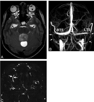
Fig 2.A 30-year-old woman with arterial hypertension and visual disturbances has bilateral papilledema.A, MR imaging of the orbit displays a flattened posterior sclera (arrowheads).
[image:4.594.132.453.43.296.2]sex-adapted lower normal limits of the pituitary height of 3 mm for children, 2.7 mm for men of all ages, 3.3 mm for women 18 – 62 years of age, and 1.8 mm for women older than 62 years of age,2322 patient and 2 control measurements were
abnormal, resulting in a sensitivity and specificity of 61% and 94%, respectively.
Flattened Posterior Sclera, Optic Papilla Protrusion, Elongation, and Edema of the Optic Nerve
The posterior sclera was flattened in 23 patients and in 8 con-trols. Protrusion of the optic disc, optic nerve edema, and optic nerve elongation were observed in 33%, 14%, and 6% of patients but in none of the controls (specificity of 100% each). Funduscopy was performed in 11 of 12 patients with protru-sion of the disc, and bilateral papilledema was found in all of them. Optic disc protrusion was found in only 12/23 patients with proved papilledema (sensitivity of 52%).
Dilation of the SOV
The mean diameter of the SOV was greater in patients with SIH than in controls (Table 2). In 11 patients and in 1
con-trol, the maximum diameter of the SOV exceeded 2.6 mm (Table 3).
Ventriculomegaly
Ventriculomegaly was noted in 7 patients. In 2 patients with obstructive hydrocephalus and 1 patient with hydrocephalus of unknown origin, periventricular edema was present. In all of these 7 patients, CVOO was present; in 7/8 patients, cross-sectional MR imaging signs of IH were seen.
Correlating CSF Pressure with MR Imaging Findings and Vision Loss
CSF pressure was significantly higher in patients with optic disc protrusion (48.3⫾13.4 cm H2O) and optic nerve edema
(54.7⫾8.1 cm H2O) than in patients without these findings
(mean, 36.1⫾10.9 cm H2O and 38.3⫾12.3 cm H2O,
respec-tively;P⬍.05 each). In patients with CVOO, CSF pressure was higher in those having CVOO type III (54.4⫾12.7 cm H2O)
than in those having either CVOO type I or II (36.7⫾11.2 cm H2O,P⬍.05). ONS diameter, height of the pituitary gland,
and diameter of the SOV did not significantly correlate with
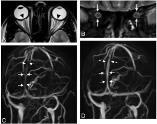
Fig 3.A 28-year-old woman with venous sinus thrombosis secondary to in vitro fertilization.A, Signal intensity typical for thrombosed blood is seen in the SSS and the RTS on axial T2w (black arrow) but not in the LTS (white arrow).B, There is corresponding signal-intensity loss on MIP of MRV in the RTS and SSS (left oblique view). There is a stenosis in the LTS (white arrow) without evidence of a thrombus.C, ONS hydrops is present (not shown), and the height of the pituitary gland is reduced to 2.5 mm (coronal STIR).DandE, CSF pressure is 40 cm H2O. Six months later, partial recanalization of the RTS and SSS occurs following therapy with low-molecular heparin seen on T2WI (black arrowinD) and on MIPs of MRV
[image:5.594.54.532.43.407.2]the degree of CSF pressure increase. There was a positive cor-relation of CSF pressure and the number of positive MR im-aging findings of IH (Spearman correlation coefficient,r⫽ 0.62;P⫽.01). At least 2 MR imaging findings could be dem-onstrated in each patient. Concerning visual impairment, no significant differences in CSF pressure values were found be-tween patients in groups 1 (little or transient impairment) and 2 (severe impairment) (mean, 37.4⫾10.7 cm H2O versus
43.3⫾14.0 cm H2O), but patients in group 2 had significantly
more frequent MR imaging features of optic disc protrusion (58%) and optic nerve edema (33%) than patients in group 1
(25% and 5%,P⬍.05 each). There were no noted differences between patients with and without direct pressure measure-ments (Tables 2 and 3).
Interobserver Reliability
Concerning quantitative signs, the interobserver reliability was best for measurements of the pituitary height, was good for measurements of the ONS diameters, and was worst for measurements of the SOVs (Table 2). Concerning qualitive signs, the interobserver reliability was excellent for CVOO, ventriculomegaly, and periventricular CSF flow; was mediocre
[image:6.594.54.528.38.544.2]for optic disc protrusion, optic nerve elongation, and “optic nerve edema; and was worst for flattened posterior sclera (Ta-ble 3).
Follow-Up Investigations
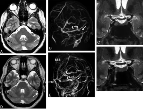
Follow-up MR images were available in 27 patients. After CSF pressure was lowered, 20 patients improved clinically with as-sociated decrease in MR imaging signs of IH (Fig 3). One tient remained stable clinically and on MR imaging. Six pa-tients improved clinically, but MR imaging findings did not change. The rate of agreement (clinical versus neuroradio-logic), therefore, was 21/27 (78%). Follow-up MR images after shunt revisions were available in 2 of 3 children with hydro-cephalus and demonstrated normalization of MR imaging fea-tures except for the presence of hydrocephalus (Fig 4).
Discussion
Our study shows that the imaging parameters CVOO, ONS hydrops, and reduction in pituitary height discriminated best between patients with IH and controls. Most other signs stud-ied did show a lesser sensitivity but a high specificity.
Intracranial Venous Morphology in IH
Thrombosis or occlusion of venous sinuses (primary CVOO) can result in IH,23-26probably by inducing venous
hyperten-sion and subsequent impairment of CSF absorption.25In
pa-tients with IIH, bilateral narrowing of the TS not associated with thrombosis has been found regularly.27-31Because stent
angioplasty can help control IH in these patients, a causative role of these stenoses is favored by some.29,30On the other
hand, lowering the intracranial pressure can result in
normal-Table 2: Differences in MRI measurements between 36 patients with IH and 36 controlsa
Diameter of the ONS (mm)b Pituitary
Height (mm)
SOV Diameter
(mm) Position 1 Position 2 Position 3 Position 4
Patients with IH (all,n⫽36) Mean 7.0 6.1 5.4 5.1 3.1 2.3
SD 0.9 0.9 1.0 0.7 1.6 0.7
95% CI 6.7–7.3 5.8–6.4 5.1–5.7 4.9–5.3 2.6–3.6 2.0–2.5
ICC 0.832 0.84 0.866 0.84 0.863 0.389
Patient subgroup I (n⫽12) Mean 7.2 6.2 5.3 5.1 3.2 2.2
CSF pressure 22–38 cm H2O SD 0.6 0.7 1.1 0.5 1.0 0.8
Patient subgroup II (n⫽10) Mean 6.7 5.9 5.2 5.1 2.8 2.4
CSF pressure 40–70 cm H2O SD 0.6 0.7 0.8 0.7 1.3 0.5
Patient subgroup III (n⫽14) Mean 7.0 6.2 5.7 5.0 3.2 2.2
No CSF pressure measurements SD 1.2 1.1 1.0 0.8 2.0 0.7
Controls (n⫽36) Mean 5.8 4.9 4.4 4.1 5.2 1.7
SD 0.6 0.6 0.5 0.3 1.5 0.5
95% CI 5.6–6.0 4.7–5.1 4.3–4.6 4.0–4.3 4.7–5.6 1.5–1.8
ICC 0.76 0.728 0.78 0.732 0.893 0.634
aResults of all measurements differed significantly between all patients and controls (P⬍.01) and also between the different patient subgroups (I~III) and controls (P⬍.05). No significant
differences existed among results of patient subgroups I, II, and III.
[image:7.594.51.545.61.235.2]bONS diameter was measured 3, 6, 10, and 20 mm behind the globe (positions 1– 4).
Table 3: MR imaging signs in 36 patients with IH and 36 control subjectsa
Patient Group Opening
CSF Pressure on LP in cm H2O
(mean)
(range) CVOO
ONS Hydropsb
Reduced Pituitary Height (⬍2.6 mm)
Flattened Posterior Sclera
Optic Disc Protrusion
Dilated SOV (⬍2.6 mm
in
diameter) Ventriculomegaly
Periventricular CSF Flow
Optic Nerve Edema
Optic Nerve Elongation
No. of Positive MRI Signs
(mean) (range)
All patients 39.8 Frequency, 34/36 33/36 20/36 23/36 12/36 11/36 7/36 5/36 5/36 2/36 4.3 (2–9)
24–70 Sensitivity, 94% 92% 56% 64% 33% 31% 19% 14% 14% 6%
CI 95%, 81%–99% 76%–98% 38%–72% 46%–79% 19%–91% 16%–48% 8%–36% 5%–29% 5%–29% 1%–19%
Specificity, 100% 89% 97% 78% 100% 97% 100% 100% 100% 100%
95% CI, 90%–100% 73%–96% 85%–100% 61%–90% 90%–100% 85%–100% 90%–100% 90%–100% 90%–100% 90%–100%
OR 1007 88 44 6 37 15 18 13 13 5
95% CI, 47–21,741 18–424 5–355 2–17 2–658 2–127 1–338 1–240 1–240 .3–114
Agreement,c
100% 100% 91.7% 63.9% 77.8% 67% 91.7% 91.7% 69.4% 80.6%
c 1 1 0.888 0.484 0.675 0.338 0.852 0.79 0.47 0.533
Patient 29.5 Frequency 10/12 11/12 4/12 5/12 2/12 4/12 1/12 1/12 0/12 0/12 3.3 (2–6)
subgroup I 22–38 83% 92% 33% 42% 17% 33% 8% 8% 0% 0%
Patient 52 Frequency 10/10 9/10 7/10 8/10 6/10 3/10 3/10 1/10 3/10 1/10 5.1 (3–7)
subgroup II 40–70 100% 90% 70% 80% 60% 30% 30% 10% 30% 10%
Patient N.T. Frequency 14/14 13/14 9/14 10/14 4/14 4/14 3/14 3/14 2/14 1/14 4.5 (2–9)
subgroup III 100% 93% 64% 71% 29% 29% 21% 21% 14% 7%
Controls N.T. Frequency 0/36 4/36 35/36 8/36 0/36 1/36 0/36 0/36 0/36 0/36 0.4 (0–1)
0% 11% 3% 22% 0% 3% 0% 0% 0% 0%
Agreementc 100% 78% 100% 69.4% 94.4% 94.4% 94.4% 97.2% 97.2% 94.4%
aFrequency of all MRI signs differed between patients and controls (P⬍.01). Frequency of the MRI signs “optic disc protrusion” and “optic nerve edema” were more often found in patient group II compared with
patient group I (P⬍.05). Frequencies of all MRI signs did not differ between patient subgroup III and the other patient groups.
bONS hydrops was assumed if the diameter was above normal limits inⱖ1 different position. Normal upper limits of ONS diameters were 6.4, 5.8, 5.1, and 4.8 mm in measurement positions 1, 2, 3, and 4 (3, 6, 10,
and 20 mm behind the globe).
[image:7.594.53.532.275.505.2]ization of venous morphology, suggesting that these stenoses might be induced by IH itself (secondary CVOO).25,26We
have shown that CVOO is not a specific feature of patients with IIH but is a universal phenomenon in most patients with IH irrespective of the underlying cause (Figs 2– 4). Also, stenoses are not restricted to both lateral TSs as in IH but can occur in a variety of locations and present with different shapes. A widespread involvement of sinuses was associated with very high CSF pressure. Most interesting, in patients with primary CVOO, additional stenoses also occur in sinuses not affected by primary obstruction. We hypothesize that IH com-presses the dural sleeve of the venous sinuses in all patients. This compression might lead to lengthy narrowing or—in the case of rather short bilateral TS stenoses— enlargement and invagination of arachnoid granulations into the sinus lumen or might induce a collapse of “vulnerable” sinus segments. The latter phenomenon might induce a vicious circle because the sinus stenoses themselves worsen IH via the mechanism of prestenotic venous hypertension. Because principally all seg-ments of the venous sinuses can be affected, failures of venous sinus stent placement can occur.11
Analysis of MRVs can be challenging because flow signal-intensity irregularities occurred in a significant number of our controls (44%). Similar results have been published by Higgins et al.32Nevertheless, by using a simple new scoring
system, stenoses were found to lead to CVOO in 94% of our patients with SIH but in none of the controls. Widening of the SOV in patients with IH has been demonstrated with T1WI16 and could also be shown in our patients by using MRV. The reason for this is not clear but could be due to increased col-lateral venous outflow.
Cross-Sectional MR Imaging Signs of IH
Cross-sectional MR imaging and CT signs of IH have been studied in patients with IIH13,14,17-22,33,34with varying results.
In a recent study, only 1 MR imaging sign (flattening of the posterior sclera) was found to be useful.21This could be due to
the different nature of IIH compared with SIH but could also be due to technical limitations of that study: Although most MR imaging signs were derived from the orbital content, no detailed orbital studies were performed and authors did not describe at which location the width of the ONS was mea-sured. However, this is an important aspect because this width varies significantly from anterior to posterior.35,36Changes of the pituitary shape were judged subjectively despite age- and sex-dependent variations of the pituitary size.35,37
With our approach of using quantitative markers and by using MR imaging sequences optimized for orbital imaging, we found several parameters to be useful for predicting IH, especially ONS hydrops and a decrease of the pituitary height. Flattening of the posterior sclera was a sign less sensitive and not specific for IH in our study, and interobserver reliability for this sign was low. In our opinion, judgment of this sign is delicate and prone to be influenced by partial volume effects. Protrusion of the optic disc indicates papilledema. This sign and optic nerve edema were associated with very high CSF pressure and with severe visual impairment. We favor a diag-nostic approach that considers multiple indirect MR imaging signs of IH, because we found that the number of positive signs correlates with the degree of pressure increase and because
pathologies of the orbit or sella might prevent using a single sign.
Making use of MR imaging signs of IH in patients with SIH has several possible implications: Because unlike in IIH, patients with SIH display a wider range of signs and symptoms due to their underlying disease, the clinical diagnosis of IH can be difficult and thus could be substantiated by MR imaging. In patients lacking obvious pathology on neuroimaging (Fig 2), MR imaging signs of IH might be the only indicators of IH and may lead to effective treatment (CSF diversion). Some patients with venous sinus thrombosis develop visual problems that can be attributed to IH. We found MR imaging to be helpful in diagnosing IH in these cases (Fig 3). In patients with hydro-cephalus with shunt surgery procedures, ventriculomegaly can persist irrespective of the actual intracranial pressure (Fig 4). To our knowledge, this is the first report to show that the presence of venous sinus narrowing on MRV can easily demonstrate an increase in intracranial pressure in these pa-tients. In the case of severe noncommunicating hydrocepha-lus, additional MR imaging features of IH probably do not influence patient treatment (Fig 1), but they may be of use on follow-up. In our study, these cases provide evidence that MR imaging signs of IH work in a variety of settings (com-municating and noncom(com-municating hydrocephalus). Our preliminary data by using follow-up MR images indicate that MR imaging signs of IH are at least partially reversible on normalization of intracranial pressure.
Larger numbers of patients and controls are needed to sub-stantiate our findings, especially with regard to children with shunt placements and patients acutely affected by IH.
Conclusions
With a standardized protocol, MR imaging is sensitive and specific in the diagnosis of patients with IH irrespective of the underlying cause. In our cohort, all patients but none of the controls displayed at least 2 MR imaging signs of increased intracranial pressure. This technique can be potentially help-ful in increasing the diagnostic reliability and in detecting pa-tients who need invasive procedures in IH. Venous sinus nar-rowings are not a specific feature of IIH. In patients with secondary IH, these narrowings occur in different locations and with different shapes than those reported for IIH. We speculate that they are caused by IH but might contribute to the increase in intracranial pressure. Venous sinus narrowing might be a sensitive sign for diagnosing ventriculoperitoneal shunt failure but larger numbers of patients are needed to confirm this possibility.
References
1. Ball AK, Clarke CE.Idiopathic intracranial hypertension.Lancet Neurol
2006;5:433– 42
2. Binder DK, Horton JC, Lawton MT, et al.Idiopathic intracranial hyperten-sion.Neurosurgery2004;54:538 –51, discussion 551–32
3. Skau M, Brennum J, Gjerris F, et al.What is new about idiopathic intracranial hypertension? An updated review of mechanism and treatment.Cephalalgia
2006;26:384 –99
4. Bershad EM, Humphreis WE, Suarez JI.Intracranial hypertension.Semin Neurol2008;28:690 –702
5. Algahtani HA, Baeesa SS, Obeid TH, et al.Idiopathic intracranial hyper-tension: atypical presentation.Saudi Med J2007;28:762– 65
7. Marcelis J, Silberstein SD. Idiopathic intracranial hypertension without papilledema.Arch Neurol1991;48:392–99
8. Owler BK, Parker G, Halmagyi GM, et al.Pseudotumor cerebri syndrome: venous sinus obstruction and its treatment with stent placement.J Neurosurg
2003;98:1045–55
9. Higgins JN, Pickard JD.Lateral sinus stenoses in idiopathic intracranial hypertension resolving after CSF diversion.Neurology2004;62:1907– 08 10. King JO, Mitchell PJ, Thomson KR, et al.Manometry combined with cervical
puncture in idiopathic intracranial hypertension.Neurology2002;58:26 –30 11. Rohr A, Dorner L, Stingele R, et al.Reversibility of venous sinus obstruction in
idiopathic intracranial hypertension.AJNR Am J Neuroradiol2007;28:656 –59 12. Farb RI, Vanek I, Scott JN, et al.Idiopathic intracranial hypertension: the prevalence and morphology of sinovenous stenosis.Neurology 2003;60: 1418 –24
13. Brodsky MC, Vaphiades M.Magnetic resonance imaging in pseudotumor cerebri.Ophthalmology1998;105:1686 –93
14. Yuh WT, Zhu M, Taoka T, et al.MR imaging of pituitary morphology in idiopathic intracranial hypertension.J Magn Reson Imaging2000;12:808 –13 15. Bono F, Messina D, Giliberto C, et al.Bilateral transverse sinus stenosis
pre-dicts IIH without papilledema in patients with migraine.Neurology2006; 67:419 –23
16. Lirng JF, Fuh JL, Wu ZA, et al.Diameter of the superior ophthalmic vein in relation to intracranial pressure.AJNR Am J Neuroradiol2003;24:700 – 03 17. Gibby WA, Cohen MS, Goldberg HI, et al.Pseudotumor cerebri: CT findings
and correlation with vision loss.AJR Am J Roentgenol1993;160:143– 46 18. Silbergleit R, Junck L, Gebarski SS, et al.Idiopathic intracranial hypertension
(pseudotumor cerebri): MR imaging.Radiology1989;170:207– 09
19. Zagardo MT, Cail WS, Kelman SE, et al.Reversible empty sella in idiopathic intracranial hypertension: an indicator of successful therapy?AJNR Am J Neuroradiol1996;17:1953–56
20. Jinkins JR, Athale S, Xiong L, et al.MR of optic papilla protrusion in patients with high intracranial pressure.AJNR Am J Neuroradiol1996;17:665– 68 21. Agid R, Farb RI, Willinsky RA, et al.Idiopathic intracranial hypertension: the
validity of cross-sectional neuroimaging signs. Neuroradiology 2006;48: 521–27
22. Weisberg LA.Computed tomography in benign intracranial hypertension.
Neurology1985;35:1075–78
23. Fuentes S, Metellus P, Levrier O, et al.Depressed skull fracture overlying the
superior sagittal sinus causing benign intracranial hypertension: description of two cases and review of the literature.Br J Neurosurg2005;19:438 – 42 24. Duke BJ, Ryu RK, Brega KE, et al.Traumatic bilateral jugular vein thrombosis:
case report and review of the literature.Neurosurgery1997;41:680 – 83 25. Karahalios DG, Rekate HL, Khayata MH, et al.Elevated intracranial venous
pressure as a universal mechanism in pseudotumor cerebri of varying etiolo-gies.Neurology1996;46:198 –202
26. Biousse V, Ameri A, Bousser MG.Isolated intracranial hypertension as the only sign of cerebral venous thrombosis.Neurology1999;53:1537– 42 27. Baryshnik DB, Farb RI.Changes in the appearance of venous sinuses after
treatment of disordered intracranial pressure.Neurology2004;62:1445– 46 28. McGonigal A, Bone I, Teasdale E.Resolution of transverse sinus stenosis in
idiopathic intracranial hypertension after L-P shunt.Neurology2004;62: 514 –15
29. Donnet A, Metellus P, Levrier O, et al.Endovascular treatment of idiopathic intracranial hypertension: clinical and radiologic outcome of 10 consecutive patients.Neurology2008;70:641– 47
30. Higgins JN, Cousins C, Owler BK, et al.Idiopathic intracranial hypertension: 12 cases treated by venous sinus stenting.J Neurol Neurosurg Psychiatry
2003;74:1662– 66
31. Fera F, Bono F, Messina D, et al.Comparison of different MR venography techniques for detecting transverse sinus stenosis in idiopathic intracranial hypertension.J Neurol2005;252:1021–25
32. Higgins JN, Gillard JH, Owler BK, et al.MR venography in idiopathic intra-cranial hypertension: unappreciated and misunderstood.J Neurol Neurosurg Psychiatry2004;75:621–25
33. Brodsky MC.MR of papilledema.AJNR Am J Neuroradiol1997;18:1592–94 34. Brodsky MC, Glasier CM.Magnetic resonance visualization of the swollen
optic disc in papilledema.J Neuroophthalmol1995;15:122–24
35. Rohr A, Riedel C, Reimann G, et al.Pseudotumor cerebri: quantitative in-vivo measurements of markers of intracranial hypertension[in German].Rofo
2008;180:884 –90
36. Hansen HC, Helmke K.The subarachnoid space surrounding the optic nerves: an ultrasound study of the optic nerve sheath.Surg Radiol Anat1996;18: 323–28