ORIGINAL RESEARCH
Widespread Microstructural White Matter
Involvement in Amyotrophic Lateral Sclerosis: A
Whole-Brain DTI Study
M. Cirillo F. Esposito G. Tedeschi G. Caiazzo A. Sagnelli G. Piccirillo R. Conforti F. Tortora M.R. Monsurro` S. Cirillo F. Trojsi
BACKGROUND AND PURPOSE: The extensive application of advanced MR imaging techniques to the study of ALS has undoubtedly improved our knowledge of disease pathophysiology, even if the actual spread of the neurodegenerative process throughout the central nervous system is not fully under-stood. The present study aimed to detect WM patterns of microstructural abnormalities to better investigate the pathologic process in ALS, within but also beyond CSTs, in a whole-brain analysis.
MATERIALS AND METHODS:DTI was performed in 19 patients with ALS and 20 matched healthy
controls, by using whole-brain TBSS and VOI analyses.
RESULTS: We observed a significant decrease of FA in the body of CC of the ALS group (P⬍.05). At the VOI level, both FA decrease and RD increase in the body of CC significantly correlated with the UMN score (P⫽.003 andP⫽.02). Additionally, significant voxelwise positive correlations between FA and the ALSFRS-R were detected in the WM tracts underneath the left premotor cortex (P⬍.05).
CONCLUSIONS:The correlations between reduction of FA and increase of RD in the body of CC with the UMN score indicate that the WM degeneration in the CC is strictly related to the ALS pyramidal impairment, while the correlation between FA and ALSFRS-R in the associative tracts underneath the left premotor cortex might reflect the progressive spread of the disease from the motor toward the extramotor areas.
ABBREVIATIONS:AD⫽axial diffusivity; ALS⫽amyotrophic lateral sclerosis; ALSFRS-R⫽revised ALS functional rating scale; CC⫽ corpus callosum; CST ⫽corticospinal tract; FA ⫽fractional anisotropy; GLM⫽ general linear model; GM⫽ gray matter; MD ⫽mean diffusivity; MNI ⫽ Montreal Neurological Institute; RD ⫽ radial diffusivity; TBSS ⫽ tract-based spatial statistics; UMN⫽upper motor neuron
T
he diagnosis of ALS, a progressive neurodegenerative dis-order affecting both upper and lower motor neurons, is mainly based on clinical criteria. Lower motor neuron injury can be detected even at a subclinical level by suitable tech-niques, such as needle electromyography. UMN pathology of-ten starts in the primary motor and premotor cortices, with secondary degeneration of motor fibers and gliosis along the CSTs.1 Nevertheless, it may often be difficult to detect the involvement of the UMN. Transcranial magnetic stimulation measurements may contribute to the diagnosis of ALS by re-vealing a clinically undetectable UMN dysfunction, and par-ticularly the triple stimulation technique can increase the sen-sitivity to UMN lesions. However, transcranial magnetic stimulation is not yet being used routinely, and its role in monitoring disease progression is unclear.2DTI is a relatively new method for structural neuroimag-ing, which allows visualizing the orientation of the fiber tracts and assessing their integrity in the WM by measuring
aniso-tropic water diffusion properties of the brain with MR imag-ing.3DTI has already produced promising results in assessing
UMN pathology in patients with ALS,2,4-9suggesting that FA,
the most sensitive DTI measure, is reduced along the CSTs due to axonal degeneration and loss of fiber integrity,2,4-6though
AD, MD, and RD changes along the CSTs and other WM structures may also be indicative of degenerative injury.8,9
Sig-nificant correlations of diffusion parameters with duration, progression, and severity of the disease also have been re-ported in some previous studies.2,9-12
Even if the clinical signs of ALS consist of motor impair-ments, recent evidence suggests that ALS is not an isolated motor neuron disorder13and that the variability in the
loca-tion and extension of FA reducloca-tion in patients with ALS is such that relying on a priori ROIs to assess DTI changes may not give a consistent and complete picture of ALS neurode-generation. Therefore, a novel approach based on whole-brain DTI analysis may result in improved accuracy in detecting subclinical microstructural disease-related WM changes. Pre-vious whole-brain DTI analyses reported regions of WM dam-age in ALS via voxel-based6,11,14,15and TBSS8,16-19techniques. Most of these studies reported changes of FA and MD mainly in the CSTs, but also in the CC6,8,15,17-19and the frontal and temporal lobes.8,9,15,17,20
Here we used TBSS and VOI DTI analyses to evaluate FA, MD, RD, and AD in whole-brain WM of a heterogeneous group of patients with ALS. These 2 quantitative methods are quite different; TBSS performs statistical analysis on the so-called “FA skeleton” derived from mean FA maps, whereas
Received July 20, 2011; accepted after revision September 23.
From the Department of Neurological Sciences (M.C., G.T., A.S., G.P., R.C., F.T., M.R.M., S.C., F.T.), Second University of Naples, Naples, Italy; Magnetic Resonance Imaging Center (M.C., F.E., G.T., G.C., R.C., F.T., S.C., F.T.), Italian Foundation for Multiple Sclerosis, Naples, Italy; Neurological Institute for Diagnosis and Care “Hermitage Capodimonte,” (G.T., F.E.) Naples, Italy; Neuroscience (F.E.), University of Naples “Federico II,” Naples, Italy; and Department of Cognitive Neuroscience (F.E.), Maastricht University, Maastricht, the Netherlands.
Please address correspondence to Gioacchino Tedeschi, MD, Department of Neurological Sciences, Second University of Naples, Piazza Miraglia 2, 80138 Naples, Italy; e-mail: gioacchino.tedeschi@unina2.it
VOI analysis is based on the Johns Hopkins University WM tractography atlas maps21,22 obtained from the direct fiber tracking. So, the results by 2 different approaches could be inconsistent. Particularly, the VOI method, which averages the values within the VOI, is prone to errors due to partial-volume effects and would, therefore, be considered inferior to the voxel-based TBSS method. However, there are also cases in which a VOI-based method can take advantage of the local averaging and produce more robust results. Therefore, in the present study, we performed both methods of analysis to cor-relate the regional DTI parameters with the degree of clinical UMN impairment, motor disability, and disease progression.
Materials and Methods
Patients and Controls
We examined 19 patients with ALS (9 men, 10 women; mean age, 61.1⫾11.2 years; ranging from 34 to 80 years), with definite (n⫽9) or probable (n⫽10) sporadic ALS, according to the revised El Esco-rial criteria of the World Federation of Neurology.23The mean disease duration, estimated from the time of symptom onset to scanning, was 4.1⫾3.6 years, ranging from 1 to 14 years. In all patients, we assessed the ALSFRS-R for evaluation and monitoring of functional status24; the rate of disease progression (calculated according to Ellis et al2by the following formula: 40⫺ALSFRS-R / disease duration); and the UMN score, a measure of pyramidal dysfunction25(for more details, see the Table). Two of the 19 patients had disease durations of⬎10 years and were receiving artificial respiratory support (home mechan-ical noninvasive ventilation; mean duration, 68 months). We admin-istered the Frontal Systems Behavior Scale to our patients and their caregivers,26a questionnaire that measures executive dysfunction; in patients with ALS, we found a frontal behavioral impairment (Table). The control group comprised 20 healthy controls (10 men, 10 women) from 46 to 78 years of age (mean age, 62.1⫾8.5 years) with no history of neurologic or psychiatric diseases and without any ab-normalities detected on conventional T1- and T2-weighted images.
All 39 subjects included in the study were right-handed. Addition-ally, before enrollment in the present study, FLAIR images of 40 pa-tients and 50 caregivers (not consanguineous of papa-tients) were re-viewed, and 39 subjects without periventricular WM disease were recruited.
Ethics approval for all procedures was obtained before the study. Written informed consent was obtained from all participants.
MR Imaging
MR imaging was acquired on a 3T scanner equipped with an 8-chan-nel parallel head coil (GE Healthcare, Milwaukee, Wisconsin). Whole-brain DTI was performed by using a gradient-recalled echo EPI sequence (TR⫽10,000 ms, TE⫽88 ms, FOV⫽320 mm, iso-tropic resolution⫽2.5 mm,b⫽1000 s/mm2, 32 isotropically distrib-uted gradients, frequency encoding right-left). DTI datasets were pro-cessed with the FSL software package (www.fmrib.ox.ac.uk/fsl). Preprocessing included eddy currents and motion correction and brain-tissue extraction. After preprocessing, we averaged and concat-enated DTI images into 33 (1b⫽0⫹32b⫽1000) volumes and a diffusion tensor model was fitted at each voxel, generating FA, MD, and eigenvalue (1,2,3) maps. The average of the second and third eigenvalues of the diffusion tensor was used for the definition of the RD, whereas AD corresponded to the first eigenvalue.
DTI group analyses included voxel-based TBSS and atlas-based VOI analyses. For these group analyses, DTI images were warped to the MNI 152 template, available as standard T1 dataset in the FSL software package.
TBSS was run with FA maps to create the “skeleton,” which rep-resented the center of all fiber bundles in common to all subjects, and which was used for all other maps. To this purpose, FA images of all subjects (N⫽39) were aligned to a common target (FMRIB 58_FA standard space) by using nonlinear registration; thereby, FA, MD, AD and RD maps were calculated by using the FSL Diffusion Toolbox tool and aligned to a 1⫻1⫻1 mm MNI 152 standard space.16A mean FA skeleton was then created with a threshold of FA⬎0.2.
Detailed patient characteristics
Patient Sex Age (yr)
Disease Duration
(yr) UMN Score ALSFRS-R
Disease Progression Rate
FrSBe Scale (t-score)a
1 M 70 4 14 45 ⫺0.104 142
2 F 65 2 10 22 0.75 89
3 M 42 4 1 44 ⫺0.083 107
4 F 80 5 3 25 0.25 106
5 F 72 1 12 18 1.83 143
6 F 60 3 4 42 ⫺0.05 90
7 F 62 13 2 35 0.03 74
8 M 34 4 9 39 0.02 106
9 F 70 2 5 22 0.75 125
10 M 66 2 3 42 ⫺0.083 106
11 M 59 14 16 29 0.083 143
12 F 68 1 8 31 0.75 60
13 M 59 4 6 44 ⫺0.083 102
14 M 59 1 7 39 0.083 112
15 F 48 3 2 47 ⫺0.194 82
16 F 66 2 11 38 0.083 119
17 M 65 1 12 21 1.583 92
18 M 69 2 3 32 0.333 106
19 F 48 4 7 36 0.083 123
61.1⫾11.2 4.1⫾3.6 7.1⫾4.4 34.2⫾9.1 0.31⫾0.57 106.6⫾22.7
Note:—FreSBe indicates Frontal Systems Behavior. a
Values represent mean⫾SD.T-score (assessed by FrSBe scale) was derived from the caregiver form, referring to the present time.
BRAIN
ORIGINAL
Individual skeleton images were smoothed before being submit-ted to a GLM analysis with appropriate design matrices and linear contrasts defined for group comparisons and correlation with the clinical scores. Voxelwise correlations were performed to compare between all diffusivity parameters (FA, RD, MD, AD) on one side and all clinical parameters (the UMN, ALSFRS-R, and rate of disease pro-gression scores) on the other side. The results were shown on the skeleton map after correction for multiple comparisons with the threshold-free cluster enhancement technique.
A VOI analysis was also performed to correlate the TBSS results with standard anatomic VOI data. VOIs were defined by anatomic marks obtained from the International Consortium of Brain Map-ping DTI-81 WM labels atlas (Johns Hopkins University, Baltimore, Maryland).21,22Individual diffusivity parameters were extracted by first aligning the specific VOI to the DTI maps of all subjects with the Nonlinear Image Registration Tool in FMRIB. Then, all subject maps were masked with this VOI, and the mean values of all diffusivity parameters were obtained. DTI values were evaluated in the CC, di-vided into splenium, body, and genu.
Pearson correlation coefficients were calculated to evaluate the relationship between some clinical variables (ALSFRS-R, UMN score, and disease duration) and the mean values of all diffusivity parame-ters within the VOIs, andPvalues⬍.05 were considered statistically significant, after correction for multiple comparisons with the Bon-ferroni method.
Results
Comparing the FA skeletons of patients with ALS and controls in the TBSS analysis, we observed reduced FA values in the anterior and middle body of the CC and bilaterally in WM tracts from the central CC to the primary motor and premotor cortices, also including, with slight prevalence in the left hemi-sphere, the corona radiata; anterior cingulate; and superior longitudinal, inferior longitudinal, inferior occipitofrontal,
and uncinate fasciculi (P⬍.05, corrected) (Fig 1). At an un-corrected level of significance, regional differences were also observed caudally in the CST at the left pontomesencephalic junction (P⬍.001, uncorrected) (Fig 1).
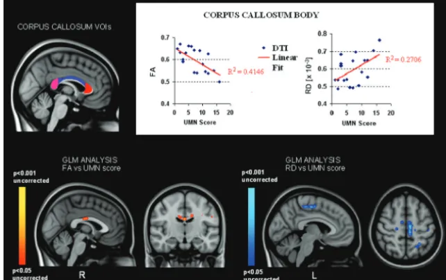
Within the patient group, significant correlations between FA and clinical measures were observed. Specifically, individ-ual FA data, extracted via the VOI analysis selectively from the TBSS results, showed a strong inverse correlation with UMN scores in the body of the CC (P⫽.003) (Fig 2). Even at the voxel level, although at an uncorrected level, GLM analyses on the group of patients showed an inverse correlation between FA and UMN scores inside the midbody of CC (P⬍.001, uncorrected) (Fig 2).
Voxel-level GLM analyses also showed that FA was posi-tively correlated with ALSFRS-R in the WM underneath the left premotor cortex. This pattern included the left paracentral lobule, the anterior cingulate and superior longitudinal fascic-ulus (P⬍.05, corrected) (Fig 3); and at an uncorrected level, FA was negatively correlated with the disease progression rate in the CSTs, the pons, and the WM underneath the middle temporal gyrus (bilaterally) and the left inferior frontal gyrus (P⬍.001, uncorrected) (Fig 3).
Trends of group differences between patients with ALS and controls were also observed in both the genu and splenium of the CC when comparing the MD and RD values (P⬍.001, uncorrected). Within the CC, this pattern of MD-RD increase in patients with ALS was strikingly complementary to the pat-tern of FA decrease.
No significant group differences were found in AD bilater-ally in the CSTs and the CC.
RD, MD, and AD did not correlate with the UMN score in the anterior and midbody of the CC according to the voxel-based GLM analysis, though in the body of the CC, mean RD data, extracted by VOI analysis of the TBSS results, showed a
[image:3.594.136.451.44.287.2]significant correlation with UMN score (P⫽.02) (Fig 2). At an uncorrected level of significance, these GLM analyses allowed identifying positive correlations between the RD and UMN score within the left paracentral lobule WM (P⬍.001, uncor-rected) (Fig 2) and between the MD and UMN score in the CSTs, mainly in the cerebral peduncles, principally at the left pontomesencephalic junction and within the genu of left in-ternal capsule and thalamus (left lateral posterior nucleus) (P⬍.001, uncorrected). Finally, the disease-progression rate showed significant positive correlation with both RD and MD bilaterally in the hippocampus and with slight left lateraliza-tion in the insular gyri and inferior longitudinal and uncinate fasciculi (P⬍.001, uncorrected) (Fig 3).
No significant correlation was observed between all diffu-sivity parameters and disease duration in both the CSTs and the CC.
Discussion
Even if currently ALS is not considered basically a motor dis-ease, the actual spread of the neurodegenerative process throughout the central nervous system is not fully understood. The present study depicted the microstructural pattern of WM impairment in ALS within but also beyond the motor system and allowed us to hypothesize the underlying mechanisms of cortical spreading of the pathologic process.
We showed a reduced anisotropy in the body of the CC, which correlated significantly with clinical UMN involvement in patients with ALS. These results are consistent with previ-ous studies of CC impairment in ALS.6,8,15,17,18Conversely, in
a recent study,19the callosal involvement in ALS resulted
in-dependent of clinical UMN involvement, and changes of FA were significantly correlated with UMN bilaterally in the CSTs but not in the CC.
Fig 2.VOI (top panel) and GLM (lower panel) correlation analyses of FA and RD measures with UMN score values. VOI analysis (top panel) shows significant inverse and positive correlations, respectively, of FA and RD values (expressed in mm2⫻s⫺1) with the UMN score in the body of CC (P⫽.003,P⫽.02). GLM analysis (lower panel) also shows an inverse correlation between FA and the UMN score in the midbody of CC (red-yellow), while RD values correlate with the UMN score mainly within the left paracentral lobule WM (blue) but not in the CC (uncorrected level of significance).
[image:4.594.133.454.45.247.2] [image:4.594.134.451.301.479.2]With respect to data reported by Filippini et al,19we
ob-served a similar anisotropic pattern in the body of CC, though it was associated with a significant inverse correlation between the FA and UMN score. We argue that these diverging results depend on a possible more advanced pathologic stage in the process of axonal loss in our patients with ALS, which showed a modest clinical UMN involvement but longer disease course. Given that the staging of ALS depends on the clinical charac-terization and the grade of disability, most DTI studies were performed in patients with mildly disabling ALS (ALSFRS-Rⱖ20) and a mean disease duration of ⱕ40 months.8,9,15
Therefore, despite making less evident the effects of reactive gliosis (typical of extremely disabled patients), the selection of mildly disabled patients could not allow depicting structural changes that characterize more advanced stages of disease. Thus, we speculated that in advanced stages of ALS, the CC diffusivity pattern and its relation with UMN involvement might more strikingly reflect a progressive cortical spread of the disease across the 2 hemispheres.
The more rostral impairment of the CSTs has already been described before17and might be interpreted as an advanced
stage of more cranial and widespread distribution of the pathologic degenerative process, similar to what has been seen and reported in postmortem examinations of patients with sporadic ALS who had survived for long periods.27,28
The reduction of FA in the midpart of the CC and mainly in the cranial parts of the CSTs is probably a result of the preva-lent axonal loss within the motor fiber pathway, as was also demonstrated by neuropathologic evidence.29Moreover,
be-cause the preferential loss of water diffusion in the fiber direc-tion (decreased FA) was not accompanied by higher diffusion perpendicular to the CC fibers (measured by RD) in the body of CC, these effects seem to reflect a wallerian degeneration (atrophy) of the axons rather than a loss of myelin integrity. We would also have expected to detect corresponding changes of AD, because an increase of AD along fiber tract bundles may be an index of axonal degeneration in ALS.8Additionally, we did not find an increase of MD within the CC or along the CSTs. We hypothesized that such a diffusivity pattern proba-bly was due to a conspicuous process of reactive gliosis, sec-ondary to tissue loss, which might have lead to a ‘‘pseudonor-malization” of MD, RD, and AD values, while still reducing FA (glial cells do not have the same anisotropic morphology as the tissue they replace).15
Previous evidence showed that both neuronal and non-neuronal elements may be involved in neurodegeration dur-ing the different phases of the disease process,30though the
specific neuropathologic substrate underlying the DTI abnor-malities reported in ALS has not yet been clarified. However, in extremely disabled patients, a prominent effect of reactive gliosis as a counterbalance of tissue loss has been detected.31
Indeed, in the postmortem examinations of patients with ALS, there was loss of pyramidal motor neurons in the primary motor cortex and axonal degeneration of the CSTs, with pro-liferation of glial cells, extracellular matrix expansion, and whole-brain intracellular abnormalities.29In terms of age and
global atrophy, our ALS population did not significantly differ from the control population. Atrophy was assessed by com-puting the average brain parenchymal fraction score (ratio of
brain parenchymal volume to the intracranial volume) ob-tained by a fully automated method (structural imaging eval-uation of normalized atrophy,32data not shown).
The significant correlations shown within the patient group between the UMN score and RD in the body of the CC at the VOI level and between the UMN score and RD and MD along the CSTs, via voxel-based GLM analysis (uncorrected level of significance), might reflect the frequently observed CST histopathologic association of axonal loss with secondary pallor and injury of the myelin sheaths throughout the extent of axonal projections.29,33
If we take these results together, our data support the hy-pothesis of an active and at least partially independent bilateral cortical process of degeneration with secondary damage to the CC, particularly in its midbody, known to be significantly in-volved in the process of axonal degeneration in ALS34and substantially formed by interhemispheric fibers connecting the motor cortices, as also demonstrated by diffusion tensor tractography.35
According to previous neuropathologic findings,27,34,36 ALS degenerative changes in the cerebral cortex mainly in-volve motor areas, though recent whole-brain voxel-based morphometry analyses have largely reported GM abnormali-ties even in extramotor regions.12,14,15,20,31,37 Thereby, the trends of MD and RD changes, which largely overlapped in our patients in both the genu and splenium of the CC, may represent an increase in the extracellular volume secondary to axonal loss associated with injury to the myelin sheaths, as previously reported in human and animal models of demyeli-nation and axonal degeneration.2,38,39Thus, our results also
suggest that in advanced stages of ALS, FA changes in the mid-dle body of the CC reflect the degenerative process of axonal damage, inducing loss of motor function, whereas the increase in RD and MD in both the genu and splenium of the CC reflect more subtle axonal damage that could affect fine motor or extramotor skills. Moreover, the mild microstructural changes that we observed in both the genu and splenium of the CC in our patients, affected by a frontal cognitive dysfunction, are highly reminiscent of what has been previously found in callosal areas of the healthy population in relation to aging40 and, to a greater extent, in the case of the compounded effect of age and alcohol abuse.41
The left lateralization of the impairment of the CSTs ob-served in our patients was probably due to the fact that all subjects were right-handed. This finding, with the recent ob-servation of concordance between handedness and laterality of the upper limb onset in ALS,42allowed us to further support
the hypothesis of an independent cortical vulnerability to the disease of the 2 hemispheres (ie, our results confirm the hy-pothesis that, despite the systemic clinical symmetry, lateral-ized motor degeneration is possible). Moreover, this is not the first study in which ALS effects were manifest as lateralized in the motor system.4,10,15,20,37,43,44 Our findings are also in
agreement with the descriptions of asymmetry in ALS pathol-ogy of the CSTs,45though a correspondence between the side
of greater MR imaging differences and the side of greater mus-cle strength deficit of the limbs was not found,10 probably
Using TBSS and voxel-based GLM analyses of the FA maps, we were able to detect significant positive correlations between FA and ALSFRS-R in the WM underneath the whole left pre-motor frontal cortex, especially the paracentral lobule (cor-rected level of significance), and between FA and the disease-progression rate along the CSTs in the pons (though at an uncorrected level of significance). These results are consistent with previous DTI correlation analyses between FA and clini-cal measure of disease severity and progression within the pyramidal system.2,10,16,46However, not all previous MR im-aging studies reported significant correlations between diffu-sivity parameters and clinical scores in ALS,4,7,11,15probably
due to the following: 1) the intrinsic heterogeneity of the pathologic process (in ALS, degeneration of fiber tracts may not be a classic wallerian degeneration in terms of the contri-bution from intra- and extracellular changes, astrocytosis, dis-orders of axonal transport, and accumulation of axonal spher-oids29); 2) the different clinical characteristics of the patients, with predominantly upper or lower motor neuron signs and different disease duration and disability; 3) the lack of clinical scores highly specific for the detection of UMN impairment (associations between clinical symptoms and pathologic find-ings are weak because it has been shown that prominent clin-ical disability may occur even in the absence of impairment of the CSTs, and vice versa)29; and 4) the different methodologies used.
Comparing our patients with ALS with controls, we de-tected reduced FA values in the WM underneath the left me-dial frontal lobe, including the anterior cingulate and superior longitudinal fasciculus, whereas we also found a positive cor-relation between FA changes and the ALSFRS-R. The DTI pat-tern of predominantly frontal WM injury clearly reflects the frontal executive dysfunction that characterizes our patients with ALS and has been also extensively described in other co-horts of patients with ALS.47,48However, we cannot directly
validate this hypothesis in our population because our patients did not show overt signs of cognitive impairment, and we did not perform an extensive neuropsychological assessment. Nonetheless, the location of the diffusivity changes in the as-sociative tracts of the frontal lobes that we report in the present study is in line with similar FA and RD changes described in patients with the behavioral variant of frontotemporal dementia.49
The correlations between the disease-progression rate and FA in WM bilaterally underneath the middle temporal gyrus and the left inferior frontal gyrus and between the disease-progression rate and RD and MD bilaterally within the hip-pocampus, the left insula, and the left inferior longitudinal and uncinate fasciculi might provide further structural evi-dence of the progressive development in ALS of a multisystem disorder. Furthermore, the highly distributed and widespread pattern of our results is in agreement with the recent findings that in ALS, the cerebral expression of a new neuropathologic marker of disease, the transactivating responsive sequence DNA– binding protein inclusions, involves the nigrostriatal system, the neocortical and allocortical areas, and the cerebel-lum, more often in case of ALS dementia than in ALS without cognitive dysfunction (eg, Geser et al13).
Conclusions
Even if longitudinal follow-up studies with larger patient groups and extensive clinical and neuropsychological testing are needed to validate the use of DTI parameters as possible biomarkers for neurodegeneration and/or disease progres-sion, our findings underline the crucial feature of CC impair-ment as neuroradiologic marker of UMN and widespread cor-tical involvement in ALS, especially to better define the different clinical phenotypes and to more accurately assess and eventually follow, in vivo, the pathologic spread of the disease within and beyond motor areas.
References
1. Davidoff RA.The pyramidal tract.Neurology1990;40:332–39
2. Ellis CM, Simmons A, Jones DK, et al.Diffusion tensor MRI assesses cortico-spinal tract damage in ALS.Neurology1999;53:1051–58
3. Basser PJ, Pierpaoli C.Microstructural and physiological features of tissues elucidated by quantitative-diffusion tensor MRI.J Magn Reson1996;111: 209 –19
4. Toosy AT, Werring DJ, Orrell RW, et al.Diffusion tensor imaging detects corticospinal tract involvement at multiple levels in amyotrophic lateral scle-rosis.J Neurol Neurosurg Psychiatry2003;74:1250 –57
5. Graham JM, Papadakis N, Evans J, et al.Diffusion tensor imaging for the as-sessment of upper motor neuron integrity in ALS.Neurology2004;63:2111–19 6. Sach M, Winkler G, Glauche V, et al.Diffusion tensor MRI of early upper motor neuron involvement in amyotrophic lateral sclerosis.Brain2004;127: 340 –50
7. Iwata NK, Aoki S, Okabe S, et al.Evaluation of corticospinal tracts in ALS with diffusion tensor MRI and brainstem stimulation.Neurology2008;70:528 –32 8. Metwalli NS, Benatar M, Nair G, et al.Utility of axial and radial diffusivity
from diffusion tensor MRI as markers of neurodegeneration in amyotrophic lateral sclerosis.Brain Res2010;1348:156 – 64
9. Agosta F, Pagani E, Petrolini M, et al.Assessment of white matter tract damage in patients with amyotrophic lateral sclerosis: a diffusion tensor MR imaging tractography study.AJNR Am J Neuroradiol2010;31:1457– 61
10. Ciccarelli O, Behrens TE, Altmann DR, et al. Probabilistic diffusion
tractography: a potential tool to assess the rate of disease progression in amyotrophic lateral sclerosis.Brain2006;129:1859 –71
11. Sage CA, Peeters RR, Gorner A, et al.Quantitative diffusion tensor imaging in amyotrophic lateral sclerosis.Neuroimage2007;34:486 –99
12. Thivard L, Pradat PF, Lehe´ricy S, et al.Diffusion tensor imaging and voxel based morphometry study in amyotrophic lateral sclerosis: relationships with motor disability.J Neurol Neurosurg Psychiatry2007;78:889 –92 13. Geser F, Martinez-Lage M, Robinson J, et al.Clinical and pathological
contin-uum of multisystem TDP-43 proteinopathies.Arch Neurol2009;66:180 – 89 14. Abe O, Yamada H, Masutani Y, et al.Amyotrophic lateral sclerosis: diffusion
tensor tractography and voxel-based analysis.NMR Biomed2004;17:411–16 15. Agosta F, Pagani E, Rocca MA, et al.Voxel-based morphometry study of brain
volumetry and diffusivity in amyotrophic lateral sclerosis patients with mild disability.Hum Brain Mapp2007;28:1430 –38
16. Smith SM, Jenkinson M, Johansen-Berg H, et al.Tract-based spatial statistics: voxelwise analysis of multi-subject diffusion data.Neuroimage2006;31:1487– 505
17. Sage CA, Van Hecke W, Peeters R, et al.Quantitative diffusion tensor imaging in amyotrophic lateral sclerosis: revisited.Hum Brain Mapp2009;30:3657–75 18. Ciccarelli O, Behrens TE, Johansen-Berg H, et al.Investigation of white matter pathology in ALS and PLS using tract-based spatial statistics.Hum Brain
Mapp2009;30:615–24
19. Filippini N, Douaud G, Mackay CE, et al.Corpus callosum involvement is a consistent feature of amyotrophic lateral sclerosis. Neurology 2010;75: 1645–52
20. Senda J, Kato S, Kaga T, et al.Progressive and widespread brain damage in ALS: MRI voxel-based morphometry and diffusion tensor imaging study. Amy-otroph Lateral Scler 2011;12:59 – 69
21. Hua K, Zhang J, Wakana S, et al.Tract probability maps in stereotaxic spaces: analysis of white matter anatomy and tract-specific quantification. Neuroim-age2008;39:336 – 47
22. Wakana S, Caprihan A, Panzenboeck MM, et al.Reproducibility of quantita-tive tractography methods applied to cerebral white matter.Neuroimage
2007;36:630 – 44
23. Brooks BR, Miller RG, Swash M, et al.El Escorial revisited: revised criteria for the diagnosis of amyotrophic lateral sclerosis.Amyotroph Lateral Scler Other Motor Neuron Disord2000;1:293–99
25. Kaufmann P, Pullman SL, Shungu DC, et al.Objective tests for upper motor neuron involvement in amyotrophic lateral sclerosis (ALS).Neurology2004; 62:1753–57
26. Grace J, Stout JC, Malloy PF.Assessing frontal lobe behavioral syndromes with the frontal lobe personality scale.Assessment1999;6:269 – 84
27. Nishihira Y, Tan CF, Toyoshima Y, et al.Sporadic amyotrophic lateral
sclerosis: widespread multisystem degeneration with TDP-43 pathology in a patient after long-term survival on a respirator.Neuropathology2009;29: 689 –96
28. Nishihira Y, Tan CF, Onodera O, et al.Sporadic amyotrophic lateral sclerosis: two pathological patterns shown by analysis of distribution of TDP-43-im-munoreactive neuronal and glial cytoplasmic inclusions.Acta Neuropathol
2008;116:169 – 82
29. Ince PG, Wharton SB.Cytopathology of the motor neuron. In: Eisen AA, Shaw PJ, eds.Motor Neuron Disorders and Related Diseases: Handbook of Clinical Neu-rology. 3rd Series. Amsterdam, the Netherlands: Elsevier; 2007:89 –119
30. Clement AM, Nguyen MD, Roberts EA, et al.Wildtype non-neuronal cells
extend survival of SOD1 mutant motor neurons in ALS mice.Science2003;302: 113–17
31. Chang JL, Lomen-Hoerth C, Murphy J, et al.A voxel-based morphometry
study of patterns of brain atrophy in ALS and ALS/FTLD. Neurology
2005;65:75– 80
32. De Stefano N, Iannucci G, Sormani MP, et al.MR correlates of cerebral atrophy in patients with multiple sclerosis.J Neurol2002;249:1072–77
33. Brownell B, Oppenheimer DR, Hughes JT.The central nervous system in mo-tor neuron disease.J Neurol Neurosurg Psychiatry1970;33:338 –57
34. Smith MC.Nerve fibre degeneration in the brain in amyotrophic lateral scle-rosis.J Neurol Neurosurg Psychiatry1960;23:269 – 82
35. Chao YP, Cho KH, Yeh CH, et al.Probabilistic topography of human corpus callosum using cytoarchitectural parcellation and high angular resolution diffusion imaging tractography.Hum Brain Mapp2009;30:3172– 87 36. Matsuoka T, Fujii N, Kondo A, et al.An autopsied case of sporadic adult-onset
amyotrophic lateral sclerosis with FUS-positive basophilic inclusions. Neuro-pathology2011;31:71–76
37. Tedeschi G, Trojsi F, Tessitore A, et al.Interaction between aging and
neuro-degeneration in amyotrophic lateral sclerosis. Neurobiol Aging 2012;33: 886-98.
38. Song SK, Sun SW, Ramsbottom MJ, et al.Dysmyelination revealed through
MRI as increased radial (but unchanged axial) diffusion of water.Neuroimage
2002;17:1429 –36
39. Song SK, Yoshino J, Le TQ, et al.Demyelination increases radial diffusivity in corpus callosum of mouse brain.Neuroimage2005;26:132– 40
40. Sullivan EV, Rohlfing T, Pfefferbaum A.Quantitative fiber tracking of lateral and interhemispheric white matter systems in normal aging: relations to timed performance.Neurobiol Aging2010;31:464 – 81
41. Pfefferbaum A, Adalsteinsson E, Sullivan EV.Dysmorphology and microstruc-tural degradation of the corpus callosum: interaction of age and alcoholism.
Neurobiol Aging2006;27:994 –1009
42. Turner MR, Wicks P, Brownstein CA, et al.Concordance between site of onset and limb dominance in amyotrophic lateral sclerosis.J Neurol Neurosurg Psy-chiatry2011;82:853–54
43. Hatazawa J, Brooks RA, Dalakas MC, et al.Cortical motor-sensory hypome-tabolism in amyotrophic lateral sclerosis: a PET study.J Comput Assist Tomogr
1988;12:630 –36
44. Rule RR, Schuff N, Miller RG, et al.Gray matter perfusion correlates with disease severity in ALS.Neurology2010;74:821–27
45. Swash M, Scholtz CL, Vowles G, et al.Selective and asymmetric vulnerability of corticospinal and spinocerebellar tracts in motor neuron disease.J Neurol Neurosurg Psychiatry1988;51:785– 89
46. Wang S, Poptani H, Woo JH, et al.Amyotrophic lateral sclerosis: diffusion-tensor and chemical shift MR imaging at 3.0 T.Radiology2006;239:831–38. Epub 2006 Apr 26
47. Murphy J, Henry R, Lomen-Hoerth C.Establishing subtypes of the continuum of frontal lobe impairment in amyotrophic lateral sclerosis.Arch Neurol2007; 64:330 –34
48. Abrahams S, Goldstein LH, Suckling J, et al.Frontotemporal white matter changes in amyotrophic lateral sclerosis.J Neurol2005;252:321–31 49. Whitwell JL, Avula R, Senjem ML, et al.Gray and white matter water diffusion