Original Article
Expression of Talin1 in tissues of ovarian cancer and its
role in invasion and migration of ovarian cancer cells
Jun Wang, Heling Zhou, Kai Wang, Xianwen Shang, Xing Chen, Lingzhi Zheng, Junhui Yu
Department of Gynecology, Tai Zhou Hospital of Zhe Jiang Province, Lin Hai, Zhe Jiang, P. R. China
Received July 8, 2016; Accepted October 29, 2016; Epub January 15, 2017; Published January 30, 2017
Abstract: Objective: To investigate the expression of Talin1 in tissues of ovarian cancer and its role in invasion and migration of ovarian cancer cells as well as the related mechanisms. Method: The expression of Talin1 in the tissues of ovarian cancer and normal ovarian tissues was detected by immunohistochemical methods; lentivirus silencing
Talin1, GFP fluorescence and Western blotting were used to detect the LV3-Talin1 silencing efficiency and efficacy;
the effect of Talin1 expression on invasion of ovarian cancer cells was detected by Transwell invasion assay; and the effect of Talin1 expression on migration of ovarian cancer cells was detected by wound scratch test; and expression
of the proteins E-Cadherin, Vimentin and Twist was detected by Western blotting. Results: Compared with normal ovarian tissues, the Talin1 expression in tissues of ovarian cancer was significantly increased; and the Talin1 ex
-pression in tissues of advanced, poorly differentiated ovarian cancer with lymph node metastasis was significantly higher than that in tissues of early, well-differentiated ovarian cancer without lymph node metastasis; LV3-Talin1
lentivirus could effectively inhibit the expression of Talin1; Talin1 silencing could inhibit invasion and migration of the A2780 cells; Talin1 silencing up-regulated the expression of E-Cadherin and down-regulated the expression of
Vimentin and Twist. Conclusion: Talin1 expression is significantly increased in ovarian cancer, and Talin1 is closely
associated with staging and grading of ovarian cancer; in addition, Talin1 silencing promotes invasion and migration of ovarian cancer cells by inhibiting epithelial-mesenchymal transition (EMT).
Keywords: Talin1, ovarian cancer, EMT, E-Cadherin, transwell
Introduction
Ovarian cancer is one of the three most com-monly seen gynecological malignancies, and its incidence is only second to cervical cancer and endometrial cancer, with a 5-year survival rate of 25%~30%. It is the gynecological malignancy with the highest mortality [1]. Since the ovaries are deep in the pelvis and ovarian cancer in the early stage is lack of specific symptoms, most patients are in the advanced stage and have abdominal cavity implantation metastasis at diagnosis [2]. Therefore, studying the mecha-nism of invasion and metastasis of ovarian can-cer and searching the ovarian cancan-cer-related molecular targets have become the focus in ovarian cancer research.
Tumor metastasis is a complex multi-step, multi-stage process, mainly including local infil -tration, invasion in blood vessels, metastasis with and survival in the blood circulation
sys-tem, emigration from blood vessels, and settle-ment and proliferation in new sites, and the cytoskeletal changes, cell adhesion and chang-es in kinetic characteristics, epithelial-mchang-esen- epithelial-mesen-chymal transition and activation of various sig-naling pathways are involved. Tumor microenvi-ronment also plays an important role in tumor metastasis process. The microenvironment includes not only the tumor cells, but also the stromal cells, inflammatory cells, vascular sys -tem, and extracellular matrix (ECM). The ECM, which contains the growth factors and cyto-kines required for tumor growth, plays a very important role in the biological behaviors of tumor [3].
interactions among ECM, integrin and cytoskel-eton and plays an important role in tumor inva-sion and metastasis [5]. Many studies have shown [6, 7] that Talin1 expression is increased significantly in tumor tissues and its expression is associated with invasion and migration of tumors. However, expression of Talin1 in the tis-sues of ovarian cancer and its role in invasion and migration of the ovarian cancer cells, and the relevant mechanisms are still unclear. In this study, we investigated the expression of Talin1 in the tissues of ovarian cancer, its role in invasion and migration of the ovarian cancer cells and the relevant mechanisms.
Materials and methods
Collection and treatment of clinical specimens
The ovarian cancer tissues of 65 patients con-firmed pathologically after surgical excision were collected in Taizhou Hospital of Zhejiang from January 2015 to January 2016, and the normal ovarian tissues of 65 patients who received hysterectomy due to uterine fibroids or other benign uterine lesions and resection of uterine appendages were collected. The patients were aged 52.45 ± 4.79 years. The patients with ovarian cancer did not receive chemotherapy and radiotherapy before sur-gery. The ovarian cancer staging was in accor-dance with FIGO2000 criteria: 4 cases in stage I, 10 cases in stage II, 36 cases in stage III, 15 cases in stage IV; 43 poorly differentiated cases, 16 moderately differentiated cases and 6 well differentiated cases. 39 cases received pelvic lymphnodedissection, 26 cases with lymph node metastasis, and 13 cases without lymph node metastasis. When the tumor tis-sues were removed from the body, they were placed into 4% paraformaldehyde for fixation after blood stain was removed.
Cell line and main reagents
The human ovarian cancer cell line HO8910 was obtained from Wuhan University cell collec-tion center, and C30, A2780, COC1, SKOV3 were from ATCC. Cell culture conditions: cul-tured in RPMI 1640 containing 10% fetal calf serum at 37°C, 5% CO2. The fetal calf serum and RPMI 1640 medium were purchased from HyClone. The primary antibodies of Talin1, E-Cadherin, Vimentin and Twist were purchased from Abcam (ab71333, ab40772, ab92547
and ab50887). Transwell chambers were obtained from Millipore (US). Talin1-silencing and control lentiviruses were purchased from Shanghai GenePharma Co., Ltd.
Immunohistochemistry
The tissues were embedded in paraffin and cut into sections of 4 μm in thickness. The immu -nohistochemical operations were performed according to the immunohistochemical S-P kit (Beijing Zhongshan Golden Bridge Biotech Co., Ltd.): dewaxing of the tissues and then hydrat-ing. Antigen retrieval was performed in micro-wave with citrate buffer solution for 30 min, cooled down to room temperature, then washed 3 times with PBS, 3 min each time, incubated with 3% H202 at 37°C for 15 min, then washed 3 times with PBS, 3 min each time, then the primary antibody was placed overnight, washed 3 times with PBS, 3 min each time; the horse-radish peroxidase-labeled donkey anti-rabbit IgG (1:200, Beijing BIOSS Bio-tech Co., Ltd.), washed 3 times with PBS, 3 min each time; the horseradish peroxidase labeled streptavidin avidin working solution (Beijing BIOSS Bio-tech Co., Ltd.) was placed in 37°C water bath for 20 min, then washed 3 times with PBS, 3 min each time, and then stained with diaminobenzidine (DAB). In the negative control group, the prima-ry antibody was replaced by PBS. All sections were reviewed independently by two patholo-gists, and 22 representative high power fields (10×40 fold) were selected by each patholo-gist. The percentage of positive cells in each specimen was counted and the positive results were judged and scored according to the meth-od described by De Falco M et al. [8]: 0 (posi-tive cells less than 1%); 1 (between 1% and 20%), 2 (between 21% and 40%); 3 (between 41% and 60%); and 4 (more than 61%).
Lentivirus transfected into the A2780 cells
One day before the experiment, 5×10³ A2780 cells were inoculated in 96-well plates, to make the cell fusion degree at 40%~60%. According to the GenePharma lentivirus operating manu-al, the appropriate MOI (multiplicity of infection) of Talin1 lentivirus was determined using the gradients of 0, 10 and 100.
first tube to the second and mixed well, to get virus solutions at three different concentra-tions: stock solution, 10x dilution, and 100x dilution.
10 μl of virus solutions at three different gradi -ents were added to three wells in each group, to calculate the MOI of three wells, which were 100, 10 and 1 respectively. The most appropri-ate MOI was determined to be 100.
Effects of Talin1 expression on invasion ability of the ovarian cancer cells detected by tran-swell invasion assay
All reagents and equipment were pre-cooled on ice. The Transwell chambers were placed in a 24-well plate. 50 μl (0.2 μg/μl) Matrigel gel was evenly applied to inner membrane of Transwell chamber, incubated for 15 min at 37°C to solid-ify the gel; when digested, centrifuged and counted, the cells were diluted with 2.5×10⁴/ mL serum-free medium to prepare cell suspen-sion; the cell suspension was added to the upper Transwell chamber at 200 μL each well, and 500 μL of 10% FBS and medium were added to the lower Transwell chamber, placed in a 37°C incubator for culture; fixed with for -malin, stained by crystal violet for 15 min, and then the cells on the inner membrane were wiped with a cotton swab, counted under a microscope, to count the cells that passed through the membrane under 4 high power fields (x40). The experiment was performed in triplicate.
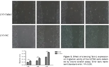
Effects of Talin1 expression on migration abil-ity of the ovarian cancer cells detected by wound scratch assay
[image:3.612.91.373.72.214.2]Wound scratch assay: The A2780 cells were inoculated to a 6-well plate, and when cell con-fluence reached 90%, scratch from up to bot -tom using a 200 μl sterile pipette tip, observe under a microscope, to measure the initial dis-tance of scratch (0 time); at 24 h, 48 h and 72 h, the distances of scratch were measured respectively and photographed, to calculate the cell migration rate. The experiment was performed in triplicate.
[image:3.612.90.289.302.355.2]Figure 1. The expression of Talin1 in ovarian cancer tissues and normal ovar-ian tissues detected by immunohistochemicalassay (10×).
Table 1. Comparison of the expression of Talin1 between ovarian cancer tissues and normal ovarian tissues
Tissue specimen Quantity Talin1 positive
N %
Ovarian cancer 65 45 69.2%* Normal ovary 65 15 23.1%
Compared to the normal ovarian tissues, *P<0.05.
The experiment consisted of two groups: the silencing gro- up, transfected with Talin1 sil- encing lentivirus (LV3-Talin1), and the control group, trans-fected with the negative con-trol lentivirus (LV3-NC). 100-fold diluted virus stock solu-tion was added to the LV3-Talin1 group, and 100-fold diluted negative control virus solution was added to the LV3-NC group. The expression of GFP fluorescence was observed 24 h later.
Table 2. Relationship between Talin1 protein expression and clinicopathological features of ovarian cancer
Clinical pathological parameters Quantity Talin1 positive Pathological stages
I-II 14 7*
III-IV 51 38
Degree of differentiation
Moderate-well 22 12*
Poor 43 33
Lymph node metastasis
Yes 26 23*
No 13 5
[image:3.612.89.292.436.571.2]Expression of the proteins Talin1, E-Cadherin, Vimentin and Twist detected by western blot-ting
The proteins were extracted from different ovarian cancer cells, and the protein concen-trations were determined by BCA method, and then loading buffer was added for protein dena-turation. 8% and 10% SDS-PAGE was prepared, and 20 μg protein sample was added into each hole, then transferred to a PVDF membrane using the electric wet transfer method, sealed 2 h with 5% skim milk, and the primary antibody (Talin1) was diluted by 1:1000 TBST, overnight at 4°C; then 1:5000 dilution of goat anti-rabbit secondary antibody was added, incubated at room temperature for 2 h; and ECL was per-formed. The experiment was performed in triplicate.
72 h after cell transfection, the total proteins were extracted from the cells in the experimen-tal and control groups, and the protein concen-trations were determined by BCA method, and then loading buffer was added for protein dena-turation. 8% and 10% SDS-PAGE was prepared, and 20 μg protein sample was added into each hole, then transferred to a PVDF membrane using the electric wet transfer method, sealed 2 h with 5% skim milk, and the primary antibody (E-Cadherin, Vimentin and Twist) was diluted by 1:1000 TBST, overnight at 4°C; then 1:5000 dilution of goat anti-rabbit secondary antibody was added, incubated at room temperature for 2 h; and ECL was performed. The experiment was performed in triplicate.
Statistical analysis
The SPSS 19.0 software was used for statisti-cal analysis, measurement data were expressed in (_x ± s), t-test was employed for comparison
of means between groups, and P<0.05 indicat-ed statistically significant difference.
Results
Increased expression of Talin1 in ovarian can-cer tissues
The immunohistochemical results showed that Talin1 was localized in cytomembrane and cyto-plasm. In 65 ovarian cancer tissues, 45 were positive for Talin1 expression; in 65 normal ovarian tissues, 15 were positive for Talin1 expression (Figure 1; Table 1). It suggested that the expression of Talin1 in ovarian cancer tissues was significantly higher than that in nor -mal ovarian tissues, with statistically significant differences (P<0.05).
Correlation between Talin1 expression and clinical-pathological characteristics of ovarian cancer
The expression of Talin1 increased with the increased pathological staging of ovarian can-cer (Table 2, P<0.05); with decrease in degree of differentiation, the expression of Talin1 increased gradually (Table 2, P<0.05); in the ovarian cancer tissues with lymph node metas-tasis, the expression of Talin1 increased signifi -cantly (Table 2, P<0.05).
Talin1 expression in different ovarian cancer cell lines
[image:4.612.93.521.78.193.2]Figure 3. A. Efficiency of transfection of Talin1
knocked down lentivirus into the ovarian cancer
[image:5.612.92.376.444.720.2]A2780 cells detected by GFP fluorescence. B. Ex -pression of Talin1 detected by Western blotting. Er-ror bars represent standard erEr-ror. *P<0.05.
Figure 4. Effect of si-lencing Talin1 expres-sion on invaexpres-sion abil-ity of the A2780 cells detected by Transwell invasion assay. Error bars represent stan-dard error. *P<0.05.
Low expression of Talin1 after LV3-Talin1 was transfected into the A2780 cells
5.16)% vs (25.80 ± 3.27)%, P<0.05], suggest-ing that Talin1 expression could be significantly decreased after the A2780 cells were trans-fected by LV3-Talin1.
Silencing Talin1 expression inhibits invasion ability of the A2780 cells
The Transwell results (Figure 4A and 4B) showed that in the LV3-Talin1 group, the num -ber of cells passing the Matrigel gel was 81.54 ± 6.76, significanlty less than that in the LV3-NC group (223.92 ± 12.88), with statistically significant differences (P<0.05). It indicated that silencing Talin1 expression could inhibit invasion ability of the A2780 cells.
Silencing Talin1 expression inhibits migration ability of the A2780 cells
The width of scratches in any three parts of cells in each group was measured under a microscope at the time points of 0 h, 24 h, 48 h and 72 h. The migration rate was calculated according to the formula: Migration rate=[D(t=24 h, 48 h, 72 h)-D(t=0 h)]/D(t=0 h). Results of the wound scratch assay (Figure 5) suggested that com-pared to the LV3-NC group, migration rate in the LV3-Talin1 group was significantly reduced at 24 h, 48 h and 72 h [24 h (0.17 ± 0.03)% vs (0.29 ± 0.04)%, P<0.05; 48 h (0.28 ± 0.04)% vs
(0.57 ± 0.05)%, P<0.05; 72 h (0.61 ± 0.04)% vs (0.87 ± 0.06)%, P<0.05], with statistically sig-nificant differences. It was revealed in the wound scratch assay that silencing Talin1 expression could inhibit migration ability of the A2780 cells.
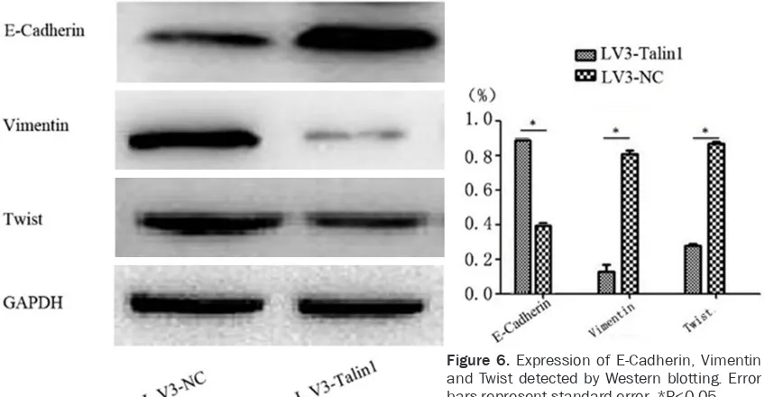
Silencing Talin1 expression could inhibit the expression of E-Cadherin, Vimentin and Twist
[image:6.612.93.520.71.336.2]Many studies have demonstrated that EMT is activated in invasion and migration of epithelial tumors, which is the critical molecular event enablingthe epithelial tumor cells to acquire invasion ability and plays an important role in invasion and migration of malignant tumors [9]. The E-Cadherin protein is the epithelial marker in the EMT process, while Twist and Vimentin are the mesenchymal markers in the process. Results of Western blotting (Figure 6) showed that compared to the LV3-NC group, the expres -sion levels of Vimentin and Twist in the LV3-Talin1 group decreased significantly [Vimentin (0.14 ± 0.01)% vs (0.81 ± 0.06)%; Twist (0.26 ± 0.02)% vs (0.88 ± 0.06)%, P<0.05]; compared to the LV3-NC group, the expression level of E-Cadherin in the LV3-Talin1 group increased significantly (0.88±0.06)% vs (0.38±0.02)%, P<0.05). It suggested that silencing the expres-sion of Talin1 could up-regulate that of
E-Cadherin, as well as down-regulate that of Vimentin and Twist. It indicated that Talin1 could promote EMT of ovarian cancer cells. Discussion
Tumor infiltration and metastasis is a compli -cated process with multiple steps. The first step is to reduce the adhesive capacity of tumor cells, enhance cell mobility and infiltrate the peripheral tissues for distant metastasis [3, 10]. Integrin is involved in all of the processes. The integrin signals are involved in tumor inva-sion and migration mainly through activating FAK, which is an important linking molecule causing different signal proteins to gather on the adhesion plaques, thereby mediating differ-ent biological behaviors.
The Talin protein contains an N-terminal globu-lar head region and C-terminal rod-shaped region, and its head region can bind with FAK. After involved in the formation of adhesion plaque complex, Talin and FAK can bind with integrin intracellular domain to activate integrin and regulate invasion and migration of the tumor cells. It hasbeen shown instudy [11] that-Talin activates integrin and the highly-expressed Talin head fragments lead to activation of inte-grin β3, thus demonstrating that Talin plays an important role in the integrin activation process.
In this study, by investigating the expression of Talin1 in ovarian cancer tissues, it has been
shown that Talin1 expression in ovarian cancer tissues is significantly higher than that in the normalovarian tissue, and Talin1 expression in the advanced, poorly-differentiated ovarian cancer tissues with lymph node metastasis is significantly higher than that in the early, well-differentiated ovarian cancer tissues without lymph node metastasis. After silencing Talin1 with lentivirus, the observations on invasion and migration of the ovarian cancer cells have shown that invasion and migration of the ovari-an covari-ancer cells are reduced after Talin1 silenc-ing. The study by Fang Kunpeng et al. [12] has found out that Talin1 is associated with the invasion and migration of hepatocellular carci-noma (HCC). The study by Sakamoto et al. [13] has shown that Talinregulates the level of activ-ity of β1 integrin, thereby prompting invasion and migration of the human prostate cancer cells, and Talin silencing results in decreased metastasis of the prostate cancer cells, consis-tent with the results in this study.
[image:7.612.97.521.75.295.2]EMT plays an important role in tumor invasion and metastasis. The precondition for metasta-sis of tumor cells is that the adjacent tissues are infiltrated, the tumor adhesion is weakened to form highly invasive tumor and at the same time, expression of E-Cadherin is down-regulat-ed. A number of studies [14] have shown that expression of E-Cadherin is down-regulated in many malignant tumors, and the malignant tumors with low expression of E-Cadherin have stronger invasion and migration abilities. The Figure 6. Expression of E-Cadherin, Vimentin
study by Bonnoment et al. [15] has revealed that metastasis and invasion of tumor cells are increased significantly in the cells with EMT process. The study by Lee et al. [16] has revealed that over-expression of Twist in the HCC cells down-regulates expression of E-Ca- dherin, thereby inducing EMT. In this study, by silencing Talin1, it has been observed that silencing Talin1 expression up-regulates the E-Cadherin expression and down-regulates the expression of Vimentin and Twist, suggesting that Talin1 promotes EMT of the ovarian cancer cells.
This study suggests that Talin1 expression is significantly increased in the ovarian cancer tis -sues and is closely related with tumor staging and grading and lymph node metastasis; in addition, Talin1 promotes invasion and migra-tion of the ovarian cancer cells by inducing EMT. It indicates that Talin1 is likely to be involved in the progression of ovarian cancer and it may evolve into the marker for predicting the progression and prognosis of ovarian can-cer and for monitoring the therapeutic effect. Disclosure of conflict of interest
None.
Address correspondence to: Junhui Yu, Department of Gynecology, Tai Zhou Hospital of Zhe Jiang Province, Xi Men Avenue 150#, Lin Hai 317000, Zhe Jiang, P. R. China. E-mail: wangjunyujun@sina.com
References
[1] Kuchenbaecker KB, Ramus SJ, Tyrer J, Lee A, Shen HC, Beesley J, Lawrenson K, Mcguffog L,
Healey S and Lee JM. Identification of six new
susceptibility loci for invasive epithelial ovarian cancer. Nat Genet 2015; 47: 164-171.
[2] Bowtell DD, Böhm S, Ahmed AA, Aspuria PJ, Jr
BR, Beral V, Berek JS, Birrer MJ, Blagden S and
Bookman MA. Rethinking ovarian cancer II: re-ducing mortality from high-grade serous ovari-an covari-ancer. Nat Rev Covari-ancer 2015; 15: 668-679.
[3] Mueller M, Zhou J, Yang L, Gao Y, Wu F, Schoeberlein A, Surbek D, Barnea ER, Paidas M and Huang Y. PreImplantation factor pro-motes neuroprotection by targeting microRNA let-7. Proc Natl Acad Sci U S A 2014; 111: 13882-13887.
[4] Jiang G, Sutton DH, Critchley DR and Sheetz MP. Talin1 is critical for force-dependent rein-forcement of initial integrin-cytoskeleton bonds
but not tyrosine kinase activation. J Cell Biol 2003; 163: 409-419.
[5] Kanamori H, Kawakami T, Effendi K, Yamazaki K, Mori T, Ebinuma H, Masugi Y, Du W,
Nagasaka K and Ogiwara A. Identification by
differential tissue proteome analysis of talin-1 as a novel molecular marker of progression of hepatocellular carcinoma. Oncology 2011; 80: 406-415.
[6] Beaty BT, Wang Y, Bravocordero JJ, Sharma VP, Miskolci V, Hodgson L and Condeelis J. Talin
regulates moesin-NHE-1 recruitment to inva-dopodia and promotes mammary tumor me-tastasis. J Cell Biol 2014; 205: 737-751. [7] Lai MT, Hua CH, Tsai MH, Wan L, Lin YJ, Chen
CM, Chiu IW, Chan C, Tsai FJ and Jinn-Chyuan
Sheu J. Talin-1 overexpression defines high
risk for aggressive oral squamous cell carcino-ma and promotes cancer metastasis. J Pathol 2011; 224: 367-376.
[8] Falco MD, Fedele V, Cobellis L, Mastrogiacomo A, Giraldi D, Leone S, Luca LD, Laforgia V and
Luca AD. Pattern of expression of cyclin D1/ CDK4 complex in human placenta during ges-tation. Cell Tissue Res 2004; 317: 187-194. [9] Thiery JP. Epithelial-mesenchymal transitions
in tumour progression. Nat Rev Cancer 2002; 2: 442-454.
[10] Heerboth S, Housman G, Leary M, Longacre M, Byler S, Lapinska K, Willbanks A and Sarkar S. EMT and tumor metastasis. Clin Transl Med 2015; 4: 1-13.
[11] Nader GP, Ezratty EJ and Gundersen GG. FAK, talin and PIPKI [gamma] regulate endocytosed integrin activation to polarize focal adhesion assembly. Nat Cell Biol 2016; 18: 491-503. [12] Farh KH, Grimson A, Jan C, Lewis BP, Johnston
WK, Lim LP, Burge CB and Bartel DP. The wide-spread impact of mammalian MicroRNAs on mRNA repression and evolution. Science 2005; 310: 1817-1821.
[13] Sakamoto S, Mccann RO, Dhir R and Kyprianou N. Talin1 Promotes Tumor Invasion and Metastasis via Focal Adhesion Signaling and Anoikis Resistance. Cancer Res 2010; 70: 1885-1895.
[14] Roy FV. Beyond E-cadherin: roles of other cad -herin superfamily members in cancer. Nat Rev Cancer 2014; 14: 121-134.
[15] Bonnomet A, Brysse A, Tachsidis A, Waltham M, Thompson EW, Polette M and Gilles C. Epithelial-to-mesenchymal transitions and cir-culating tumor cells. J Mammary Gland Biol Neoplasia 2010; 15: 261-273.