Original Article
Expression level of miR-146b-5p via miRNA sequencing
and its potential targets in papillary thyroid cancer
Lin Shi
1,2*, Peng Lin
3*, Dongyue Wen
3, Li Gao
1, Liang Liang
4, Yihuan Luo
4, Yichen Wei
1, Yu He
3, Hong Yang
3,
Wei Ma
1Departments of 1Pathology, 3Medical Ultrasonography, 4Gastrointestinal Surgery, The First Affiliated Hospital of Guangxi Medical University, Nanning, People’s Republic of China; 2Department of Pathology, The First Affiliated Hospital of Guangxi University of Science and Technology, Liu Zhou, People’s Republic of China. *Equal contribu-tors.
Received September 5, 2017; Accepted January 4, 2018; Epub March 15, 2018; Published March 30, 2018
Abstract:Background and objective: MiR-146b-5p is one of the deregulated miRNAs in papillary thyroid carcinoma (PTC) that promotes the malignant potential of cancer cells. To interpret the clinical significance and underlying mo-lecular mechanism of miR-146b-5p in PTC, a comprehensive analysis combining The Cancer Genome Atlas (TCGA) data and in silico investigation was conducted. Methods: Expression and clinical data for PTC were obtained from TCGA, and the relationships between miR-146b and clinicopathological parameters as well as the prognosis were later analyzed. Putative target genes of miR-146-5p were acquired by intersecting the differentially expressed genes of GSE76050 with genes predicted by twelve online software programs. Subsequently, Gene Ontology (GO) enrich-ment, Kyoto Encyclopedia of Genes and Genomes (KEGG) pathway, and PPI network analyses were performed using the chosen target genes to analyze the probable molecular mechanisms of PTC. Finally, several hub genes were validated via GEPIA and The Human Protein Atlas. Results: MiR-146b was strongly overexpressed in PTC tissues as evidenced by TCGA data. MiR-146b levels were also significantly associated with the progression of PTC. In total, 6273 and 4228 genes were identified as potential targets from GSE chip data and online prediction, respectively. Ultimately, 994 genes were chosen as the most probable targets from the intersection of the two gene sets. Accord-ing to the GO enrichment analysis, ‘intracellular signalAccord-ing cascade’, ‘regulation of programmed cell death’, ‘positive regulation of cellular biosynthetic process’, ‘insoluble fraction’, ‘cell fraction’, ‘membrane fraction’, ‘transcription regulator activity’ and ‘transcription activator activity’ were the most significant GO terms for the target genes. In regard to KEGG analysis, the targets were significantly clustered into cancer, apoptosis, and calcium signaling pathways. Two prospective targets, TRAF1 and PML, were both down-regulated at the mRNA and protein level in PTC tissues. Conclusions: MiR-146b-5p may play an essential role in the progression of PTC and influence the biological processes of cancer cells by regulating downstream targets involved in multiple signaling pathways.
Keywords: Papillary thyroid carcinoma, miR-146b-5p, the Cancer Genome Atlas, hub genes
Introduction
Papillary thyroid carcinoma (PTC) is the
pre-dominant type of thyroid cancer (TC),
constitut-ing approximately 80% of all TCs [1, 2]. PTCs
have been classified into several subtypes
according to the histological and morphological
characteristics, among which PTCs frequently
present as multifocal intra-thyroid tumors (65%
of all cases) [3, 4]. Despite the fact that current
treatments for PTC, such as surgical resection
and adjuvant radioactive iodine (RAI) therapy,
can provide patients with good prognosis,
tumor recurrence accompanied by lymph node
metastasis still occurs in some patients [5, 6]
and an increasing morbidity of PTC has been
reported over the last 40 years [7, 8]. Therefore,
better targets are needed to improve their
survival.
BRAF, RAS, PTEN, and TP53 [12], the molecular
pathogenesis of PTC is far from fully elucidated.
Recent studies have revealed that abnormally
expressed microRNAs (miRNAs) are actively
involved in PTC [13, 14]. miRNAs are a class of
endogenous non-coding RNAs that suppress
the expression of downstream targets by
bind-ing to the 3’-untranslated regions of mRNAs
[15, 16]. Abundant evidence has suggested
that miRNAs play pivotal roles in various human
cancers by affecting the growth, proliferation,
differentiation, apoptosis, and metastasis of
cancer cells [17, 18]. The deregulation of
miR-NAs has also been observed in PTC [13, 19-22].
Among the miRNAs studied to date,
miR-146-5p has been a research hot spot. Prior studies
indicated that miR-146b-5p contributes to the
metastasis, migration and invasion of cancer
cells by interacting with different molecular
tar-gets in PTC [18, 23-29]. Nevertheless, the
molecular mechanism of miR-146-5p in PTC
remains unknown. Furthermore, no study has
mined miRNA-seq data to explore the clinical
role of miR-146a-5p. Thus, we carried out this
study to investigate the clinical role of
miR-146-5p in PTC by combining miRNA-seq data and
bioinformatics methods to identify its target
genes.
Material and methods
MiRNA-seq data mining based on TCGA
Since only the data for precursor miRNA was
provided by the miRNA-seq from TCGA, the
expression level of precursor miR-146b was
downloaded and re-calculated
(https://gdc-portal.nci.nih.gov/). Differences in miR-146b
levels between PTC tissues and their
non-can-cerous counterparts, as well as between
differ-ent groups based on clinical parameters were
assessed with a Student’s t-test. The
diagnos-tic value of miR-146b was examined by using
the receiver operating characteristic (ROC). The
prognostic value of miR-146b was evaluated by
using Kaplan-Meier analysis. All statistical
analysis was conducted with SPSS 22.0 and
P<0.05 was considered significant.
Optimal candidate target genes of
miR-146b-5p
Differentially expressed mRNAs targeted by
miR-146b-5p were identified by GSE76050
from the Gene Expression Omnibus (GEO)
data-set, an expression profiling array. For this
analy-sis, a control sample of untreated BCPAP cells
and a sample transfected with miR-146b-5p
mimic in PTC cell lines were included in a
two-condition experiment. The processed data
(nor-malized by log10 ratio) were transformed into
fold changes, and down-regulated mRNAs with
changes of less than 0.5-fold were gathered.
Additionally, to obtain the predicted target
mRNAs of miR-146b-5p, twelve online
predic-tion programs, including miRWalk, Microt4,
miRanda, mirbridge, miRDB, miRMap,
miRNA-Map, Pictar2, PITA, RNA22, RNAhybrid and
Targetscan, were employed in this study. Genes
that appeared in the results of at least four of
the platforms were selected as potential
tar-gets of miR-146b-5p. Next, genes overlapping
in the lists from both GSE microarray and
pre-diction software were considered as optimal
candidate targets of miR-146b-5p in PTC.
PPI network of target genes
To visualize the interactions between the tar-
get genes of miR-146b-5p, a Protein-Protein
Interaction network was drawn using STRING
(http://www.string-db.org/). Genes with the
highest degree values were considered to be
hub genes.
Functional annotation of miR-146b-5p target
genes
To evaluate the function of the selected target
genes, we carried out Gene Ontology (GO)
enrichment analysis and KEGG pathway
anno-tation in DAVID (http://david.abcc.ncifcrf.gov/).
GO terms classified as biological process (BP),
cellular component (CC), molecular function
(MF) with a modified Fisher Exact
P
-value less
than 0.01 and pathways with
p
-values less
than 0.05 were considered to be significant.
The GO enrichment analysis was visualized by
Cytoscape v3.5.0. Protein-protein interaction
(PPI) maps were also plotted to illustrate the
network of target genes in the three most
sig-nificant KEGG signaling.
Validation of the hub target genes of
miR-146b-5p
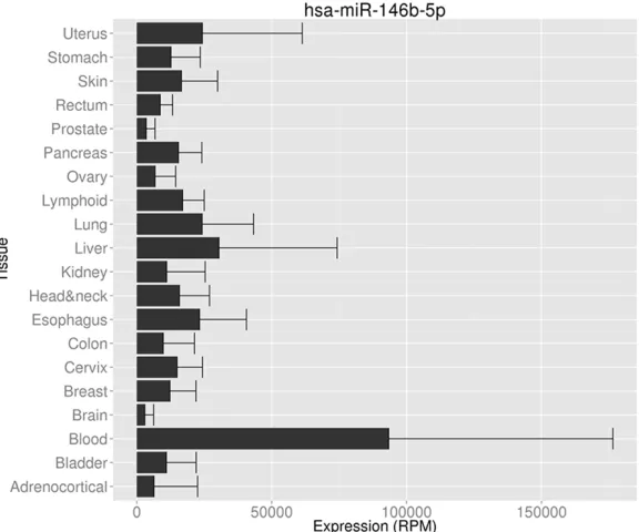
Figure 1. miR-146a-5p expression in different organs and tumors. miR-146a-5p expression data was downloaded from Human MiRNA Expression Database (HMED) (http://bioinfo.life.hust.edu.cn/smallRNA/index.php).
Figure 2. miR-146a-5p expression in different organs. miR-146a-5p expres-sion data were downloaded from YM500v3 (http://driverdb.tms.cmu.edu.tw/ ym500v3/knownmir_sample.php).
Simultaneously, protein levels of the genes
were displayed via The Human Protein Atlas.
levels were significantly higher in cases with
lymph node metastasis than without. Patients
Results
Characteristics of patient
cohort and expression of
miR-146b in PTC tissues
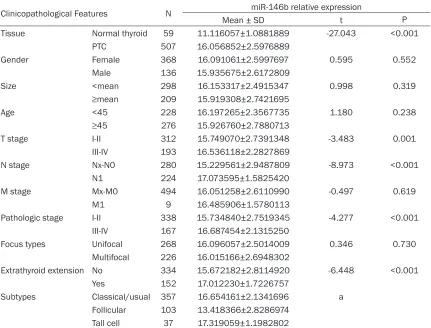
[image:3.612.93.381.398.638.2]Table 1.
Relationship between miR-146b expression and clinicopathological parameters in PTC
pa-tients
Clinicopathological Features N miR-146b relative expression
Mean ± SD t P
Tissue Normal thyroid 59 11.116057±1.0881889 -27.043 <0.001
PTC 507 16.056852±2.5976889
Gender Female 368 16.091061±2.5997697 0.595 0.552
Male 136 15.935675±2.6172809
Size <mean 298 16.153317±2.4915347 0.998 0.319
≥mean 209 15.919308±2.7421695
Age <45 228 16.197265±2.3567735 1.180 0.238
≥45 276 15.926760±2.7880713
T stage I-II 312 15.749070±2.7391348 -3.483 0.001
III-IV 193 16.536118±2.2827869
N stage Nx-N0 280 15.229561±2.9487809 -8.973 <0.001
N1 224 17.073595±1.5825420
M stage Mx-M0 494 16.051258±2.6110990 -0.497 0.619
M1 9 16.485906±1.5780113
Pathologic stage I-II 338 15.734840±2.7519345 -4.277 <0.001
III-IV 167 16.687454±2.1315250
Focus types Unifocal 268 16.096057±2.5014009 0.346 0.730
Multifocal 226 16.015166±2.6948302
Extrathyroid extension No 334 15.672182±2.8114920 -6.448 <0.001
Yes 152 17.012230±1.7226757
Subtypes Classical/usual 357 16.654161±2.1341696 a
Follicular 103 13.418366±2.8286974
Tall cell 37 17.319059±1.1982802
LSD test: Classical/usual vs. Follicular P<0.001, Classical/usual vs. Tall Cell P=0.086, Follicular vs. Tall Cell P<0.001.
Figure 3. Expression level of precursor miR-146b in thyroid cancer from TCGA. A: NT: non-tumorous tissues; T: tumor. B: ROC curve.
in advanced stages showed higher levels of
miR-146b than those in early stages. Moreover,
cases with extrathyroid extension also had
markedly higher levels of miR-146b compared
[image:4.612.93.520.455.642.2]Figure 4. Prognostic value of miR-146b in thyroid cancer from TCGA. A: Disease-free survival; B: Overall survival.
[image:5.612.92.523.264.673.2]1575
Int J Clin Exp Med 2018;11(3)
:1570-1586
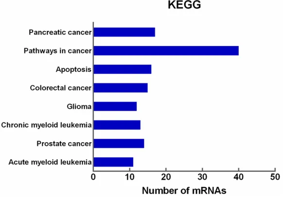
Figure 7. Functional annotation of DAVID KEGG pathways enriched by the putative target mRNAs of miR-146b-5p. The eight most significant signaling pathways are shown.
Optimal candidate targets of miR-146b-5p in
PTC
From the GSE76050 dataset, 6273
differen-tially expressed genes were identified as
poten-tial targets with the analysis described in the
methods. With respect to the twelve miRNA
databases, 4228 genes were identified as
pre-dicted targets of miR-146b-5p. Taking the
inter-section of the two sets of candidates, 994
genes were considered to be optimal candidate
targets of miR-146b-5p in PTC.
Functional annotation of target genes
The PPI network consisted of a total of 249
nodes and 180 edges. The following genes with
a degree value greater than six were identified
as hub genes: CBL (degree=14), CRK (degree=
9), FURIN (degree=9), JAK2 (degree=8), FLT3
(degree=8), PLCG1 (degree=8), DOK1 (degree=
7), CDK5 (degree=7), CSK (degree=7), and PIA
S4 (degree=7).
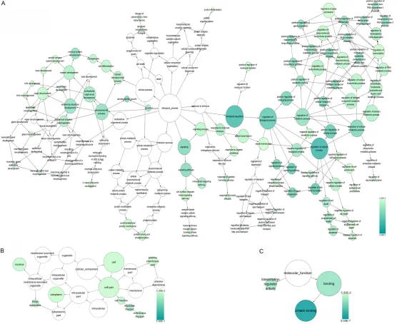
According to the results of GO analysis, the
tar-gets of miR-146b-5p were significantly involved
in biological processes including intracellular
signaling cascade, regulation of programmed
cell death, and positive regulation of cellular
biosynthetic processes. Among the cellular
components, the targets were most enriched in
the insoluble fraction, cell fraction, and
mem-brane fraction. In terms of molecular function,
[image:7.612.91.382.74.277.2]the targets were significantly
associated with
transcrip-tional regulator activity and
transcriptional activator ac-
tivity (
Figures 5
and
6
).
Moreover, KEGG pathway
analysis indicated that the
targets of miR-146b-5p were
closely associated with 36
signaling pathways,
specifi-cally Pancreatic cancer, Pa-
thways in cancer, and Apo-
ptosis (
Figure 7
). These po-
tential targets of
miR-146b-5p from the top three KEGG
pathways were shown in
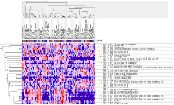
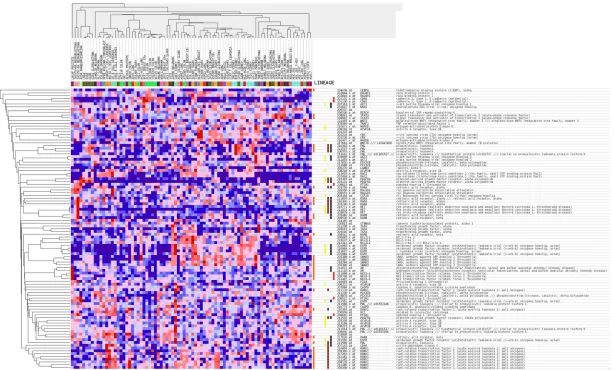
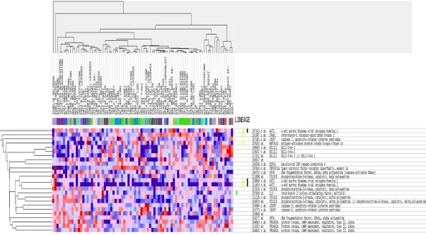
heatmaps by GSEA (
Figures
8
-
10
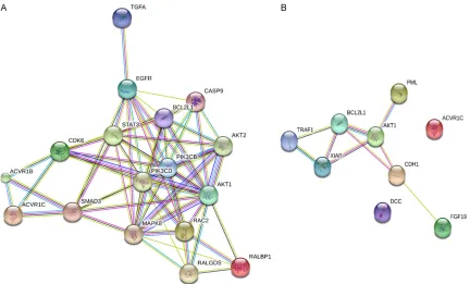
). The PPI maps in
Figure 11
showed the target
genes within the two most
significant pathways from
KEGG analysis. We next
fur-ther validated the nine genes in the PPI of
‘Pathways in cancer’, as this is the key pathway
associated with all malignancies. Interestingly,
two genes, TRAF1 (
Figure 12
) and PML (
Figure
13
), showed down-regulation in their mRNA
lev-els as compared to non-cancerous controls.
Consistently, their protein levels were also
decreased as shown by The Human Protein
Atlas (
Figures 14
and
15
). However, due to the
limited number of cases from The Human
Protein Atlas, these differences were not
statis-tically significant.
Discussion
compre-1577
Int J Clin Exp Med 2018;11(3)
:1570-1586
1579
Int J Clin Exp Med 2018;11(3)
:1570-1586
Figure 11. Protein-protein interaction (PPI) network of selected miR-146b-5p target genes. A: Genes from the path-way ‘Pancreatic cancer’; B: Genes from the pathpath-way ‘Pathpath-ways in cancer’.
hensive understanding of the molecular
mech-anisms behind miR-146b-5p in PTC [37-40].
Our set of 994 probable candidate target genes
of miR-146b-5p originated from the
intersec-tion of genes found in the GSE76050 dataset
comparing miR-146b-5p and mock-transfected
cells, with genes identified by the twelve online
prediction programs, to increase the probability
that these genes were truly target genes of
miR-146b-5p in PTC. Among the numerous target
genes of miR-146b-5p, hub genes with
exten-sive connections with other target genes might
be the most crucial downstream molecules,
thus focusing on the hub genes of miR-146b-5p
targets will allow us to dive deeper into the
underlying mechanism. The hub genes
identi-fied from the PPI network revealed potential
key targets in the pathogenesis of
miR-146b-5p-related PTC. Some of the hub genes,
includ-ing CBL, CrK, Furin, and CDK5, play pivotal
roles in regulating multiple biological processes
through signaling transduction or interactions
with their corresponding substrates. The CBL
family, a class of ubiquitin ligases, might act as
tumor suppressors in human cancers by
ubiqui-tinating active RTKs, promoting their
subse-quent degradation [41]. CrK is a signaling
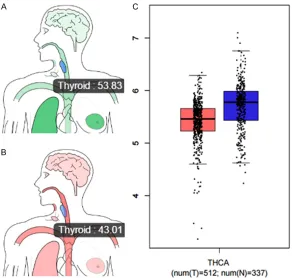
Figure 12. mRNA levels of the hub gene TRAF1 from TCGA data. Median ex-pression of TRAF1 in the normal thyroid gland (A) and tumor samples (B) in bodymap. (C) Gene expression of TRAF1 across all tumor samples (Red) and paired normal tissues (Blue) of thyroid cancer. Bar heights represent the medi-an expression from tumors or normal tissue. (Bar plot, Log2(TPM + 1) Scale). Data were downloaded from GEPIA.
Figure 13. mRNA levels of the hub gene PML from TCGA data. Median expres-sion of PML in the normal thyroid gland (A) and tumor samples (B) in
[image:12.612.90.382.427.705.2]Figure 14. Protein level of the hub gene TRAF1 from The Human Protein At-las data. TRAF1 protein was detected with the antibody HPA001852, and the staining pattern indicated cytoplasmic/membranous localization. A-D: Thyroid gland (T-96000), Male, age 61, Normal tissue, NOS (M-00100), Patient ID: 2072. Staining: Medium, Intensity: Moderate. E-H: Thyroid gland (T-96000), Male, age 20, Papillary adenocarcinoma, NOS (M-82603), Patient ID: 688. Staining: Not detected, Intensity: Negative. A, C, E, G: x100; B, D, F, H: x400.
degrees (degree≥1), which
suggests that these genes
may play key roles in their
corresponding signaling pa-
thways to contribute to PTC
development. It is necessary
to highlight two key genes,
TRAF1 and PML, which have
great potential to be real
tar-gets of miR-146b-5p in PTC
as shown by the
down-regu-lation of their mRNA and
pro-tein levels as assessed by
TCGA and The Human Pro-
tein Atlas data. This
identifi-cation of relevant pathways
and hub genes may help us
to achieve a deeper
under-standing of the underlying
molecular mechanisms of
miR-146b-5p in PTC.
In conclusion, miR-146b-5p
may play an essential role in
PTC by regulating specific
target genes and signaling
pathways related to cancer,
promoting the malignant
potential of PTC. Future ex-
perimental work is needed
to validate these hub genes
and signaling pathways.
Acknowledgements
Figure 15. Protein level of the hub gene PML from The Human Protein At-las data. PML protein was detected with the antibody HPA008312, and the staining pattern indicated cytoplasmic/membranous localization. A-D: Thyroid gland (T-96000), Female, age 44, Normal tissue, NOS (M-00100), Patient ID: 3005. Staining: High, Intensity: Strong. E-H: Thyroid Thyroid gland (T-96000), Male, age 75, Follicular adenoma carcinoma, NOS (M-83303), Patient ID: 3107. Staining: Low, Intensity: Weak. A, C, E, G: x100; B, D, F, H: x400.
Disclosure of conflict of
interest
None.
Address correspondence to:
Wei Ma, Department of Pa- thology, The First Affiliated Hos- pital of Guangxi Medical Uni- versity, 6 Shuangyong Road, Nanning, Guangxi Zhuang Auto- nomous Region, People’s Re- public of China. E-mail: mawei_ [email protected]; Hong Yang, Department of Medical Ultra- sonography, The First Affiliated Hospital of Guangxi Medical University, 6 Shuangyong Road, Nanning, Guangxi Zhuang Auto- nomous Region, People’s Re- public of China. E-mail: [email protected]
References
[1] Yin DT, Yu K, Lu RQ, Li X, Xu J and Lei M. Prognostic impact of minimal extra-thyroidal extension in papillary thyroid carcino-ma. Medicine (Baltimore) 2016; 95: e5794.
[2] Dong S, Meng X, Xue S, Yan Z, Ren P and Liu J. mi-croRNA-141 inhibits thy-roid cancer cell growth and metastasis by target-ing insulin receptor sub-strate 2. Am J Transl Res 2016; 8: 1471-1481. [3] Fagin JA and Wells SA Jr.
Biologic and clinical per-spectives on thyroid can-cer. N Engl J Med 2016; 375: 1054-1067.
[4] Pradhan D, Sharma A and Mohanty SK. Cribriform-morular variant of papil-lary thyroid carcinoma. Pathol Res Pract 2015; 211: 712-716.
[image:14.612.93.383.68.636.2]Clini-cal significance of prophylactic central compartment neck dissection in the treatment of clinically node-negative papillary thyroid cancer patients. World J Surg Oncol 2016; 14: 247.
[6] Shen J, Wang S, Zhao X, Shao X, Jiang X, Dai Y, Xu S and Pan X. Skull metastasis from follicular thyroid carcinoma: report of three cases and review of literature. Int J Clin Exp Pathol 2015; 8: 15285-15293.
[7] Zhao S, Li L, Wang S, Yu C, Xiao B, Lin L, Cong W, Cheng J, Yang W, Sun W and Cui S. H2O2 treatment or serum deprivation induces au-tophagy and apoptosis in naked mole-rat skin fibroblasts by inhibiting the PI3K/Akt signaling pathway. Oncotarget 2016; 7: 84839-84850. [8] Sun W, Lan X, Zhang H, Dong W, Wang Z, He L,
Zhang T and Liu S. Risk factors for central lymph node metastasis in CN0 papillary thy-roid carcinoma: a systematic review and meta-analysis. PLoS One 2015; 10: e0139021. [9] Lee YS, Kim Y, Jeon S, Bae JS, Jung SL and
Jung CK. Cytologic, clinicopathologic, and mo-lecular features of papillary thyroid carcinoma with prominent hobnail features: 10 case re-ports and systematic literature review. Int J Clin Exp Pathol 2015; 8: 7988-7997.
[10] Prescott JD and Zeiger MA. The RET oncogene in papillary thyroid carcinoma. Cancer 2015; 121: 2137-2146.
[11] Cong D, He M, Chen S, Liu X, Liu X and Sun H. Expression profiles of pivotal microRNAs and targets in thyroid papillary carcinoma: an anal-ysis of the cancer genome atlas. Onco Targets Ther 2015; 8: 2271-2277.
[12] Penna GC, Vaisman F, Vaisman M, Sobrinho-Simoes M and Soares P. Molecular markers involved in tumorigenesis of thyroid carcino-ma: focus on aggressive histotypes. Cytogenet Genome Res 2016; 150: 194-207.
[13] Saiselet M, Pita JM, Augenlicht A, Dom G, Tara-bichi M, Fimereli D, Dumont JE, Detours V and Maenhaut C. miRNA expression and function in thyroid carcinomas: a comparative and criti-cal analysis and a model for other cancers. Oncotarget 2016; 7: 52475-52492.
[14] Hua K, Jin J, Zhang H, Zhao B, Wu C, Xu H and Fang L. MicroRNA-7 inhibits proliferation, mi-gration and invasion of thyroid papillary cancer cells via targeting CKS2. Int J Oncol 2016; 49: 1531-1540.
[15] Tang W, Liao Z and Zou Q. Which statistical sig-nificance test best detects oncomiRNAs in can-cer tissues? An exploratory analysis. Oncotar-get 2016; 7: 85613-85623.
[16] Sekhon K, Bucay N, Majid S, Dahiya R and Saini S. MicroRNAs and epithelial-mesenchy-mal transition in prostate cancer. Oncotarget 2016; 7: 67597-67611.
[17] Micolucci L, Akhtar MM, Olivieri F, Rippo MR and Procopio AD. Diagnostic value of microR-NAs in asbestos exposure and malignant me-sothelioma: systematic review and qualitative meta-analysis. Oncotarget 2016; 7: 58606-58637.
[18] Titov SE, Ivanov MK, Karpinskaya EV, Tsivlikova EV, Shevchenko SP, Veryaskina YA, Akhmerova LG, Poloz TL, Klimova OA, Gulyaeva LF, Zhimu-lev IF and Kolesnikov NN. miRNA profiling, de-tection of BRAF V600E mutation and RET-PTC1 translocation in patients from novosibirsk oblast (Russia) with different types of thyroid tumors. BMC Cancer 2016; 16: 201.
[19] Vitiello M, Valentino T, De Menna M, Crescenzi E, Francesca P, Rea D, Arra C, Fusco A, De Vita G, Cerchia L and Fedele M. PATZ1 is a target of miR-29b that is induced by Ha-Ras oncogene in rat thyroid cells. Sci Rep 2016; 6: 25268. [20] Li JH, Zhang SQ, Qiu XG, Zhang SJ, Zheng SH
and Zhang DH. Long non-coding RNA NEAT1 promotes malignant progression of thyroid car-cinoma by regulating miRNA-214. Int J Oncol 2017; 50: 708-716.
[21] Li Z, Huang X, Xu J, Su Q, Zhao J and Ma J. miR-449 overexpression inhibits papillary thyroid carcinoma cell growth by targeting RET kinase-beta-catenin signaling pathway. Int J Oncol 2016; 49: 1629-1637.
[22] Panebianco F, Mazzanti C, Tomei S, Aretini P, Franceschi S, Lessi F, Di Coscio G, Bevilacqua G and Marchetti I. The combination of four mo-lecular markers improves thyroid cancer cyto-logic diagnosis and patient management. BMC Cancer 2015; 15: 918.
[23] Lima CR, Geraldo MV, Fuziwara CS, Kimura ET and Santos MF. MiRNA-146b-5p upregulates migration and invasion of different Papillary Thyroid Carcinoma cells. BMC Cancer 2016; 16: 108.
[24] Zhang Y, Xu D, Pan J, Yang Z, Chen M, Han J, Zhang S, Sun L and Qiao H. Dynamic monitor-ing of circulatmonitor-ing microRNAs as a predictive biomarker for the diagnosis and recurrence of papillary thyroid carcinoma. Oncol Lett 2017; 13: 4252-4266.
[25] Ma W, Zhao X, Liang L, Wang G, Li Y, Miao X and Zhao Y. miR-146a and miR-146b promote proliferation, migration and invasion of follicu-lar thyroid carcinoma via inhibition of ST8SIA4. Oncotarget 2017; 8: 28028-28041.
[26] Wang S, Chen Y and Bai Y. p21 participates in the regulation of anaplastic thyroid cancer cell proliferation by miR-146b. Oncol Lett 2016; 12: 2018-2022.
thyroid cancer with lymph node metastasis. PeerJ 2016; 4: e2119.
[28] Czajka AA, Wojcicka A, Kubiak A, Kotlarek M, Bakula-Zalewska E, Koperski L, Wiechno W and Jazdzewski K. Family of microRNA-146 regulates RARbeta in Papillary Thyroid Carci-noma. PLoS One 2016; 11: e0151968. [29] Xu E, Zhao J, Ma J, Wang C, Zhang C, Jiang H,
Cheng J, Gao R and Zhou X. miR-146b-5p pro-motes invasion and metastasis contributing to chemoresistance in osteosarcoma by target-ing zinc and rtarget-ing ftarget-inger 3. Oncol Rep 2016; 35: 275-283.
[30] Zhu Y, Wu G, Yan W, Zhan H and Sun P. miR-146b-5p regulates cell growth, invasion, and metabolism by targeting PDHB in colorectal cancer. Am J Cancer Res 2017; 7: 1136-1150. [31] Cinegaglia NC, Andrade SC, Tokar T, Pinheiro
M, Severino FE, Oliveira RA, Hasimoto EN, Cat-aneo DC, CatCat-aneo AJ, Defaveri J, Souza CP, Marques MM, Carvalho RF, Coutinho LL, Gross JL, Rogatto SR, Lam WL, Jurisica I and Reis PP. Integrative transcriptome analysis identifies deregulated microRNA-transcription factor networks in lung adenocarcinoma. Oncotarget 2016; 7: 28920-28934.
[32] Li C, Miao R, Liu S, Wan Y, Zhang S, Deng Y, Bi J, Qu K, Zhang J and Liu C. Down-regulation of miR-146b-5p by long noncoding RNA MALAT1 in hepatocellular carcinoma promotes cancer growth and metastasis. Oncotarget 2017; 8: 28683-28695.
[33] Ding HY, Qian WQ and Xu J. MicroRNA-146b acts as a potential tumor suppressor in human prostate cancer. J BUON 2016; 21: 434-443. [34] Yang W, Yu H, Shen Y, Liu Y, Yang Z and Sun T.
MiR-146b-5p overexpression attenuates stem-ness and radioresistance of glioma stem cells by targeting HuR/lincRNA-p21/beta-catenin pathway. Oncotarget 2016; 7: 41505-41526. [35] Chou CK, Liu RT and Kang HY. MicroRNA-146b:
a novel biomarker and therapeutic target for human papillary thyroid cancer. Int J Mol Sci 2017; 18.
[36] Deng X, Wu B, Xiao K, Kang J, Xie J, Zhang X and Fan Y. MiR-146b-5p promotes metastasis and induces epithelial-mesenchymal transi-tion in thyroid cancer by targeting ZNRF3. Cell Physiol Biochem 2015; 35: 71-82.
[37] Liu X, Song B, Li S, Wang N and Yang H. Identi-fication and functional analysis of the risk mi-croRNAs associated with cerebral low-grade glioma prognosis. Mol Med Rep 2017; 16: 1173-1179.
[38] Li CY, Xiong DD, Huang CQ, He RQ, Liang HW, Pan DH, Wang HL, Wang YW, Zhu HW and Chen G. Clinical value of miR-101-3p and bio-logical analysis of its prospective targets in breast cancer: a study based on the cancer
genome atlas (TCGA) and bioinformatics. Med Sci Monit 2017; 23: 1857-1871.
[39] Fan Q and Liu B. Identification of a RNA-Seq based 8-Long Non-Coding RNA signature pre-dicting survival in esophageal cancer. Med Sci Monit 2016; 22: 5163-5172.
[40] Zhang YH, Chu C, Wang S, Chen L, Lu J, Kong X, Huang T, Li H and Cai YD. The Use of gene on-tology term and KEGG pathway enrichment for analysis of drug Half-Life. PLoS One 2016; 11: e0165496.
[41] Lv K, Jiang J, Donaghy R, Riling CR, Cheng Y, Chandra V, Rozenova K, An W, Mohapatra BC, Goetz BT, Pillai V, Han X, Todd EA, Jeschke GR, Langdon WY, Kumar S, Hexner EO, Band H and Tong W. CBL family E3 ubiquitin ligases control JAK2 ubiquitination and stability in hematopoi-etic stem cells and myeloid malignancies. Genes Dev 2017; 31: 1007-1023.
[42] Dong G, Kalifa R, Nath PR, Gelkop S and Isakov N. TCR crosslinking promotes Crk adaptor pro-tein binding to tyrosine-phosphorylated CD3ze-ta chain. Biochem Biophys Res Commun 2017; 488: 541-546.
[43] Jaaks P and Bernasconi M. The proprotein con-vertase furin in tumour progression. Int J Can-cer 2017; 141: 654-663.
[44] Pan DH, Zhu ML, Lin XM, Lin XG, He RQ, Ling YX, Su ST, Wickramaarachchi MM, Dang YW, Wei KL and Chen G. Evaluation and clinical sig-nificance of cyclin-dependent kinase5 expres-sion in cervical leexpres-sions: a clinical research study in Guangxi, China. Eur J Med Res 2016; 21: 28.
[45] Yushan R, Wenjie C, Suning H, Yiwu D, Tengfei Z, Madushi WM, Feifei L, Changwen Z, Xin W, Roodrajeetsing G, Zuyun L and Gang C. In-sights into the clinical value of cyclin-depen-dent kinase 5 in glioma: a retrospective study. World J Surg Oncol 2015; 13: 223.
[46] Wei K, Ye Z, Li Z, Dang Y, Chen X, Huang N, Bao C, Gan T, Yang L and Chen G. An immunohisto-chemical study of cyclin-dependent kinase 5 (CDK5) expression in non-small cell lung can-cer (NSCLC) and small cell lung cancan-cer (SCLC): a possible prognostic biomarker. World J Surg Oncol 2016; 14: 34.
[47] Zhang X, Zhong T, Dang Y, Li Z, Li P and Chen G. Aberrant expression of CDK5 infers poor outcomes for nasopharyngeal carcinoma pa-tients. Int J Clin Exp Pathol 2015; 8: 8066-8074.
[48] Costa V, Esposito R, Ziviello C, Sepe R, Bim LV, Cacciola NA, Decaussin-Petrucci M, Pallante P, Fusco A and Ciccodicola A. New somatic muta-tions and WNK1-B4GALNT3 gene fusion in papillary thyroid carcinoma. Oncotarget 2015; 6: 11242-11251.
Identifica-tion of novel oncogenic mutaIdentifica-tions in thyroid cancer. J Am Coll Surg 2016; 222: 11036-1043, e1032.
[50] Kong X, Sun H, Pan P, Li D, Zhu F, Chang S, Xu L, Li Y and Hou T. How does the L884P muta-tion confer resistance to Type-II inhibitors of JAK2 kinase: a comprehensive molecular mod-eling study. Sci Rep 2017; 7: 9088.
[51] Metts J, Bradley HL, Wang Z, Shah NP, Kapur R, Arbiser JL and Bunting KD. Imipramine blue sensitively and selectively targets FLT3-ITD positive acute myeloid leukemia cells. Sci Rep 2017; 7: 4447.
[52] Zhu GZ, Yang YL, Zhang YJ, Liu W, Li MP, Zeng WJ, Zhao XL and Chen XP. High expression of AHSP, EPB42, GYPC and HEMGN predicts fa-vorable prognosis in FLT3-ITD-Negative acute myeloid leukemia. Cell Physiol Biochem 2017; 42: 1973-1984.
[53] Daver N and Kantarjian H. FLT3 inhibition in acute myeloid leukaemia. Lancet Oncol 2017; 18: 988-989.
[54] Zhu S, Zhang C, Weng Q and Ye B. Curcumin protects against acute renal injury by sup-pressing JAK2/STAT3 pathway in severe acute pancreatitis in rats. Exp Ther Med 2017; 14: 1669-1674.
[55] Kim DH, Park JE, Chae IG, Park G, Lee S and Chun KS. Isoliquiritigenin inhibits the prolifera-tion of human renal carcinoma Caki cells through the ROS-mediated regulation of the Jak2/STAT3 pathway. Oncol Rep 2017; 38: 575-583.
[56] Luo LN, Xie Q, Zhang XG and Jiang R. Osthole decreases renal ischemia-reperfusion injury by suppressing JAK2/STAT3 signaling activa-tion. Exp Ther Med 2016; 12: 2009-2014. [57] Chu YH, Li H, Tan HS, Koh V, Lai J, Phyo WM,
Choudhury Y, Kanesvaran R, Chau NM, Toh CK, Ng QS, Tan PH, Chowbay B and Tan MH. Asso-ciation of ABCB1 and FLT3 polymorphisms with toxicities and survival in asian patients receiving sunitinib for renal cell carcinoma. PLoS One 2015; 10: e0134102.