Original Article
Effects of MAPK/ERK pathway on
learning and memory in sleep deprivation rats
Suyun Miao1,2, Yan Liu3, Ling Zhang4, Min Shan2, Zaijian Miao5
1Department of Neurology, Qianfoshan Hospital Affiliated to Shandong University, Ji’nan, Shandong Province, China; Departments of 2Neurology, 3Nephrology, 4Clinical Oncology, Taian City Central Hospital, Taian, Shandong Province, China; 5Department of Blood Transfusion, Zhucheng People’s Hospital, Zhucheng, Shandong Province, China
Received May 23, 2018; Accepted July 28, 2018; Epub September 15, 2018; Published September 30, 2018
Abstract: Objective: This study aims to analyze the relationship between changes in MAPK/ERK signaling pathway and learning/memory in sleep deprivation rats, and explore the mechanism of memory impairment in those rats. Methods: Sixty male rats were randomly divided into two groups: control group and sleep deprivation group. The sleep deprivation model was prepared using a multi-platform water environment method. The Morris water maze was employed to observe the changes on spatial navigation and spatial search abilities of rats in control group and sleep deprivation group, respectively. Meanwhile, western blot was used to detect MAPK/ERK signaling pathway-related protein changes. Results: With the increasing number of training, the frequency of the search platform was improved gradually. The search time and exploration distance also were decreased in both groups. The escape latency of rats in the sleep deprivation group was significantly longer than that in the control group (P<0.05), and the exploration distance was significantly increased than that in the control group (P<0.05). Comparing the ability of space exploration between the two groups, the percentages of exploration time in the sleep deprivation group on the original platform (1, 2, and 4 quadrants) were significantly lower than those in the control group (all P<0.05). The total number of crossed times on the platform in the control group (12.51±2.14) was significantly higher than that in the sleep deprivation group (6.03±3.07, P<0.001). Western blot results showed that the expression levels of Ras, Raf-1, MEK1/2, ERK1/2, p-ERK1/2, and p-ERK/ERK in the control group were significantly higher than those in the sleep deprivation group. B-Raf expression in the control group was significantly lower than that in the sleep deprivation group. Conclusion: Sleep deprivation can affect learning and memory in rats, and its mechanism may be related to the MAPK/ERK signaling pathway.
Keywords: Sleep deprivation, MAPK/ERK pathway, learning, memory
Introduction
Sleep deprivation (SD) usually refers to the state or process of abnormal sleep rhythm or severe lack of sleep (less than 4 hours per day) [1]. Sleep deprivation can easily lead to the decreased levels of behavior, lack of concentra-tion, memory loss, and decreased learning ability, which can result in serious death [2]. In recent years, studies have found that sleep deprivation can affect body health to varying degrees, but research on the mechanism of the effect of sleep deprivation on learning and memory ability is still limited [3, 4].
The mitogen-activated protein kinase (MAPK) superfamily is a kind of highly evolved enzyme, that is able to associate with intracellular and
extracellular information pathway. The main members of the MAPK family, ERK1/2 and MEK, produce effects when they receive exter-nal stimuli. Then these proteins can transduce extracellular signals into the nucleus and com-plete the regulation of cell functions. MAPK/ ERK signaling pathway is one of the most wi- dely studied signal transduction pathways. It participates in various physiological and pat- hological processes such as cell proliferation, growth, and differentiation [5].
inhibit the generation of LTP, thereby affect the brain’s ability to learn and memory [8]. Therefore, we believe that MAPK/ERK plays an important role in brain learning and memory. In this study, a sleep deprivation rat model was established to observe the behavioral changes in the Morris water maze after sleep deprivation in rats. Simultaneously, the chang-es of the related protein in the hippocampus of the rats were examined to investigate the mechanism of the effects in sleep deprivation on learning and memory abilities in rats.
Materials and methods
Animal information
This study was approved by Experimental Ani- mal Ethics Committee of Zhucheng People’s Hospital. Adult SD male rats (198-274 g, pur-chased from Kay Science and Technology, China) were selected. Adaptive feeding was used during one week, and the rats could eat and drink freely. Then the rats were randomly divided into the control group and the sleep deprivation group using a random sampling method. Each group contained 30 animals. The control group was treated with a platform con-trol system (no rotation of the breeding plat-form). The rats in the sleep deprivation group were subjected to 6 days of sleep deprivation based on the requirements.
Sleep deprivation rat model preparation
Using a sleep deprivation device (SA107, SANS Biological Technology, China), a rat sleep
depri-vation model was prepared according to the instrument operating instructions, and 6 days of continuous sleep deprivation was perform- ed as follows. The experimental rats were pla- ced in a sleep deprivation apparatus to pro- vide adequate food and water. The platform was set to rotate forward and backward, and the running speed was 100 rpm/min. When the rats entered into rapid eye movement (REM) sleep and the whole body muscle tension decreased, the standing platform of the rats moved randomly. Thus, the rats could not enter into out-of-phase sleep at all times. The phenomenon of unresponsiveness, apa- thy, and decreased alertness in rats indicates that the rat model of sleep deprivation was successfully prepared [9].
Morris water maze experiment
The Morris water maze tracking system (WMT-100S, Taimeng, China) was used for position- ing navigation and space exploration experi-ments [10]. Positioning navigation experiment was performed as follows. The experimental training period was 4 days, which was divided into morning and afternoon periods. In each period, the rats were trained 4 times. The plat-form quadrant was randomly selected as the water entry point to record the time that the rats reached the platform from the water (the escape latency). If the rats stayed in the water for more than 90 s, they were guided to the platform artificially, and the escape latency was recorded as 90 s.
After the training, the rats in the control group were reared on the platform, and the rats in the sleep deprivation group were subjected to sleep deprivation for 1, 2, 3, 4 and 5 days. The positioning navigation experiments were performed daily to record the escape latency of the control group and the sleep deprivation group. Space exploration experiment was per-formed as follows. After withdrawing the sleep deprivation platform (6 days), the original quad-rant of the platform was recorded as the target area. The number of times that the rat crossed the original platform in 120 s was measured. The ratio of the time in the original platform’ quadrant to the total time was also recorded.
Western blot determination
[image:2.612.92.288.69.234.2]The expression of key proteins in the MAPK/ ERK pathway in the rat hippocampus was Figure 1. Comparison of escape latency between two
detected [11]. After the completion of the spa- ce exploration experiment in rats, they were sacrificed, and the whole brain were quickly extracted. The bilateral hippocampal tissues were peeled off on ice, frozen in liquid nitro- gen and ground. The total protein was extract-ed with RIPA lysate (P0013D, Beyotime Bio- technology, China).
Protein was quantified according to the proce-dures of BSA protein quantitation kit (PA115-01, Tiangen, China). SDS-PAGE gel electropho-resis was used for 80 min. Then the protein was transferred to membrane. It was blocked with 5% skim milk powder at room tempera- ture for 1 h, and incubated with primary anti-bodies (anti-Ras: SAB4301113, Merck; anti-B-Raf: 7H30L21, Invitrogen; anti-Raf-1: ab1735- 39, Abcam; anti-MEK1/2: ab178876, Abcam; anti-ERK1/2: ab17942, Abcam; anti-p-ERK1/2, 4377, CST; anti-β-actin: ab8226, Abcam) over-night at 4°C. After washing with 1* TBST on the next day (3 times for 5 min each time), the corresponding resistant secondary antibodies (goat anti-rabbit H&L antibody ab6721, Abcam;
J software was performed to evaluate the pro-tein by gray-scale analysis.
Statistical analysis
SPSS 21.0 software was used for statistical analysis. The measurement data was express- ed as mean ± standard deviation (_x ± sd). The measurement data with normal distribution was conducted with t test. The comparison of incubation period and exploration distance between the control group and the sleep de- prived rats were performed using multivaria- te analysis of variance. The difference in the relative gray value between the two groups was determined by t test and expressed by t. P<0.05 indicates statistically significant diffe- rence.
Results
The effect of sleep deprivation on positioning navigation
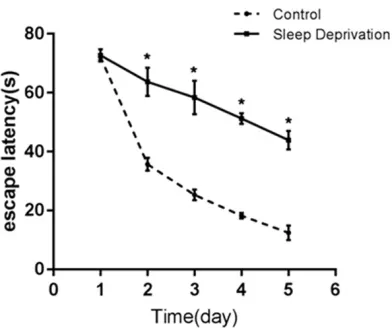
In the positioning navigation experiment, the escape latency of rats in the sleep deprivation group was significantly extended with the pro-longed sleep deprivation time, and the explora-tion distance was also longer. Moreover, the sleep latency of rats in the sleep deprivation group was longer than that in the control group on the 2nd, 3rd, 4th, and 5th day, respectively (all P<0.05, Figure 1). After analyzing and com-paring the exploration distances between the two groups of rats, we found that as the time of sleep deprivation was increased, the explo-ration distances by the rats were decreased. However, the exploration distances in the sleep deprivation group were longer than that in the control group (all P<0.05, Table 1).
The effect of sleep deprivation on space explo -ration
In the space exploration experiment, the per-centage of exploration time in the original
plat-Table 1. Comparing the exploration distances between the control group and the sleep deprivation group
Day Control group (cm) Sleep Deprivation group (cm) t P
1 364.27±290.58 382.76±213.39 -0.281 0.780
2 286.36±74.93 374.83±190.74 -2.365 0.021
3 237.82±82.73 358.23±109.09 -4.817 <0.001
4 176.53±23.96 281.61±37.85 -12.848 <0.001
5 125.73±31.65 193.06±42.12 -7.000 <0.001
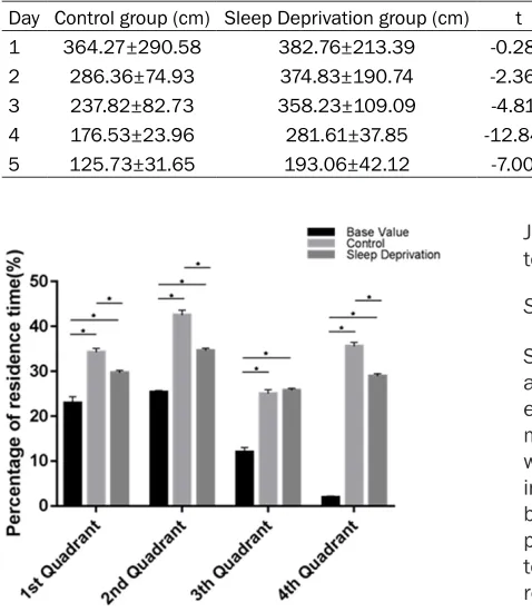
Figure 2. Comparison of the percentages of explo-ration time in the original platform’s quadrants be-tween two groups of rats. *P<0.05.
[image:3.612.89.328.96.371.2]form quadrant of the rats in the control group and the sleep deprivation group were higher than the baseline value before the training. The percentages of exploration time of the rats in the sleep deprivation group on the original platform 1, 2, and 4 quadrants were signifi- cantly lower than that in the control group (all P<0.05). There was no significant difference in the percentage of exploration time between the two groups in the third quadrant (Figure 2). The total number of crossing platforms in the control group (12.51±2.14) was significant-ly higher than that in the sleep deprivation group (6.03±3.07). The difference was statisti-cally significant (t=9.484, P<0.001).
Ras, Raf-1, B-Raf protein expression in rat hip-pocampus
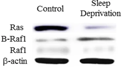
The Ras, Raf-1, and B-Raf proteins in the hip-pocampus of rats in the control and sleep deprivation groups were detected by western blot. The results showed that the expression levels of Ras (t=5.667, P=0.005) and Raf-1
(t=12.41, P<0.001) in the hippocampus of rats in the sleep deprivation group were signifi-cantly lower than the control group. Moreover, the expression level of B-Raf protein was sig-nificantly increased (t=5.024, P=0.007, Fi- gures 3, 4).
Expression of MEK1/2, ERK1/2, p-ERK1/2 proteins in rat hippocampus
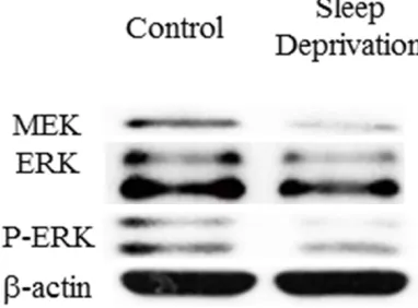
The protein expressions of MEK1/2, ERK1/2 and p-ERK1/2 in hippocampus of rats were detected by western blot. The results show- ed that the expression levels of MEK1/2 (t= 4.114, P=0.015), ERK1/2 (t=5.364, P=0.006), p-ERK1/2 (t=7.538, P=0.002) and, p-ERK1/2 and ERK1/2 ratio (t=3.888, P=0.018) in the hippocampus of rats in the sleep deprivation group were significantly lower than the control group (Figures 5, 6).
Discussion
[image:4.612.88.521.73.209.2]Sleep deprivation usually refers to a state formed by the process of the surrounding environment and its own factors leading to incapable or inadequate sleep [12]. At pre- sent, people’s living rhythm continues to ac- celerate, and the work pressure is gradually increasing. More and more people are being threatened by lack of sleep, sleep restriction, and long-term potential sleep deprivation state [13]. In recent years, studies on epidemiology, human sleep deprivation tests, and animal sleep deprivation experiments have found that people or animals that were in a state of sleep deprivation for a long time can cau- se fatigue, irritability, and sub-healthiness. It Figure 3. Ras, Raf-1, and B-Raf protein expression in hippocampus of rat. A: Expression of Ras protein in two groups; B: Expression of B-Raf protein in two groups; C: Expression of Raf-1 protein in two groups (n=30, experiment re -peated three times). RGV: Relative grey value. *P<0.05.
[image:4.612.90.288.274.384.2]would lead to confusion in thinking, learning and memory, impaired performance and ot- her phenomena [14-18]. The degree of impair-ment in learning and memory ability of humans and animals is proportional to the duration of sleep deprivation [19]. However, the mecha-nism of the effects of sleep deprivation on learning and memory is not yet clear.
As an important extracellular signal transduc-tion pathway, MAPK can transduce signals into
[image:5.612.92.376.70.335.2]As a classic experiment, the Morris water maze was mainly used to analyze the spatial and memory changes of animals [25]. Zhu et al. found that sleep deprivation rats could be studied using Morris water maze test [26]. In that study, the escape latency and exploration distance in sleep deprivation rats were signifi-cantly higher than that in the control group. The percentage of search time on the original plat-form was significantly lower than that of the control group. They confirmed that the spatial memory of rats was significantly reduced after sleep deprivation. This study explored the effect of sleep deprivation on learning and memory in rats through the Morris water maze test. The analysis of rats’ navigation and spa- tial exploration abilities showed that, with the prolonged duration of sleep deprivation, the escape latency of rats in the sleep deprivation group was significantly prolonged than that in the control group. The explored distance was significantly increased than that in the control group. Comparing the ability of space explora-tion, the percentages of exploration time in the original platform’s quadrant of the rats in the control and sleep deprivation group were high-er than the baseline value before the training. Figure 5. Expression of MEK1/2, ERK1/2, p-ERK1/2 proteins in
hippocam-pus of rat. A: MER protein expression in the two groups of rats; B: ERK protein expression in the two groups; C: p-ERK protein expression in the two groups; D: p-ERK and ERK ratio in the two groups (n=30, experiment repeated three times). RGV: Relative grey value. *P<0.05.
Figure 6. Expression of MEK1/2, ERK1/2, p-ERK1/2 proteins in hippocampus of rat on western blot.
[image:5.612.93.284.421.562.2]The percentages of the rats in the sleep depri-vation group on the original platform 1, 2, and 4 exploration time were lower than that in the control group. The total number of crossing the platform was significantly lower than that of the control group. The difference was statisti-cally significant and consistent with the above studies. Therefore, sleep deprivation can lead to a decrease in learning and memory of rats. Ras, the promoter of the MAPK/ERK pathway, is a GTP-binding protein. Ras is activated by signal transduction receptors and can bind with Raf-1 and B-Raf. Then it will activate their functions [27]. Raf-1 and B-Raf can bind to- gether and activate MEK1/2. Then the acti- vated MEK can bind with ERK1/2. Activated ERK1/2 transmits signals into the nucleus and affects the related transcription factors to achieve cell regulation. Zhang et al. used inhibitors to block the MAPK/ERK signaling pathway in rats. In that study, western blot results showed that decreased levels of ERK phosphorylation can influence learning and memory in rats [28].
This study found that the expression of Ras, Raf-1, MEK1/2, ERK1/2, and p-ERK1/2 pro-teins in the MAPK/ERK pathway, the ratio of relative expression level of p-ERK1/2 and ERK1/2 in the sleep-deprived rats were signifi-cantly lower than that in the control group. These results indicated that the MAPK/ERK signaling pathway was inhibited in the sleep deprivation group. The protein expression level of B-Raf in the sleep deprivation group was significantly increased. This was considered as its widespread distribution. Therefore, we believe that the declination of learning and memory in sleep deprivation rats may be relat-ed to inhibition of MAPK/ERK signaling path-way. However, there were still limitations in this study. The sample size was relative small, and we only performed western blot on MAPK/ERK related proteins in this study. Thus, we will examine the expression of MAPK/ERK protein in rat brain tissue by immunohistochemical staining to determine the change.
In conclusion, the effects of sleep deprivation on learning and memory in rats may be relat- ed to the changes of expression of key proteins in the MAPK/ERK pathway in the hippocam- pus.
Disclosure of conflict of interest
None.
Address correspondence to: Zaijian Miao, Depart- ment of Blood Transfusion, Zhucheng People’s Hospital, No.59 Nanhuan Road, Zhucheng 262200, Shandong Province, China. Tel: +86-0536-6212678; E-mail: [email protected]
References
[1] Walker MP. The role of sleep in cognition and emotion. Ann N Y Acad Sci 2010; 1156: 168-197.
[2] Whitney P, Hinson JM, Satterfield BC, Grant DA, Honn KA, Van Dongen HPA. Sleep deprivation diminishes attentional control effectiveness and impairs flexible adaptation to changing conditions. Sci Rep 2017; 7: 214-223.
[3] Touitou Y, Reinberg A, Touitou D. Association between light at night, melatonin secretion, sleep deprivation, and the internal clock: health impacts and mechanisms of circadian disruption. Life Sci 2017; 173: 94-106. [4] Stanojevic C, Simic S, Milutinovic D. Health
ef-fects of sleep deprivation on nurses working shifts. Med Pregl 2016; 69: 183-188.
[5] Zhang G, Cheng Y, Zhang Q, Li X, Zhou J, Wang J, Wei L. ATX-LPA axis facilitates estro-gen-induced endometrial cancer cell proli- feration via MAPK/ERK signaling pathway. Mo-lecular Medicine Reports 2018; 17: 4245-4252.
[6] Yuan LL, Adams JP, Swank M, Sweatt JD, John-ston D. Protein kinase modulation of dendritic K+ channels in hippocampus involves a mito-gen-activated protein kinase pathway. J Neuro-sci 2002; 22: 4860-4868.
[7] Shankar GM, Li S, Mehta TH, Garcia-Munoz A, Shepardson NE, Smith I, Brett FM, Farrell MA, Rowan MJ, Lemere CA, Regan CM, Walsh DM, Sabatini BL, Selkoe DJ. Amyloid-beta protein dimers isolated directly from Alzheimer’s brains impair synaptic plasticity and memory. Nat Med 2008; 14: 837-842.
[8] Watabe AM, Zaki PA, O’Dell TJ. Coactivation of beta-adrenergic and cholinergic receptors en-hances the induction of long-term potentiation and synergistically activates mitogen-activated protein kinase in the hippocampal CA1 region. J Neurosci 2000; 20: 5924-5931.
[9] Gessa GL, Pani L, Fadda P, Fratta W. Sleep de-privation in the rat: an animal model of mania. Eur Neuropsychopharmacol 1995; 5 Suppl: 89-93.
forms of learning and memory. Nat Protoc 2006; 1: 848-858.
[11] Roberts PJ, Der CJ. Targeting the Raf-MEK-ERK mitogen-activated protein kinase cascade for the treatment of cancer. Oncogene 2007; 26: 3291-3310.
[12] Krause AJ, Simon EB, Mander BA, Greer SM, Saletin JM, Goldstein-Piekarski AN, Walker MP. The sleep-deprived human brain. Nat Rev Neu-rosci 2017; 18: 404-418.
[13] Samatra DPGP, Kesanda IMP, Adnyana IMO, Widyadharma E. The effect of partial sleep de-privation in decrease of cognitive function in resident doctors of Udayana University/San-glah general hospital. International Journal of Science & Research 2017; 6: 215-218. [14] Institute of Medicine (US) Committee on Sleep
Medicine and Research, Colten HR, Altevogt BM. Sleep disorders and sleep deprivation: an unmet public health problem. Journal of the American Academy of Child & Adolescent Psy-chiatry 2008; 47: 473-474.
[15] Ben Simon E, Maron-Katz A, Lahav N, Shamir R, Hendler T. Tired and misconnected: a breakdown of brain modularity following sleep deprivation. Hum Brain Mapp 2017; 81: 3300-3314.
[16] Wadhwa M, Kumari P, Chauhan G, Roy K, Alam S, Kishore K, Ray K, Panjwani U. Sleep depriva-tion induces spatial memory impairment by altered hippocampus neuroinflammatory re -sponses and glial cells activation in rats. J Neuroimmunol 2017; 312: 38-48.
[17] Qiu H, Zhong R, Liu H, Zhang F, Li S, Le W. Chronic sleep deprivation exacerbates learn-ing-memory disability and Alzheimer’s dis- ease-like pathologies in AβPP(swe)/PS1(ΔE9) mice. J Alzheimers Dis 2016; 50: 669-685. [18] Chaput JP, Dutil C. Lack of sleep as a
contribu-tor to obesity in adolescents: impacts on eat-ing and activity behaviors. Int J Behav Nutr Phys Act 2016; 13: 103.
[19] Hagewoud R, Whitcomb SN, Heeringa AN, Havekes R, Koolhaas JM, Meerlo P. A time for learning and a time for sleep: the effect of sleep deprivation on contextual fear condition-ing at different times of the day. Sleep 2010; 33: 1315-1322.
[20] Hatzivassiliou G, Song K, Yen I, Brandhuber BJ, Anderson DJ, Alvarado R, Ludlam MJ, Stokoe D, Gloor SL, Vigers G, Morales T, Aliagas I, Liu B, Sideris S, Hoeflich KP, Jaiswal BS, Seshagiri S, Koeppen H, Belvin M, Friedman LS, Malek S. RAF inhibitors prime wild-type RAF to activate the MAPK pathway and enhance growth. Na-ture 2010; 464: 431-435.
[21] Kolch W. Coordinating ERK/MAPK signalling through scaffolds and inhibitors. Nat Rev Mol Cell Biol 2005; 6: 827-837.
[22] Visochek L, Grigoryan G, Kalal A, Milshtein-Pa-rush H, Gazit N, Slutsky I, Yeheskel A, Shain-berg A, Castiel A, Seger R, Langelier MF, Dantzer F, Pascal JM, Segal M, Cohen-Armon M. A PARP1-ERK2 synergism is required for the induction of LTP. Sci Rep 2016; 6: 24950. [23] Chen X, Wang Y, Xu F, Wei X, Zhang J, Wang C,
Wei H, Xu S, Yan P, Zhou W, Mody I, Xu X, Wang Q. The rapid effect of bisphenol-A on long-term potentiation in hippocampus in-volves estrogen receptors and ERK activation. Neural Plast 2017; 2017: 5196958.
[24] Tanasic S, Mattusch C, Wagner EM, Eder M, Rupprecht R, Rammes G, Di Benedetto B. Desipramine targets astrocytes to attenuate synaptic plasticity via modulation of the ep- hrin A3/EphA4 signaling. Neuropharmacology 2016; 105: 154-163.
[25] D’Hooge R, De Deyn PP. Applications of the Morris water maze in the study of learning and memory. Brain Res Brain Res Rev 2001; 36: 60-90.
[26] Zhu C, Yao X, Zhang W, Song Y, Hou Y. Progres-sive paradoxical sleep deprivation impairs par-tial memory following learning tasks in rats. Neural Regen Res 2008; 3: 598-603.
[27] Athuluri-Divakar SK, Vasquez-Del Carpio R, Dutta K, Baker SJ, Cosenza SC, Basu I, Gupta YK, Reddy MV, Ueno L, Hart JR, Vogt PK, Mul-holland D, Guha C, Aggarwal AK, Reddy EP. A small molecule RAS-mimetic disrupts RAS association with effector proteins to block sig-naling. Cell 2016; 165: 643-655.