Original Article
Correlation analysis of spontaneous lumbar curve
correction with cross-sectional rotational
deformity after fusion surgery for Lenke
1 adolescent idiopathic scoliosis (AIS)
Yong Cao1, Dong Jiang1, Lidong Li1, Jianwei Zhu2, Shujun Lu1
1Department of Orthopedics, Hai’an Hospital Affiliated to Nantong University, Hai’an, Nantong, Jiangsu, China; 2Department of Orthopedics, Affiliated Hospital to Nantong University, Nantong, Jiangsu, China
Received November 2, 2017; Accepted April 4, 2018; Epub August 15, 2018; Published August 30, 2018
Abstract: We aimed to investigate the relationship between rotational deformity and spontaneous correction of lumbar curve in cases of Lenke 1 adolescent idiopathic scoliosis (AIS), when position relation between the lowest in-strumented vertebra and stable vertebra remains relatively unchanged. A total of 53 patients with Lenke 1 AIS, with right thoracic curve, were diagnosed in our hospital from March 2010 to March 2014. These were retrospectively reviewed. Before surgery, antero-posterior (AP) lateral and side-bending (SB) radiographs of full-length spines in the standing position were collected. More than 2 years of follow up was conducted for all patients. Coronal balance parameters presenting as center sacral vertical line (CSVL), thoracic and lumbar apical vertebral translation (AVT), Cobb angles of thoracic and lumbar curves, sagittal plane parameters, and other indexes of patients were recorded. Perdriolle method was used to measure total rotations of all vertebrae of thoracic and lumbar curves in full-length spine radiographs, in the standing position, and SB radiographs. Thoracic and lumbar curve corrections and other indexes relating to balance, at 2 years after surgery, were calculated. Lenke 1 AIS was divided into two types: Lenke 1-L type and Lenke 1-R type. Furthermore, Lenke 1-L and Lenke 1-R AIS were divided into subgroup A and subgroup B, respectively. Pearson’s correlation analysis was performed for spontaneous lumbar curve correction and other parameters, at 2 years after surgery, in each group. In this research, it was found that among patients with Lenke 1-L AIS, lumbar curve correction (%) in subgroup B was significantly higher than in subgroup A. Among patients with Lenke 1-R AIS, lumbar vertebra derotation percentage in SB radiographs of subgroup B was obviously increased compared with that of subgroup A. In Lenke 1-L A and B subgroups, spontaneous lumbar curve correction at 2 years after surgery was negatively correlated with preoperative lumbar vertebra rotation (LVR) and positively related to the main thoracic curve correction. Among patients with Lenke 1-R AIS, it was only found in subgroup A that total LVR was associated with spontaneous correction. Additionally, spontaneous lumbar curve correction had a significant correlation with thoracic curve correction in subgroups A and B. Among patients with Lenke 1-L AIS, spontaneous lumbar curve correction was negatively correlated with rotational deformity degree of lumbar curve. However, as fused segments got close to and exceeded SV, negative correlation gradually weakened. Among patients with Lenke 1-R AIS, there was no definite correlation of rotational deformity of lumbar curve with spontaneous correction but spontaneous lumbar curve correction was more easily affected by thoracic curve correction.
Keywords:Adolescent idiopathic scoliosis, lumbar curve, thoracic curve, rotational deformity
Introduction
Adolescent idiopathic scoliosis (AIS) is the most common type of scoliosis among patients, ac- counting for 80%. Moreover, most deformities are located in the thoracic spine of patients. Although idiopathic scoliosis seldom causes obvious symptoms in adolescents, chest and back pain and other symptoms occur in
adult-hood because of decompensation of the three-dimensional structural balance of the spine, es- pecially sagittal decompensation. This can seri-ously affect patient quality of life [1].
et al. [3] proposed a new typing system. This system included sagittal plane parameters allowing surgeons to formulate fusion strate-gies, before surgery, that are more consistent with individual requirements and decreasing errors caused by incorrect identification of sco-liosis type in the process of selective fusion. Lenke reviewed postoperative correction effe- cts in patients receiving selective fusion and then proposed that selective fusion was safe when the ratio of the thoracic apical vertebral rotation to lumbar apical vertebral rotation was higher than 1.0 [4]. Other researchers have also put forward the concept of neutral verte-bra, holding that spinal stability after fusion was correlated with degree of rotation and dis-tortion of the fused lower intervertebral disc [5]. However, among AIS patients receiving se- lective fusion, the impact of rotational deformi-ty and derotation effect of lumbar vertebra on spontaneous lumbar curve correction still re- mains unclear, when lumbar curve Cobb angles and lumbar apical vertebral translation (LAVT) are the same.
In order to discuss influence of selective fusion on the spine within the same typing system, Lenke 1 AIS patients were divided into Lenke
1-L type and Lenke l-R type, according to meth-ods put forward by Miyanji et al. [6]. The rela-tionship between degree of axial rotational de- formity of the lumbar curve and its spontane-ous correction was studied in the two types, as the relationship between the lowest instru-mented vertebra (LIV) and stable vertebra (SV) was fixed.
Patients and methods
General information
In this retrospective analysis, 53 patients with Lenke 1 adolescent idiopathic scoliosis, with right thoracic curve, were diagnosed in our ho- spital from March 2010 to March 2014. This study was approved by the Ethics Committee of Hai’an Hospital Affiliated to Nantong University. Signed written informed consent was obtained from all participants. Before surgery, conven-tional antero-posterior (AP) lateral and side-bending (SB) radiographs of full-length spines, in standing position, were collected. All patients with abnormalities of the nervous system (such as diastematomyelia and Chaffs deformity) in spinal magnetic resonance imaging (MRI) were excluded. All patients underwent posterior-only pedicle screw fixation, of which, 8 patients had the operated segment ended at T11, 25 pa- tients at T12, and 20 patients at L1. The dura-tion of follow up was more than 2 years (27-132 months, with an average of 39.5 months) and average age of patients at the time of the surgery was 15.2 years old. Coronal balance parameters, presenting as distance from C7 vertebra to center sacral vertical line (CSVL), thoracic and lumbar apical vertebral transla-tion (AVT), thoracic Cobb angle, lumbar Cobb angle, sagittal plane parameters, and other in- dexes of the patients, were recorded. Thoracic and lumbar curve corrections and other index-es relating to balance were measured at 3 months and 2 years after surgery, respectively. Grouping
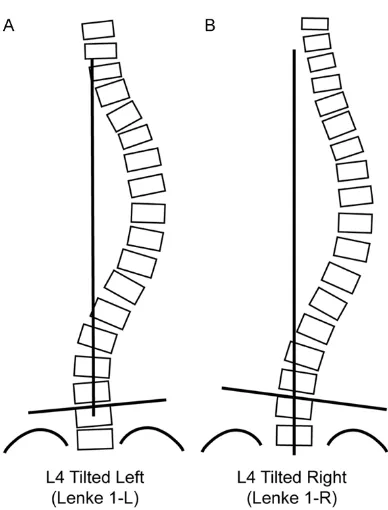
[image:2.612.91.285.65.320.2]According to methods described by Miyanji et al. [6], among Lenke 1 AIS patients, if the L4 vertebra tilted to the left, the curve would be defined as left lateral bending and was present-ed as Lenke l-L. If the L4 vertebra tiltpresent-ed to the right, the curve would be defined as Lenke l-R (Figure 1). When L4 vertebra was in the neutral position without tilt, tilt direction was assessed
based on the the proximal vertebra. Patients with Lenke 1A-R AIS were identical to those with King/Moe IV AIS. There were 33 patients with Lenke 1A-LAIS and 20 with Lenke 1A-RAIS. In the two groups, patients were further divided into subgroup A and subgroup B, respectively, in accordance with the relationship between LIV and SV. In subgroup A of Lenke 1-L, LIV= SV-l; in subgroup B: LIV=SV/SV+1. In subgroup A of Lenke 1-R, LIV=SV-2; in subgroup B: LIV= SV/SV-1.
Rotation measurement
Among all patients with Lenke l-L and Lenke l-R AIS, the Perdriolle method was used, before surgery, to measure total rotations of all verte-brae of thoracic and lumbar curves in full-length spine radiographs, in the standing position, and SB radiographs. Postoperative radiogra- phs, in the standing position [7], were used as well. Meanwhile, total axial lumbar vertebrae derotation in side-bending radiographs were measured and presented as 100 - [100 × SB]/ AP.
Statistical analysis
Parameters in subgroup A and subgroup B of patients with Lenke l-L and Lenke l-R AIS were recorded and analyzed, respectively. Indepen-
subgroup A and the difference was statistically significant (P=0.003). However, there were no statistically significant differences in preopera-tive Cobb angles and correction of thoracic and lumbar curves, total lumbar vertebra rotations (LVRs), CSVLs before and after surgery, AVTs, and sagittal balance parameters between the two groups (Table 1).
Lenke 1-R group
Among patients with Lenke l-R AIS, 13 (65%) had LIV=SV-2 (R-A group) and 7 (35%) had LIV=SV/SV-1 (R-B group). It was indicated that lumbar vertebra derotation percentage in SB radiograph of subgroup B was obviously incre- ased compared with subgroup A and the differ-ence was statistically significant (P=0.028). However, there were no statistically significant differences in preoperative Cobb angles and correction of thoracic and lumbar curves, total LVRs, CSVLs before and after the surgery, AVTs, and sagittal balance parameters between the two groups (Table 2).
Correlation analysis
[image:3.612.92.371.83.322.2]In order to determine the relationship between rotational deformity of lumbar curve and spon-taneous lumbar curve correction, correlation analyses were performed on the ratio of lumbar Table 1. Imaging results of Lenke 1-L group
Parameters Group L-A (n=11) Group L-B (n=22) P Mean SD Mean SD
Preop thoracic 48 8 53 6 NS
Post 2 year thoracic 16 10 19 10 NS
Thoracic curve correction (%) 65 13 61 10 NS
Preop lumbar 25 8 30 7 NS
Lumbar curve correction (%) 23 12 34 13 0.003
Total LVR AP 11 23 16 28 NS
Total LVR SB 9 21 14 26 NS
Total LVR derotation (%) 20 6 15 12 NS
Preop CSVL -0.7 0.8 -0.6 0.7 NS
Post CSVL -1.1 1.5 -0.5 0.8 NS
Preop lumbar AVT 1.3 0.5 1.7 1.2 NS
Post lumbar AVT 0.6 0.3 0.7 0.3 NS
Preop sagittal T5-12 20 8 22 16 NS
Post sagittal T5-12 19 5 17 9 NS
Preop sagittal L1-5 38 9 40 10 NS
Post sagittal L1-5 39 7 35 7 NS
Abbreviation: LVR: Lumbar vertebral rotation. AP: anteroposterior. SB: Sagittal bal-ance. CSVL: Center sacral vertical line. AVT: Apical vertebrate tilt.
dent-samples t-test was used for comparison between sub-group A and subsub-group B. Pear- son’s correlation analysis was performed for spontaneous lumbar curve correction and other parameters, at 2 years after surgery. All data were analyzed using Statistical Pro- duct and Service Solutions (SPSS) 22.0. P<0.05 suggest-ed a statistically significant difference.
Results
Lenke 1-L group
curve correction, in the follow-up 2 years after surgery, to preoperative Cobb angle, total LVR AP, total LVR SB, total LVR derotation in SB radiographs, and other indexes.
Among patients with Lenke l-L AIS, after group-ing in accordance with relationship between fused segments and SV, it was discovered that
with thoracic curve correction (%) was more remarkable than in subgroup A (r=0.609, P< 0.001 VS. r=0.469, P<0.001) (Table 4).
Discussion
[image:4.612.92.374.84.296.2]Idiopathic scoliosis is a complex three-dimen-sional deformity where spinal deformity occurs Table 3. Relationship between spontaneous correction rate of
lum-bar curvature and imaging parameters in Lenke 1-L group after 2-year follow up
Parameters Group L-A Group L-B
Preop thoracic NS NS
Post thoracic NS NS
Thoracic curve correction (%) r=0.381 (P=0.002) r=0.603 (P<0.001)
Preop lumbar NS NS
Total LVR AP r=-0.571 (P<0.001) r=-0.318 (P=0.012) Total LVR SB r=-0.409 (P<0.001) NS Total LVR derotation (%) NS NS
Preop CSVL NS NS
Post CSVL NS NS
Preop lumbar AVT NS NS
Post lumbar AVT NS NS
Preop sagittal T5-12 NS NS Post sagittal T5-12 NS NS Preop sagittal L1-5 NS NS Post sagittal L1-5 NS NS
Abbreviation: LVR: Lumbar vertebral rotation. AP: Anteroposterior. SB: sagittal bal-ance. CSVL: Center sacral vertical line. AVT: Apical vertebrate tilt.
3 patients had trunk decom-pensation. Two years after surgery, in subgroup A (of which the fused segments were located above SV), while total LVR (including total LVR AP + total LVR SB) had sig- nificant correlation with spon-taneous lumbar curve correc-tion (r=-0.571, P<0.001; r= -0.409, P<0.001). Although total LVR AP was significantly correlated with spontaneous lumbar curve correction in su- bgroup B (of which the fus- ed segments were located at or below the SV), the correc-tion degree was decreased compared with that in sub-group A (r=-0.318, P=0.012). This indicates that such cor-relativity was weakened grad-ually as fused segments mo- ved downward along the SV. In subgroup B of Lenke l-L AIS, correlation of spontane-ous lumbar curve correction with thoracic curve correct- ion (%) was more remarkable than that in subgroup A (r= 0.603, P<0.001 VS. r=0.381, P=0.002) (Table 3).
[image:4.612.92.372.376.588.2]Among patients with Lenke l-R AIS, the relationship be- tween total LVR (including total LVR AP + total LVR SB) and spontaneous lumbar cur- ve correction was discovered only in subgroup A, where the fusion was located 2 segme- nts above the SV (r=0.433, P<0.001; r=0.298, P=0.021). In subgroup B of Lenke l-R AIS, correlation of spontane-ous lumbar curve correction Table 2. Imaging results of Lenke 1-R group
Parameters Group R-A (n=13) Group R-B (n=7) P Mean SD Mean SD
Preop thoracic 48 6 50 9 NS
Post 2 year thoracic 18 7 20 6 NS
Thoracic curve correction (%) 60 10 56 7 NS
Preop lumbar 22 9 28 7 NS
Lumbar curve correction (%) 38 11 50 6 NS
Total LVR AP 21 17 13 30 NS
Total LVR SB 21 15 9 27 NS
Total LVR derotation (%) 17 8 33 7 0.028
Preop CSVL -1.2 0.5 -0.8 0.7 NS
Post CSVL -0.7 1.3 -0.4 1.3 NS
Preop sagittal T5-12 18 6 21 10 NS
Post sagittal T5-12 16 7 19 7 NS
Preop sagittal L1-5 38 10 39 13 NS
Post sagittal L1-5 39 10 40 12 NS
in a three-dimensional plane. With constant aggravation of the deformity, wedge-shaped changes are generated in the vertebrae. Many scholars believe that along with development of primary segments of the lateral curve, lateral curve also occurs in compensatory segments of the spine, developing along with the advance of the thoracic curve. It is believed that such lateral curves may be an approach that the spine attempts in order to restore balance. Therefore, if selective fusion is conducted for the thoracic spine when correcting scoliosis, spontaneous correction will occur in the lum-bar spine [8, 9].
In the history of surgical treatment of the spine, fused segments during surgery were first deter-mined to be in accordance with correction in preoperative antero-posterior and lateral radio-graphs. Although it was emphasized at that time that fusion strategies were formulated according to differences in every vertebra [10], postoperative trunk decompensation still can-not be avoided. In 1983, King and Moe intro-duced a kind of typing system specific to AIS, based on imaging. For the first time, this sys-tem helped spine surgeons formulate fusion strategies [2]. They proposed that it was not necessary to fuse to the lower vertebra of the compensatory lumbar curve in scoliosis sur-gery. As for King II scoliosis, it is suitable to merely fuse to the SV of the thoracic curve. The King typing system and its fusion strategies
[11-13]. For this reason, Lenke et al. [3] review- ed cases of the disease and concluded a new kind of AIS typing system. In the Lenke typ- ing system, physicians can determine different Lenke types mainly according to morphology and main curve location in coronal and sagittal planes of the scoliosis, as well as lumbar apical vertebra and thoracic kyphosis correction typ-ing. Patients with Lenke l AIS are the most com-mon. They are utilized to study and analyze se- lective fusion strategies. Lenke defined Lenke 1A scoliosis as the combination of main tho-racic curve with compensatory lumbar curve, of which the pedicle of the apical vertebra does not exceed the opposite side of the CSVL. This includes King III type (type C thoracic curve, of which the compensatory lumbar curve does not exceed the CSVL) and King IV type (long tho-racic curve complicated with the L4 vertebral inclination, entering into the scoliosis), in origi-nal King typing system. The Lenke typing sys-tem not only considers coronal deformity but also includes the measurement of sagittal plane parameters. Lenke typing system has a better intra-observer and inter-observer reli-ability than the King typing system, but there are still a lot of factors that cannot explain the postoperative imbalance in patients for selec-tion of fused segments [14, 15]. As a result, many scholars still currently use the King typing system to investigate selective fusion on idio-pathic scoliosis [16]. In order to integrate the two typing systems into one and make the prep-Table 4. Relationship between compensatory lumbar Cobb Angle
correction and imaging parameters in Lenke 1-R group
Parameters Group R-A Group R-B
Preop thoracic NS NS
Post thoracic r=0.412 (P<0.001) NS
Thoracic curve correction (%) r=0.469 (P<0.001) r=0.609 (P<0.001)
Preop lumbar NS NS
Total LVR AP r=-0.433 (P<0.001) NS
Total LVR SB r=-0.298 (P=0.021) NS
Total LVR derotation (%) NS NS
Preop CSVL NS NS
Post CSVL NS NS
Preop sagittal T5-12 NS NS
Post sagittal T5-12 NS NS
Preop sagittal L1-5 NS NS
Post sagittal L1-5 NS NS
Abbreviation: LVR: Lumbar vertebral rotation. AP: anteroposterior. SB: sagittal balance. CSVL: Center sacral vertical line.
[image:5.612.91.367.96.284.2]aration of selective fusion strategies more pre-cise, Lenke 1 scoliosis was divided into L type and R type, in accordance with methods des- cribed by Miyanji et al. [6]. L type consisted of King II and III type scoliosis and R type includ- ed a part of King IV and V type scoliosis. Rotational deformity of the spine is also an important factor influencing balance during selective fusion. Lenke et al. [17] put forward that selective fusion is safe if the ratio of lum-bar apical vertebral rotation to thoracic capital vertebral rotation is lower than 1.0. Meanwhile, some scholars [18] found in Lenke 3C selective fusion that, compared with other indexes, cor-rection of lumbar apical vertebral rotation in SB radiographs can better predict occurrence of postoperative decompensation.
In this research, we found, among patients with Lenke 1-L AIS, that spontaneous lumbar curve correction was negatively correlated with rota-tional deformity degree of lumbar curve, indi-cating that axial rotational deformity of the spine affects restoration of coronal balance. Moreover, the more severed axial rotational deformity was, the more difficult spontaneous correction would be. However, as fused seg-ments exceeded the SV, such a negative corre-lation was gradually weakened. As early as the days of Harrington, it was found that a vast majority of spinal derotation correction occurs outside fused segments [19]. Although current pedicle screw techniques can have a powerful control on the spine in a three-dimensional plane, the effect of correction with convertible rods on spinal derotation remains fairly limited. Therefore, Suk et al. [20] proposed that rotating neutral vertebrae is a vital factor in selection of distally fused segments to decrease impact of the rotational deformity of the spine on balance restoration. However, when the direct vertebral derotation technique is used to correct thoracic curve, it can be directly ended at the rotated neutral vertebrae [21]. In this research, we found that thoracic vertebrae play a role in direct derotation and axial rotation of the lum-bar curve at the same time, thus lowering the degree of rotational deformity of the lumbar curve and then alleviating impact of rotational deformity on spontaneous correction. However, among patients with Lenke 1A-R AIS, relation-ship between rotational deformity of lumbar curve and spontaneous correction was not de-
tected but spontaneous lumbar curve correc-tion was more affected by thoracic curve cor-rection. Therefore, this group of AIS patients may need different fusion strategies.
Conclusion
Dividing Lenke 1 scoliosis into L type and R type can replace the King typing system more precisely in formulating functions of fused seg-ments in the thoracic curve. In Lenke 1-L AIS, the lower vertebra shall be selected at or below SV when there is a serious rotational deformity. However, in Lenke 1-R AIS, rotational deformity of the lumbar curve has no significant correla-tion with spontaneous correccorrela-tion and sponta-neous lumbar curve correction is more affected by thoracic curve correction. As a result, more attention should be paid to thoracic curve cor-rection and overall balance during fusion.
Disclosure of conflict of interest
None.
Address correspondence to: Dr. Shujun Lu, Depart- ment of Orthopedics, Hai’an Hospital Affiliated to Nantong University, 17 Zhongba Middle Road, Hai’an, Nantong 226000, Jiangsu, China. Tel: +86- 013814615960; E-mail: 764431069@qq.com
References
[1] Lenke LG, Betz RR, Harms J, Bridwell KH, Cle-ments DH, Lowe TG, Blanke K. Adolescent idio-pathic scoliosis: a new classification to deter -mine extent of spinal arthrodesis. J Bone Joint Surg Am 2001; 83-A: 1169-1181.
[2] King HA, Moe JH, Bradford DS, Winter RB. The selection of fusion levels in thoracic idiopa- thic scoliosis. J Bone Joint Surg Am 1983; 65: 1302-1313.
[3] Lenke LG, Betz RR, Haher TR, Lapp MA, Merola AA, Harms J, Shufflebarger HL. Multisurgeon assessment of surgical decision-making in ad-olescent idiopathic scoliosis: curve classifica -tion, operative approach, and fusion levels. Spine (Phila Pa 1976) 2001; 26: 2347-2353. [4] Lenke LG, Edwards CN, Bridwell KH. The Lenke
classification of adolescent idiopathic scolio -sis: how it organizes curve patterns as a tem-plate to perform selective fusions of the spine. Spine (Phila Pa 1976) 2003; 28: S199-S207. [5] Suk SI, Lee SM, Chung ER, Kim JH, Kim SS.
follow-up. Spine (Phila Pa 1976) 2005; 30: 1602-1609.
[6] Miyanji F, Pawelek JB, Van Valin SE, Upasani VV, Newton PO. Is the lumbar modifier useful in surgical decision making?: defining two dis -tinct Lenke 1A curve patterns. Spine (Phila Pa 1976) 2008; 33: 2545-2551.
[7] Perdriolle R, Vidal J. Thoracic idiopathic scolio-sis curve evolution and prognoscolio-sis. Spine (Phila Pa 1976) 1985; 10: 785-791.
[8] Kalen V, Conklin M. The behavior of the un-fused lumbar curve following selective thoracic fusion for idiopathic scoliosis. Spine (Phila Pa 1976) 1990; 15: 271-274.
[9] Large DF, Doig WG, Dickens DR, Torode IP, Cole WG. Surgical treatment of double major scolio-sis. Improvement of the lumbar curve after fu-sion of the thoracic curve. J Bone Joint Surg Br 1991; 73: 121-124.
[10] Goldstein LA. The surgical management of sco-liosis. Clin Orthop Relat Res 1971; 77: 32-56. [11] Mielke CH, Lonstein JE, Denis F, Vandenbrink
K, Winter RB. Surgical treatment of adolescent idiopathic scoliosis. A comparative analysis. J Bone Joint Surg Am 1989; 71: 1170-1177. [12] King HA. Analysis and treatment of type II
idio-pathic scoliosis. Orthop Clin North Am 1994; 25: 225-237.
[13] McCall RE, Bronson W. Criteria for selective fu-sion in idiopathic scoliosis using Cotrel-Du-bousset instrumentation. J Pediatr Orthop 1992; 12: 475-479.
[14] Suk SI, Lee SM, Chung ER, Kim JH, Kim WJ, Sohn HM. Determination of distal fusion level with segmental pedicle screw fixation in single thoracic idiopathic scoliosis. Spine (Phila Pa 1976) 2003; 28: 484-491.
[15] Sheng WB, Zheng XF, Guo HL, Zhan YL, Mai ED, Jin GL, Pu LT, Sheng J, Deng Q. [Intraobserver and interobserver reliability of the King, Lenke, and PUMC classification systems for idiopathic scoliosis]. Zhonghua Yi Xue Za Zhi 2009; 89: 1047-1052.
[16] Parisini P, Di Silvestre M, Lolli F, Bakaloudis G. Selective thoracic surgery in the Lenke type 1A: King III and King IV type curves. Eur Spine J 2009; 18 Suppl 1: 82-88.
[17] Lenke LG, Bridwell KH, Baldus C, Blanke K. Preventing decompensation in King type II curves treated with Cotrel-Dubousset instru-mentation. Strict guidelines for selective tho-racic fusion. Spine (Phila Pa 1976) 1992; 17: S274-S281.
[18] Behensky H, Cole AA, Freeman BJ, Grevitt MP, Mehdian HS, Webb JK. Fixed lumbar apical vertebral rotation predicts spinal decompensa-tion in Lenke type 3C adolescent idiopathic scoliosis after selective posterior thoracic cor-rection and fusion. Eur Spine J 2007; 16: 1570-1578.
[19] Marchesi DG, Transfeldt EE, Bradford DS, Heithoff KB. Changes in vertebral rotation af-ter Harrington and Luque instrumentation for idiopathic scoliosis. Spine (Phila Pa 1976) 1992; 17: 775-780.
[20] Suk SI, Lee SM, Chung ER, Kim JH, Kim WJ, Sohn HM. Determination of distal fusion level with segmental pedicle screw fixation in single thoracic idiopathic scoliosis. Spine (Phila Pa 1976) 2003; 28: 484-491.