Original Article
PiggyBac-modified CD19-expressing 4T1
cell line for the evaluation of CAR construct
Han Hu1*, Runyang Wang1*, Ziyi Zhang1, Haixiao Duan1, Yang Wang1, Jing Jin2, Ying Chen2, Zhen Wu2, Zhizheng Fang2, Binlei Liu1,2
1National “111” Center for Cellular Regulation and Molecular Pharmaceutics, Key Laboratory of Fermentation Engineering (Ministry of Education), Hubei Provincial Cooperative Innovation Center of Industrial Fermentation, College of Bioengineering, Hubei University of Technology, Wuhan, P. R. China; 2Wuhan Binhui Biotechnology Co. Ltd., Wuhan, P. R. China. *Equal contributors.
Received March 20, 2019; Accepted April 23, 2019; Epub July 1, 2019; Published July 15, 2019
Abstract: Reliable and stable target cell lines are required for evaluating the efficiency and studying the mechanism
of chimeric antigen receptor T (CAR-T) immunotherapy both in vitro and in vivo. Jurkat cells can be used as an alter-native for human primary lymphocytes to evaluate the constructs and function of the “CAR”. This study established a murine 4T1-CD19 cell line that stably expressed a cd19 gene. The 4T1-CD19 cells had similar growth kinetics to its parent cell 4T1. The protein CD19 expression of the 4T1-CD19 was detected by reverse transcription-polymerase chain reaction (RT-PCR) and western blot. The second-generation CAR was constructed and transfected into
Jur-kat cells. The expression of CAR protein was analyzed by flow cytometry and western blot. Finally, the interaction between the CAR and CD19 was confirmed by the upregulation of the IL-2 mRNA level of Jurkat-CAR stimulated by 4T1-CD19. Therefore, the 4T1-CD19 cell line and Jurkat-CAR have been successfully established, and may be used to access the function of various CAR constructs both in vitro and in vivo.
Keywords: 4T1, Jurkat, IL-2, murine, CAR-T
Introduction
Autologous T cells are usually modified to
express chimeric antigen receptors (CARs), and these T cells are named CAR-T. These cells are
redirected to killing specific tumor cells. This is
a novel and revolutionary anti-cancer strategy
for tumor immunotherapy and it is specifically efficient against CD19-positive B cell malignan -cies [1-3]. CARs usually consist of an extracel-lular part responsible for the recognition of
specific tumor antigen, and an intracellular seg -ment that can improve cytotoxic and prolifera-tion activity of CAR-T. The binding properties of
CARs to the chosen specific tumor antigen are
normally determined by a single-chain variable fragment (scFv), which is derived from a mo- noclonal antibody. These scFv-derived
frag-ments acquire different binding affinity, and
major histocompatibility complex (MHC)-inde- pendent binding with its ligands are usually
with specific tumor antigens. The clinical trials
of the first-generation CARs were disappointing.
Then, second-generation CARs were develop- ed, with the scFv part combining with a co-stim-ulatory part (often 4-1BB or CD28) and a T cell pro-activator cytotoxic fragment (CD3z) [4, 5]. Recent clinical trials based on anti-CD19 CAR-T cells of second-generation exhibited astonish-ing results in patients with acute lymphoid leu-kemia (ALL), non-Hodgkin’s lymphoma, and chronic lymphocytic leukemia. These clinical results have been summarized by Lorentzen
However, despite these positive data, this T cell-based treatment can cause severe toxici-ties. These engineered CAR-T cells harm non-tumor cells because of on-target/off-non-tumor effect. If this effect occurs in the important organs like heart, lung or liver, then the CAR-T cells with strong cytotoxicity could threaten the
life of the patient [7]. Besides, the killing effi -ciency of CAR-T cell remains disappointing in the settings of solid tumors [8, 9]. Some condi-tions exist that may mitigate the killing capaci- ty of CAR-T against solid tumors; for example, differently expressed tumor-associated anti-gens could cause tumor escape during treat-ment [10]. Thus, the CAR construct should be
optimized to increase its efficiency while simul -taneously decreasing the toxicity. Wu et al. designed the “ON-switch” CARs that enable small molecules to control T cell functions
with-out affecting the antigen specificity [11]. This
method would help physicians to precisely con-trol the timing, distribution, and dosage of
CAR-T, which could improve the efficiency with miti -gated toxicity. In this kind of research, corre-
sponding cancer cells expressing a specific
ligand of the CARs should be established. Us-
ually, the efficiency of the CAR-T cells is evalu -ated in an NSG mice model which lacks autolo-gous immunity. This kind of in vivo model can-not study the involvement of the host immune system during the CAR-T treatment process. So, murine-origin cancer cells expressing cor-responding ligand should be developed as a tool for the evaluation of the CAR construct. We therefore built a strategy to generate a CD19 expressing murine 4T1 cell line, which is a highly metastatic murine adenocarcinoma with rapid growth, derived from a BALB/c mouse with spontaneous mammary tumor [12] and the interaction between CD19 and CAR
was confirmed by the 4T1-CD19 induced acti -vation of Jurkat-CAR. This 4T1-CD19 cell line and Jurkat-CAR reported here will be further used in our lab as a tool to evaluate the CAR construct in a pre-clinical study.
Materials and methods
Construction of anti-CD19 CAR lentiviral vector and PBDP-CD19 vector
The anti-CD19 scFv derived from FMC63 was synthesized (GenScript Biotech, China) and inserted into a lentiviral vector plvx-acgfp-N1. The human CD19 gene was synthesized and
sub-cloned into PiggyBac Dual promoter. The
ultima vectors were confirmed by gene
sequ-encing (Tsingke Biotech, China).
Cell lines
4T1 cells and Human embryonic kidney (HEK) 293T cells were purchased from the Cell Bank of Chinese Academy of Sciences (SGST, China) and maintained in DME/F12 (Hyclone, USA) containing 10% fetal bovine serum (FBS) (Zh- ejiang Tianhang Biotechnology, China). Jurkat cells were obtained from the Cell Bank of Ch- inese Academy of Sciences (SGST, China) and maintained in RPMI-1640 medium (Hyclone, USA) containing 10% FBS. The different kinds of cells were cultured in a cell incubator at 37°C and 5% CO2.
Generation of 4T1-CD19 cell line and CAR expressing Jurkat cells
4T1 cells were transfected with the plasmid PBDP-CD19 and plasmid Super PiggyBac trans-posase using Lipofectamine 3000 (Invitrogen, USA) following the protocol introduction.
CD19-expressing cells were selected with 6 μg/ml of
puromycin (BioFroxx, Germany) diluted in DME/ F12 containing 10% FBS and the selected cul-ture medium was replaced every 2-3 days until most of the 4T1 cells were GFP-positive. Fo- llowing this stage, the cells were digested and seeded in 96-well culture plate at a density 1-2 cells/well to obtain monoclonal 4T1-CD19 ce- lls. The lightest cells were selected for further culture. For the generation of Jurkat-CAR cells, lentivirus was added to the cultures at a mu-
ltiplicity of infection (MOI) of 5, and 6 μg/ml
transduction enhancer polybrene (Solar Bio, China) was added. The culture medium was replaced every 2-3 days and the cell density was maintained at 1-3 × 10⁶ cells/ml.
Production of lentivirus
In the stage of lentivirus’ packaging, 293T ce- lls were transfected with plvx-acgfp-h1928z, psPAX2, and pMD2.G. The packaging plasmids were added at a ratio of 2:1:2. The day before transfection, 293T cells were seeded into a
T75 cell culture flask and maintained in DME/
F12 (Hyclone, USA) containing 10% FBS (Zhe- jiang Tianhang Biotechnology, China). When the
cultured in a cell incubator at 37°C, and 5% CO2. The supernatants containing viral parti-cles were harvested after 48 hours and mixed with Lenti-X™ concentrator (Takara Bio, Japan) overnight at a temperature of 4°C. The next day, the supernatants containing viruses were centrifuged at 1,500 × g for 45 minutes at 4°C, suspended in phosphate buffered saline (PBS) (Jet Bio-Filtration, China), and stored at -80°C for a later experiment.
Flow cytometry
Monoclonal 4T1-CD19 cells were incubated
with fluorescence labeled antibody directed
against human CD19 (BD Pharmingen, USA) for 20 minutes at 4°C and washed once with PBS. The sample analysis was conducted on a BD
C6 flow cytometer and analyzed with FlowJo VX software. Expression of CD19-CAR was verified
according to the GFP positive rate. The untreat-ed Jurkat cells were usuntreat-ed as a control group.
Jurkat-CAR cells function assay
An in vitro assay was conducted to evaluate the
biologic function of Jurkat-CAR cells. Briefly,
4T1-CD19 cells were seeded in a 96-well plate at a density of 104 cells per well. Effector cells and medium were added at an equal volume at an E/T ratio of 8:1 and 16:1. After 24 hour incubation, cells were obtained, the mRNA was extracted and reverse transcribed to cDNA,
and finally stored at -20°C.
Quantitative real-time PCR
To further investigate the expression level of CD19 and IL-2, qPCR was conducted through a StepOne™ Real-Time PCR system (Applied Bio- systems). RNA was extracted with RNAsimple Total RNA Kit (TianGen, China) and reverse tr-
anscribed with HiScript Q RT SuperMix
(Vazy-me, China). The human CD19 gene was detect-ed through the following primers: 5’-TACCT- GATCTTCTGCCTG-3’ and 5’-TCATCCTCTTCCTCT- TCC-3’. The human IL-2 gene was detected th- rough the following primers: 5’-GGACTTAATC- AGCAATATCAA-3’ and 5’-AAGGTAATCCATCTGTT- CA-3’.
Statistical analysis
All results are presented as the mean ± stan-dard deviation (SD). Statistical analysis was performed with GraphPad Prism software ver-sion 5.0. For comparison of the two groups,
two-tailed unpaired t tests were used and P-
values <0.05 were considered significant.
Results
Generation of the CD19 expression 4T1-CD19 cell line
The eukaryotic expression plasmid PBDP-CD- 19, containing puromycin expression cassette, was constructed and the amino acid sequen-
ce was confirmed to have 100% identity with
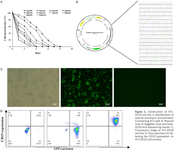
CD19 sequence (NCBI Reference Sequence: NP_001761.3). In order to screen the CD19 expressing 4T1-CD19 cells, the killing curves of 4T1 cell under different concentrations of
puromycin ranging from 1 to 10 μg/ml were
determined (Figure 1A). Following 8 days of puromycin disposal, complete cell death was
observed at 6 μg/ml on the 4th day. The plas -mid PBDP-CD19 and PiggyBac transposase
were both transfected into 4T1 cells, and 6 μg/
ml of puromycin was used for the recombinant cell selection. After 7 days of selection, only 6-9% of 4T1 cells had survived and most of them were GFP positive (Figure 1B). Then, the cells were digested and seeded in 96-wells culture plate to obtain monoclonal 4T1-CD19 cells. After about 4 rounds of passage, the high-ly CD19 expression of 4T1-CD19 cell line was selected with the positive rate being 99.7% (Figure 1C).
Characterization of the 4T1-CD19 cell line
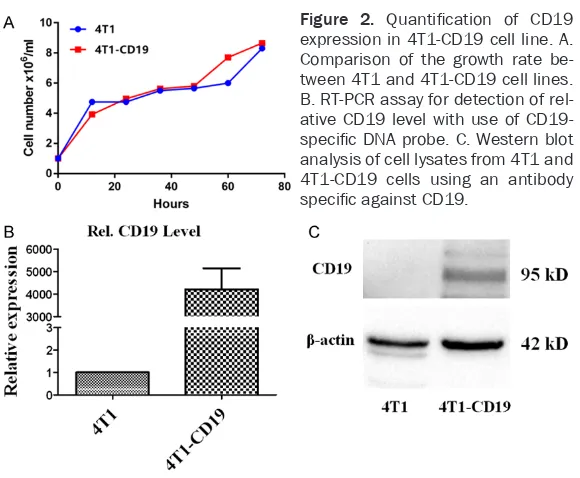
Cell numbers of 4T1 and 4T1-CD19 were deter-mined at different time points. The growth curves of the 4T1-CD19 and 4T1 were depicted according to the results. The curves indicated that CD19 insertion did not affect the prolifera-tion activity of 4T1 (Figure 2A). The expression
of CD19 was further confirmed through RT-PCR and western blotting. CD19 specific primer was
applied to detect the exogenous CD19 mRNA level. The relative expression level of CD19 in
4T1-CD19 was ~4,200 (4,210.86±938.65) fo-ld, which was significantly higher than that of
the control 4T1 (Figure 2B). Immunoblotting
Figure 1. Construction of
4T1-CD19 cell line. A. Identification of
[image:4.792.117.686.67.554.2]Construction and identification of Jurkat-CAR cells
Compared with the first-generation of chimeric
antigen receptor T cells, the second-generation cells additionally possess an anti-CD19 specif-ic scFv linked to a hinge domain, a part of the CD28 costimulatory molecule and cytoplasmic
portion of the TCR-ζ molecule (Figure 3A). The fragment was inserted into a lentiviral vector
system, plvx-acgfp-N1. A green fluorescence
protein (GFP) was added after the CAR to verify
the transfection efficiency. The expression of CD19-CAR was characterized by flow cytometry and western blot. The flow cytometry showed
expression of CD19-CAR on transfected Jurkat cells, but not on the control Jurkat (Figure 3C).
The transfection efficiency of Jurkat-CAR was
over 60% (Figure 3C). Immunoblotting of the Jurkat-CAR lane showed a band between 55-70 kD (calculated molecular weight of CAR), where-as the control Jurkat lane exhibited no band (Figure 3B).
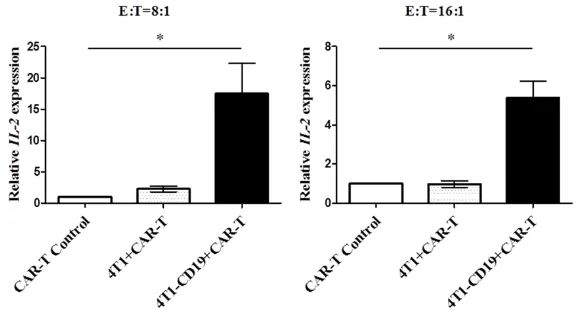
Jurkat-CAR cells produced IL-2 after stimula-tion by 4T1-CD19 cells
To verify the biologic function of CAR and CD- 19 construct, Jurkat cells were incubated with 4T1-CD19 cells at different effector-to-target (E/T) ratio of 8:1 and 16:1 for 12 h, separately. After incubation, the Jurkat cells were collected to conduct RNA extraction. The relative
expres-tion of Jurkat-CAR. 4T1-CD19 could be further used in vitro and in vivo to study both the
func-tional and structural properties of a specific
construct of CARs and its ligands.
CD19 (95 kDa), the B lymphocyte antigen re- ceptor, is an important co-receptor for mem-brane immunoglobulin (mIg). It belongs to the immunoglobulin super family that is express- ed in almost all growth stages of B-cells and
also could be identified on most acute
B-ly-mphoblastic leukemia (ALL) cells, chronic B-ly- mphocytic leukemia (CLL), and non-Hodgkin lymphoma [13]. These properties determined CD19 as the best target for CAR-T therapy against B-lymphoid malignancies. According to the published results, CART-CD19 cells show-
ed specific, effective, and persistent killing
activity against CD19+ tumor cells [14]. Thus, we tried to develop a solid murine tumor 4T1 expressing CD19, which could offer an ideal cell line to study the killing mechanism of CAR-T cells against solid tumors both in vitro and in immunocompetent mice model. PiggyBac (PB),
an efficiently transposon system, has always
been used to generate induced pluripotent stem cells from somatic cells, without gene alterations [15]. It is a highly active transposon derived from the cabbagelooper moth that can provide sustained transgene expression of human T lymphocytes [16]. Thus, we used Pig- gyBac transposon systems as a methodology
for stable genetic modification. The plasmid Figure 2. Quantification of CD19
expression in 4T1-CD19 cell line. A. Comparison of the growth rate be-tween 4T1 and 4T1-CD19 cell lines. B. RT-PCR assay for detection of rel-ative CD19 level with use of
CD19-specific DNA probe. C. Western blot
analysis of cell lysates from 4T1 and 4T1-CD19 cells using an antibody
specific against CD19.
sion level of cytokine IL-2 was detected. Compared with the control group, the expression of IL-2 mRNA was upregulated in Jurkat-CAR co-treated with CD19-4T1, with correspond-ing 17.48 and 5.37-fold in- crease, respectively under dif-ferent E/T ratios (Figure 4).
Discussion
In this study, we established a monoclonal murine cancer cell line, 4T1-CD19. Furth- ermore, the membrane local-ization of CD19 was observed
by immunofluorescence and flow cytometry. Then, the
in-teraction of CD19 and CAR
[image:5.612.90.380.66.305.2]-PBDP-CD19 was introduced into 4T1 cells and positive cells were selected based on the resis-tance to puromycin; the highest level of CD19-expressing cell clone was selected and the 4T1-CD19 cell line was established. The integration
of CD19 into 4T1 was confirmed by the RT-PCR
on mRNA level and western blot on protein level. The passaged 4T1-CD19 cells exhibited similar proliferation characteristics when com-pared with the parent 4T1 cells according to the growth curves.
The 4T1 mammary carcinoma, originally ob- tained by Fred Miller and colleagues, is a me- tastatic tumor cell line [17]. Unlike other tu- mor models, the 4T1 tumor is very tumorige-
Jurkat was obtained from a patient with acute lymphoblastic leukemia [19]. The Jurkat cell line could be used to study the effects of vari-ous natural or synthetic compounds on CD4+ T-cell activation, proliferation, and apoptosis. Wu et al. also reported that Jurkat T cell could be used to evaluate the construction of CAR
and its specific ligand [11]. Jurkat-CAR secretes IL-2 upon its ligand-specific stimulation. In this
study, lentivirus was used to introduce a sec-ond-generation CAR gene into Jurkat cells.
Compared with the first-generation CAR, the
[image:6.612.90.522.67.253.2]second-generation CAR incorporates classical costimulatory cytoplasmic domains, like CD28, 4-1BB (CD137), OX40 and ICOS, either sepa-rately or in combination, which can boost the
Figure 3. h1928z CAR gene structure and characterization of Jurkat-CAR cells. A. Schematic illustration of CAR
containing gene. B. Detection of CAR by immunoblotting assay using an antibody specific to CD3z. CAR presented at ~59 kD. C. Flow cytometry assay to determine positive ratio of Jurkat-CAR cells.
Figure 4. Study of Jurkat-CAR cell function. Jurkat-CAR cell incubation with the 4T1-CD19 cell line in different E:T ratio; IL-2 expression level determined
with IL-2 specific probe.
nic, invasive, and can initial- ly metastasize from the pri-mary sites of mampri-mary gla- nd to different distant sites including blood, liver, lymph nodes, brain, lung, and bo- ne [18]. The 4T1 tumor has several features which mak- es it an appropriate cell line for the study of human mam-mary cancer. To the best of our knowledge, there are lim-ited reports about the ex-
pression of specific protein from gene modification of
[image:6.612.88.376.313.468.2]biologic function and proliferation of CAR-T
[20]. In this study, transfection efficiency of CAR gene into Jurkat cells was analyzed by flow
cytometry. After 20 day culture of Jurkat cells, the GFP could still be detected, which indicates stable expression of CAR gene in Jurkat cells. Then, the Jurkat-CAR was incubated with 4T1-CD19 and 4T1, separately. The real time PCR results of IL2 proved that the CD19 cognate ligand and the CAR had been successfully
con-structed and it was sufficient to trigger IL-2
expression of Jurkat-CAR.
In summary, the monoclonal murine 4T1-CD19 cell line established here not only provided plat-forms for evaluation of a CAR construct in vitro
but in the future could also be used to study the activity of CAR-T against solid tumor in an immu-nocompetent murine model.
Acknowledgements
This work was supported by General Program of Hubei Province Basic Science Foundation
(2015CFB470); National Major Scientific and Technological Special Project for “Significant
New Drugs Development”
(2015ZX09501007-004); National Major Scientific and Technolo-gical Special Project for Significant New Drugs
Development (2018ZX09733002); and Nati- onal Science and Technology Major Project of China (2018ZX09201017-001) awarded to Binlei Liu.
Disclosure of conflict of interest
None.
Abbreviations
CD19, Cluster of Differentiation 19.
Address correspondence to: Dr. Binlei Liu, National “111” Center for Cellular Regulation and Molecular Pharmaceutics, Key Laboratory of Fermentation Engineering (Ministry of Education), Hubei Provincial Cooperative Innovation Center of Industrial Fermen- tation, College of Bioengineering, Hubei University of Technology, Wuhan 430068, P. R. China. Tel: +86-27-87326962; Fax: +86-27-87643065; E-mail: liubl@ hbut.edu.cn
References
[1] Barrett DM, Nathan S, Porter DL, Grupp SA, June CH. Chimeric antigen receptor therapy for cancer. Annu Rev Med 2013; 65: 333-47.
[2] Turtle CJ, Hanafi LA, Berger C, Hudecek M,
Pender B, Robinson E, Hawkins R, Chaney C, Cherian S, Chen X. Immunotherapy of
non-Hodgkin’s lymphoma with a defined ratio of CD8+ and CD4+ CD19-specific chimeric anti
-gen receptor-modified T cells. Sci Transl Med
2016; 8: 355ra116.
[3] Wang X, Popplewell LL, Wagner JR, Naranjo A, Blanchard MS, Mott MR, Norris AP, Wong CW, Urak RZ, Chang WC, Khaled SK, Siddiqi T, Budde LE, Xu J, Chang B, Gidwaney N, Thomas SH, Cooper LJ, Riddell SR, Brown CE, Jensen MC, Forman SJ. Phase 1 studies of central memory-derived CD19 CAR T-cell therapy fol-lowing autologous HSCT in patients with B-cell NHL. Blood 2016; 127: 2980-90.
[4] Chang ZL, Chen YY. CARs: synthetic immunore-ceptors for cancer therapy and beyond. Trends Mol Med 2017; 23: 430-50.
[5] Michel S. CAR therapy: the CD19 paradigm. J Clin Invest 2015; 125: 3392-400.
[6] Lorentzen CL, Straten PT. CD19-chimeric anti-gen receptor T cells for treatment of chronic lymphocytic leukaemia and acute lymphoblas-tic leukaemia. Scand J Immunol 2015; 82: 307-19.
[7] Evripidis L, Mathilde P, Klattenhoff AW, Degang S, Raphael S, June CH, Powell DJ. Chimeric an-tigen receptor T cells with dissociated signal-ing domains exhibit focused antitumor activity with reduced potential for toxicity in vivo. Cancer Immunol Res 2013; 1: 43-53.
[8] Beatty GL, O’Hara M. Chimeric antigen
recep-tor-modified T cells for the treatment of solid tumors: defining the challenges and next
steps. Pharmacol Ther 2016; 166: 30-9. [9] Newick K, Moon E, Albelda SM. Chimeric
anti-gen receptor T-cell therapy for solid tumors. Mol Ther Oncolytics 2016; 3: 16006.
[10] O’Rourke DM, Nasrallah MP, Desai A,
Melen-horst JJ, Mansfield K, Morrissette JJD, Marti -nez-Lage M, Brem S, Maloney E, Shen A, Isaa-cs R, Mohan S, Plesa G, Lacey SF, Navenot J, Zheng Z, Levine BL, Okada H, June CH, Brogdon
JL, Maus MV. A single dose of peripherally in -fused EGFRvIII-directed CAR T cells mediates antigen loss and induces adaptive resistance in patients with recurrent glioblastoma. Sci Transl Med 2017; 9.
[11] Wu CY, Roybal KT, Puchner EM, Onuffer J, Lim WA. Remote control of therapeutic T cells through a small molecule-gated chimeric re-ceptor. Science 2015; 350: aab4077.
[12] Zhang Y, Sun X, Nan N, Cao KX, Ma C, Yang GW, Yu MW, Yang L, Li JP, Wang XM. Elemene inhibits the migration and invasion of 4T1 mu-rine breast cancer cells via heparanase. Mol Med Rep 2017; 16: 794-800.
[13] Ghosh A, Smith M, James SE, Davila ML, Ve
Kreines FM, Levy ER. Donor CD19-CAR T cells exert potent graft-versus-lymphoma activity with diminished graft-versus-host activity. Nat Med 2017; 23: 242-9.
[14] Song Y, Tong C, Wang Y, Gao Y, Dai H, Guo Y, Zhao X, Wang Y, Wang Z, Han W. Effective and persistent antitumor activity of HER2-directed CAR-T cells against gastric cancer cells in vitro and xenotransplanted tumors in vivo. Protein Cell 2018; 9: 867-78.
[15] Manuri PV, Wilson MH, Maiti SN, Mi T, Singh H,
Olivares S, Dawson MJ, Huls H, Lee DA, Rao PH, Kaminski JM, Nakazawa Y, Gottschalk S, Kebriaei P, Shpall EJ, Champlin RE, Cooper LJ. PiggyBac transposon/transposase system to
generate CD19-specific T cells for the treat -ment of B-lineage malignancies. Hum Gene Ther 2010; 21: 427-37.
[16] Nakazawa Y, Saha S, Galvan DL, Huye L, Roll-ins L, Rooney CM, Wilson MH. Evaluation of long-term transgene expression in
piggyBac-modified human T lymphocytes. J Immunother
2013; 36: 3-10.
[17] Aslakson CJ, Miller FR. Selective events in the
metastatic process defined by analysis of the
sequential dissemination of subpopulations of a mouse mammary tumor. Cancer Res 1992; 52: 1399-405.
[18] Lelekakis M, Moseley JM, Martin TJ, Hards D, Williams E, Ho P, Lowen D, Javni J, Miller FR, Slavin J. A novel orthotopic model of breast cancer metastasis to bone. Clin Exp Metastas 1999; 17: 163-70.
[19] Schneider U, Schwenk HU, Bornkamm G.
Char-acterization of EBV-genome negative “null”
and “T” cell lines derived from children with acute lymphoblastic leukemia and leukemic transformed non-Hodgkin lymphoma. Int J Cancer 2010; 19: 621-6.