Original Article
Expression of NF-κB and PTEN in primary epithelial
ovarian carcinoma and the correlation with
chemoresistance
Lili Wang1, Chenxu Wang2,3, Shanshan Jin4, Donghui Qu5, Huanchun Ying1
1Department of Obstetrics and Gynecology, Shengjing Hospital Affiliated to China Medical University, Shenyang 110004, P. R. China; 2Changchun Institute of Applied Chemistry, Chinese Academy of Sciences, Changchun 130000, P. R. China; 3University of Chinese Academy of Sciences, Beijing 100000, P. R. China; 4Shenyang Women’s and Children’s Hospital, Shenyang 110011, P. R. China; 5Chengde Medical University Affiliated Hospital, Chengde 067000, P. R. China
Received July 9, 2015; Accepted August 28, 2015; Epub September 1, 2015; Published September 15, 2015
Abstract: The present study aims to investigate the relationship of NF-κB p65 and PTEN protein with chemotherapy
resistance in ovarian cancer by measuring their expression in primary epithelial ovarian cancer, and to explore the
correlation of the expression of these two proteins with ovarian carcinoma and their clinical significance. Ovarian
cancer patients (n = 161) were divided into two groups: sensitive group (n = 82) and resistant group (n = 79).
Ex-pression of NF-κB p65 and PTEN protein in the ovarian cancer tissues was determined using immunohistochemistry
to assess the relationship and correlation between the expression levels of these two proteins and chemotherapy resistance of ovarian carcinoma. The Cox model was used to analyze the independent risk factors associated with
ovarian cancer prognosis. The expression of NF-κB p65 in the sensitive group (68.29%) was lower than that of the resistant group (94.94%). In contrast, the expression of PTEN protein in the sensitive group (50.00%) was higher than that of the resistant group (17.72%). Expression of NF-κB p65 was negatively correlated with that of PTEN pro -tein in ovarian cancer tissue (rs = -0.246, P = 0.002). Expression of NF-κB p65 or PTEN protein and surgical stage
of ovarian cancer were independent risk factors associated with chemoresistance (all P < 0.05). Low expression
of PTEN and high expression of NF-κB are significant risk factors for chemotherapy resistance of ovarian cancer
patients.
Keywords: PTEN, NF-κB, immunohistochemistry, chemoresistance, ovarian epithelial cancer
Introduction
As one of the three most commonly occurring malignant tumors of female genitalia, the inci-dence rate of ovarian cancer is only second to cervical cancer and endometrial cancer, but its mortality rate ranks first [1]. Ovarian cancer is mainly treated by cytoreductive surgery com-bined with platinum-based chemotherapy. Chemotherapy has become an important part of ovarian cancer treatment, with cisplatin ter-minating the growth of tumor cells through inhi-bition of DNA replication and transcription. However, resistance against platinum-based drugs develops easily [2, 3], which limits its therapeutic effects, and leads to poor progno-sis of ovarian cancer; the 5-year survival rate is
approximately 30%. Therefore, chemotherapy resistance has become an urgent problem for ovarian cancer treatment that needs to be solved.
decrease the apoptosis and autophagy of tumor cells, promote neovascularization in tumor cells and enhance the local invasion and distant metastasis of tumor cells [4-6]. Recent studies have found that changes in the NF-κB signaling pathway also play an important role in the chemoresistance of tumor cells [7, 8]. The phosphatase and tension homolog deleted on chromosome ten (PTEN) gene is the first tumor suppressor gene found in humans with protein phosphatase and lipid phosphatase bispecific activity, which mainly inhibits tumor growth by suppressing the dephosphorylation of the PI3K/AKt signaling pathway. PTEN also plays an important role in regulation of the cell cycle and induction of cell apoptosis and autophagy, involving angiogenesis and transduction of multiple signaling pathways of cells [9-12]. Deletion and mutation of the PTEN gene is found in many human tumors [13], and dele -tion, mutation or inactivation of its expression product promotes the occurrence and develop-ment of various tumors [14]. Recent studies have also suggested that deletion of the PTEN gene may be involved in the formation of drug-resistant tumors [15, 16].
The expression of NF-κB p65 and PTEN protein in primary epithelial ovarian carcinoma was measured using immunohistochemistry to eval-uate the relationship and correlation of the expression of these two proteins and chemo-therapy resistance in ovarian cancer. The role of NF-κB and PTEN in the chemotherapy resis -tance of ovarian cancer was revealed in order to explore the mechanism of chemoresistance in ovarian cancer and seek solutions to over-come such resistance, which bears great sig-nificance for the improvement of the survival rate of ovarian cancer patients.
Material and methods
Clinical samples
This study was approved by the Research Ethics Committee of the Shengjing Hospital Affiliated to China Medical University. Written informed consent was obtained from all of the patients. Tumor tissue was obtained during operation. All specimens were collected in full accordance with the ethical and legal standards. The ovari-an covari-ancer tissue samples were collected from 161 patients who received cytoreductive sur-gery at Shengjing Hospital of China Medical
University between May 2006 and September 2012. The average age of the patients was 51 years (19-73 years). Only patients with com -plete clinical pathological data and follow-up data were included. All cases were primary can-cer with no preoperative chemotherapy, and were divided into the sensitive group (82 cases) and resistant group (79 cases) according to clinical data. The expression levels of NF-κB p65 and PTEN protein in ovarian cancer tissue were detected using immunohistochemistry, and their relationship and correlation with che-motherapy resistance in ovarian cancer were explored. The pathological types included serous carcinoma (100 cases), mucinous carci-noma (11 cases), clear cell carcicarci-noma (19 cases), poorly differentiated adenocarcinoma (20 cases), and 11 cases of other carcinoma endometrioid carcinoma (8 cases), transitional cell carcinoma (1 case) and mixed cell carcino-ma (2 cases). Among the 161 patients, there were stage I-II (73 cases) and stage III-IV (88 cases) based on surgical pathology staging according to the International Federation of Gynecology and Obstetrics (FIGO). Based on pathological classification, there were well-dif -ferentiated (15 cases), moderately differentiat-ed (54 cases), poorly differentiatdifferentiat-ed (73 cases), and unknown (19 cases).
The patients were divided, according to NCCN guidelines, into a resistant or sensitive group. Patients in the resistant group exhibited clinical remission at an early stage of chemotherapy, with recurrence at late stage of chemotherapy or within 6 months after chemotherapy; recur-rence 6-12 months after chemotherapy was defined as partial sensitivity; and recurrence beyond 12 months after chemotherapy was defined as drug sensitivity. The main clinical features of recurrent ovarian cancer include: (1) persistent increase of CA125, (2) mass found during gynecological examination, (3) tumor found during imaging examination, (4) ascites and (5) intestinal obstruction with unknown causes.
Immunohistochemical analysis
embedded in paraffin, with consecutive 5 μm slices obtained from the same section. The slices were dewaxed by xylene, followed by gra-dient hydration with ethanol, and then pro-cessed in 3% hydrogen peroxide solution for 30 min to block endogenous peroxidase activity. Slices were finally rinsed with phosphate buffer saline (PBS) for 3 min three times. Antigen retrieval was conducted with citrate buffer solution, which was then cooled to room tem-perature, and rinsed with PBS solution for 3 min three times. The slices were then incubat-ed in 5% normal goat serum for 30 min to block the non-specific binding sites. After removal of the serum, 50 μl diluted NF-κB p65 primary antigen (Abcam) or PTEN primary antigen (Abcam) was added to the slices, which were then placed in a humidity chamber overnight at 4°C. The samples were rinsed with PBS, fol -lowed by addition of 50 μl goat anti-rabbit IgG secondary antigen, and then placed in a wet box and incubated for 30 min at 37°C. After addition of 50 μl streptavidin biotin peroxidase, samples were returned to the wet box and incu-bated for another 30 min at 37°C. Fifty microli-ters of newly prepared DAB solution was added and coloration was monitored under a micro-scope. The reaction was terminated by rinsing with tap water, followed by nuclear staining with hematoxylin. Samples were then dehydrated with gradient ethanol, followed by transparen-tizing by xylene. The samples were then fixed in neutral resin. For each batch of experiments, tissue slices from the sensitive group and
resis-tant group were chosen, along with both nega-tive and posinega-tive controls. For the neganega-tive con-trol, PBS was used instead of primary antibody, while breast cancer tissue was used as the positive control.
A semi-quantitative approach was employed for the result determination. Cells with brownish-yellow staining observed in the cytoplasm and cell nucleus were considered positive. According to the staining intensity, no color, light yellow, brownish-yellow and brown were labeled as 0, 1, 2 and 3, respectively. The per-centage of stained cells was the average value of five consecutive fields under 400 power for each slice. The score for the percentage of pos-itive cells < 5% was 0, and 1 for 5-25%, 2 for 26-50%, 3 for 51-75%, and 4 for > 75%. The final result was calculated as the mean of the product of two scores of five fields: 2 or below was considered negative (-), 3-4 was consid-ered weakly positive (+), 5-8 was considconsid-ered moderately positive (++), and 9-12 was consid-ered strongly positive (+++). To control for errors, two observers reviewed each sample slice separately.
Statistical analysis
[image:3.612.92.523.70.255.2]SPSS19.0 software was employed for statisti-cal analysis. The categoristatisti-cal data were ana-lyzed using a Chi-square (χ2) test, while the con-tinuous data were analyzed using a t-test Multivariate logistic regression analysis was Figure 1. Representative photographs of NF-κB p65 and PTEN protein in primary epithelial ovarian carcinoma. A, B: NF-κB p65 expression in the sensitive group. C, D: PTEN expression in the sensitive group. E, F: NF-κB p65 expres
used to analyze the relevant factors of chemo-resistance, with the degree of closeness assessed using Spearman’s rank correlation coefficient. Furthermore, the Cox model was used to analyze the relationship with the prog-nosis. P < 0.05 was considered statistically significant.
Results
Expression of NF-κB p65 and PTEN protein in ovarian cancer sensitive group and resistant group
In the ovarian cancer tissues, NF-κB p65 was mainly expressed in the nucleus and cyto-plasm, the positive rate of NF-κB p65 in the sensitive group (65.45) is significantly lower than the resistant group (94.94%). PTEN is mainly expressed in the cytoplasm, with occa-sionally stained nuclei observed, the positive rate of the PTEN in the sensitive group (50.00%)
is significantly higher than the resistant group (17.72%) (Figure 1).
Correlation between NF-κB and PTEN expres-sion in ovarian cancer tissues
In 161 cases of ovarian cancer tissues, Spearman rank correlation test demonstrated a negative correlation between the NF-κB p65 and PTEN expression (rs = -0.246, P = 0.002) (Figure 2; Table 1).
Univariate analysis of chemoresistance in ovarian cancer
Univariate analysis of the chemoresistance -related factors in both the sensitive and resis-tant groups showed statistically significant dif -ferences in surgical stage and lymph node metastasis between the two groups (P < 0.05). No statistical significance in age, histological grade and pathological types was demonstrat-ed between the two groups (P > 0.05) (Table 2). Multivariate analysis of chemoresistance in ovarian cancer
[image:4.612.92.524.71.256.2]The surgical stage of the ovarian cancer patients, the positive expression of NF-κB p65 and PTEN protein, and occurrence of lymph node metastasis were used as dependent vari-ables in the multivariate logistic regression analysis. The results suggested that the surgi-cal stage, NF-κB and PTEN were independent risk factors that closely associated with the Figure 2. Representative photographs NF-κB p65 expression and PTEN expression in the consecutive sections. A-D: NF-κB p65 expression in ovarian cancer tissues; E-H: PTEN expression in the same tissue of A-D.
Table 1. Correlation between NF-κB p65 and PTEN expression in ovarian cancer tissues
N NF-κB p65
- + ++ +++
PTEN
- 107 13 19 53 22
+ 41 9 4 23 5
++ 5 3 2 0 0
+++ 8 5 1 2 0
[image:4.612.91.289.332.439.2]chemoresistance of ovarian cancer (P < 0.05) (Table 3).
Prognosis analysis
The Cox model was used to carry out univariate and multivariate analysis of the factors that affect the prognosis of ovarian cancer. The results showed that NF-κB, surgical stage and groups were independent risk factors associ-ated with ovarian cancer prognosis (P < 0.05) (Table 4).
All 161 cases of ovarian cancer were followed up (up to December 2014), with a survival time of 7-85 months (mean: 42 months). The results of Kaplan-Meier analysis and log-rank tests
group, with surgical stage of III-IV, lymph node metastasis, negative expression of PTEN and positive expression of NF-κB. When the patho -logical grading and patho-logical types were used as the grouping criteria, there was no sig-nificant difference in survival rates between the groups (P > 0.05) (Figure 3).
Discussion
[image:5.612.91.521.86.389.2]Nuclear transcription factor κB (nuclear factor-κB, NF-κB) is an important multifunctional tran -scription factor, with a wide range of biological activities. Upon activation, NF-κB promotes the transcription of cellular factors, adhesion mol-ecules, chemokines, etc. The NF-κB/Rel family consists of five subunits, c-Rel NF-κB1 (p50/
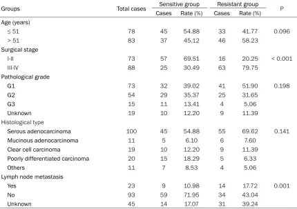
Table 2. Univariate analysis of chemoresistance in ovarian cancer
Groups Total cases Sensitive group Resistant group P
Cases Rate (%) Cases Rate (%)
Age (years)
≤ 51 78 45 54.88 33 41.77 0.096
> 51 83 37 45.12 46 58.23
Surgical stage
I-II 73 57 69.51 16 20.25 < 0.001
III-IV 88 25 30.49 63 79.75
Pathological grade
G1 73 32 39.02 41 51.90 0.198
G2 54 29 35.37 25 31.65
G3 15 11 13.41 4 5.06
Unknown 19 10 12.20 9 11.39
Histological type
Serous adenocarcinoma 100 45 54.88 55 69.62 0.141
Mucinous adenocarcinoma 11 5 6.10 6 7.60
Clear cell carcinoma 19 10 12.20 9 11.39
Poorly differentiated carcinoma 20 15 18.29 5 6.33
Others 11 7 8.53 4 5.06
Lymph node metastasis
Yes 23 9 10.98 14 17.72 0.001
No 93 59 71.95 34 43.04
Unknown 45 14 17.07 31 39.24
Table 3. Multivariate analysis of ovarian cancer chemoresistance
Factors β-value SE Wald χ2 sig Exp (β)
95% CI for Exp (β) lower
limit upper limit
NF-κB p65 1.296 0.634 4.18 0.041 3.655 1.055 12.66
PTEN -1.387 0.432 10.313 0.001 0.25 0.107 0.582
Surgical stage 1.793 0.424 17.838 < 0.001 6.005 2.614 13.799
[image:5.612.90.382.427.503.2]p105), NF-κB2 (p52/p100), RelA (p65) and ReIB, and the most common dimer of NF-κB is the p50-p65 dimer. In the resting state, the p50-p65 dimer usually directly binds with its inhibitor IκB to form an inactive trimmer, which is present in the cytoplasm of almost all cells. When subjected to stimulation by external fac-tors, NF-κB firstly dissociates with IκB, exposing its nuclear localization sequence. The p50-p65 dimer then rapidly translocates from the cyto-plasm to the nucleus, and binds with its target-ing sequence on the DNA, so as to regulate the transcription of the related gene [17]. Therefore, the expression of p65 can reflect the activity of NF-κB, and the expression of nuclear NF-κB protein can be considered a marker of NF-κB activation. NF-κB is the key in various transduc -tion pathways, and is involved in the genetic regulation of various physiological and patho-logical processes, including immunity, inflam -mation, the occurrence and development of tumors, cell proliferation, apoptosis and autophagy, and angiogenesis. Studies have shown high expression of NF-κB in various malignant tumors, such as pancreatic cancer, breast cancer, colon cancer, and gastric cancer [18-21]. The activation of NF-κB plays an impor -tant role in increasing the proliferation of tumor cells, reducing apoptosis and autophagy of tumor cells, promoting the formation of new blood vessels within the tumor, enhancing the
[image:6.612.93.522.85.322.2]local invasion and distant metastasis of tumor cells, and promoting chemotherapy chemore-sistance of tumor cells. The role of NF-κB in tumors may be related to the following aspects: (1) through upregulation of cyclin D1 transcrip-tion and regulatranscrip-tion of the cell cycle, NF-κB pro -motes cell proliferation, eventually leading to malignant transformation, and canceration of the cells [22]; (2) NF-κB regulates the transcrip -tion of inhibitors of apoptosis proteins (IAP), anti-apoptotic gene Bcl-2, Bcl-XL, XIAP, etc., and inhibits cell apoptosis [23, 24]; (3) by affecting the transcription of the autophagy-related gene Beclin-1/Bcl-2, NF-κB regulates autophagy [6]; (4) consistent activation of NF-κB could promote the abnormally high expression of important factors of angiogene-sis, vascular endothelial growth factor (VEGF), IL-6 and IL-8, promoting angiogenesis in tumor tissues and the growth of tumors [25]; (5) in promoting the invasion and expansion of tumors, NF-κB not only regulates the expres -sion of various chemotactic factors, which pro-motes cell migration, but also propro-motes the expression of matrix metalloproteinases (MMP) and urokinase plasminogen activator (u-PA), which facilitates invasion and metastasis of tumor cells [26]. Furthermore, recent studies have found that NF-κB also plays an important role in the development of tumor cell chemore-sistance [7, 8]. Many studies have found high
Table 4. Cox model analysis of the prognosis of ovarian cancer patients
β-value SE Wald χ2 sig Exp (β) 95% CI for Exp (β)
lower limit upper limit Univariate analysis
Age 0.549 0.241 5.188 0.023 1.732 1.080 2.779
Surgical stage 2.047 0.308 44.257 < 0.001 7.748 4.239 14.163
Pathological grade -0.258 0.191 1.822 0.177 0.773 0.531 1.124
Histological type -0.176 0.098 3.226 0.072 0.839 0.692 1.016
Lymph node metastasis 0.462 0.125 13.659 < 0.001 1.587 1.242 2.027
NF-κB p65 2.050 0.490 17.525 < 0.001 7.765 2.974 20.271
PTEN -0.756 0.278 7.378 0.007 0.470 0.272 0.810
Groups 3.565 0.470 57.465 < 0.001 35.348 14.061 88.857
Multivariate analysis
Age -0.061 0.263 0.053 0.817 0.941 0.563 1.575
Surgical stage 0.837 0.365 5.265 0.022 2.310 1.130 4.721
Lymph node metastasis 0.094 0.141 0.445 0.505 1.099 0.833 1.448
NF-κB p65 1.193 0.567 4.431 0.035 3.296 1.086 10.006
PTEN 0.556 0.311 3.199 0.074 1.744 0.948 3.210
expression of NF-κB in drug-resistant cancer cells. Eichholtz-Wirth H et al [27] found that the expression of NF-κB in the cervical carcinoma drug-resistant cell line Hela/B was higher than that in the sensitive cell line both at baseline and after cisplatin induction. The study by Mabuchi et al [28] showed that, in ovarian can -cer cell lines, blocking NF-κB activation by its inhibitor SN50 promotes the apoptosis of
[image:7.612.95.522.74.551.2]motherapy chemoresistance in ovarian cancer, which, for the first time, demonstrated that NF-κB plays an important role in chemoresis -tance in ovarian carcinoma at the histological level. In addition, the survival rates of patients with positive expression of NF-κB in ovarian cancer tissues were lower than that of patients with negative expression of NF-κB. COX model analysis showed that NF-κB was an indepen -dent risk factor for chemoresistance in ovarian cancer.
PTEN is a tumor-suppressing gene with bispe-cific activity towards phosphatase and lipid phosphatase. Many studies have found that PTEN may be involved in the conduction of mul-tiple signaling pathways, regulation of cell cycle, apoptosis and autophagy, cell adhesion and migration, be involved in angiogenesis and also play an important role in the transduction of multiple signaling pathways in cells. Studies have demonstrated genetic mutation and abnormal expression of PTEN in glioma, breast cancer, prostate cancer and other tumors, which play an important role in the occurrence and development of these tumors [29]. On the other hand, the expression level of PTEN in nor-mal ovarian tissue, benign ovarian tumor, bor-derline ovarian tumors and ovarian cancer gradually decreased [30, 31], indicating that down-regulation of PTEN may be involved in the occurrence and development of ovarian can-cer. A number of in vitro studies showed that gene alterations of PTEN may be involved in the development of chemoresistance in tumors. Selvendiran et al found that the chemothera-peutic drug EF24 sensitizes cells to chemother-apy by upregulating PTEN protein expression in ovarian cancer cells [32]. Wu et al increased the PTEN protein expression in a cisplatin-resistant C13K ovarian cancer cell line by in vitro liposomal transfection of the PTEN gene, and the resultant cell lines showed higher sen-sitivity towards cisplatin compared with the cells transfected with empty plasmid [33]. The in vitro cancer cell study by Yan et al [34] dem -onstrated that PTEN protein can make the cis-platin-resistant ovarian cancer cell lines OV20028, CI3* and A2780 sensitive to chemo -therapy by upregulating the expression of P53, rather than through inhibiting the activation of Akt protein. Compared with the primary ovarian cancer cell line OVCAR-3, the expression level of PTEN in drug-resistant ovarian cancer cell
The study by Gustin et al [40] found that PTEN can inhibit the function of phosphatidylinositol-3-hydroxyl kinase, reduce the activation of ser-ine/threonine protein kinase (Akt) and IκB kinase complex (IκB kinase, complex, IκK) by tumor necrosis factor (TNF), and inhibit the DNA binding and transcription of NF-κB. Another study [41] showed that PTEN can directly inhibit the transcription activating activity of NF-κB p65 to suppress cell proliferation and transcrip-tion of the anti-apoptosis gene, which inhibits cell proliferation and promotes cell apoptosis. Our study demonstrated that the positive expression rate of PTEN in the sensitive group was significantly higher than that in the resis -tant group. On the contrary, the positive expres -sion of NF-κB p65 in the sensitive group was significantly lower than that in the resistant group. Both NF-κB and PTEN were independent risk factors for chemotherapy resistance in ovarian cancer, the expression levels of which in ovarian carcinoma were negatively correlat-ed. Therefore, the down-regulation of PTEN may directly or indirectly activate NF-κB p65 involve -ment in the occurrence and develop-ment of the chemoresistance mechanism in ovarian can-cer, which can both be used as predictors of multichemoresistance in ovarian carcinoma.
Acknowledgements
This work is financially supported by the National Natural Science Foundation of China (81372486).
Disclosure of conflict of interest
None.
Address correspondence to: Dr. Huanchun Ying,
Department of Obstetrics and Gynecology,
Shengjing Hospital of China Medical University, 36 Heping District Sanhao Street, Shenyang 110004, P. R. China. Tel: + 86-18940254998; E-mail: ying-hc@sj-hospital.org
References
[1] Furukawa S, Soeda S, Kiko Y, Suzuki O, Hashi -moto Y, Watanabe T, Nishiyama H, Tasaki K, Hojo H, Abe M, Fujimori K. MCP-1 promotes in-vasion and adhesion of human ovarian cancer cells. Anticancer Res 2013; 33: 4785-90.
[2] Lee S, Choi EJ, Jin C, Kim DH. Activation of PI3K/Akt pathway by PTEN reduction and
PIK-3CA mRNA amplification contributes to cispla
-tin resistance in an ovarian cancer cell line.
Gynecol Oncol 2005; 97: 26-34.
[3] Yunos NM, Mutalip SS, Jauri MH, Yu JQ, Huq F. Anti-proliferative and pro-apoptotic effects from sequenced combinations of androgra-pholide and cisplatin on ovarian cancer cell lines. Anticancer Res 2013; 33: 4365-71.
[4] Morishita N, Tsukahara H, Chayama K, Ishida
T, Washio K, Miyamura T, Yamashita N, Oda M,
Morishima T. Activation of Akt is associated with poor prognosis and chemotherapeutic
re-sistance in pediatric B-precursor acute lym
-phoblastic leukemia. Pediatr Blood Cancer
2012; 59: 83-9.
[5] Karin M, Cao Y, Greten FR, Li ZW. NF-kappaB in
cancer: from innocent bystander to major cul-prit. Nat Rev Cancer 2002; 2: 301-10.
[6] Djavaheri-Mergny M, Amelotti M, Mathieu J,
Besançon F, Bauvy C, Souquère S, Pierron G, Codogno P. NF-kappaB activation represses
tumor necrosis factor-alpha-induced
autopha-gy. J Biol Chem 2006; 281: 30373-82. [7] Montagut C, Tusquets I, Ferrer B, Corominas
JM, Bellosillo B, Campas C, Suarez M, Fabregat X, Campo E, Gascon P, Serrano S, Fernandez
PL, Rovira A, Albanell J. Activation of nuclear
factor-kappa B is linked to resistance to neoad -juvant chemotherapy in breast cancer pa-tients. Endocr Relat Cancer 2006; 13: 607-16.
[8] Holcomb B, Yip-Schneider M, Schmidt CM. The role of nuclear factor kappaB in pancreatic
cancer and the clinical applications of targeted therapy. Pancreas 2008; 36: 225-35.
[9] Gil A, Andres-Pons A, Fernandez E, Valiente M, Torres J, Cervera J, Pulido R. Nuclear localiza-tion of PTEN by a Ran-dependent mechanism enhances apoptosis: Involvement of an N-ter-minal nuclear localization domain and multiple
nuclear exclusion motifs. Mol Biol Cell 2006;
17: 4002-13.
[10] Tang Y, Eng C. PTEN autoregulates its expres-sion by stabilization of p53 in a phosphatase-independent manner. Cancer Res 2006; 66: 736-42.
[11] Ogier-Denis E, Codogno P. Autophagy: a barrier or an adaptive response to cancer. Biochim Biophys Acta 2003; 1603: 113-28.
[12] Gozuacik D, Kimchi A. Autophagy as a cell
death and tumor suppressor mechanism. On -cogene 2004; 23: 2891-906.
[13] Maehama T, Dixon JE. PTEN: a tumour sup-pressor that functions as a phospholipid
phos-phatase. Trends Cell Biol 1999; 9: 125-8. [14] Mutter GL, Lin MC, Fitzgerald JT, Kum JB, Baak
[15] Song MS, Salmena L, Pandolfi PP. The func -tions and regulation of the PTEN tumour
sup-pressor. Nat Rev Mol Cell Biol 2012; 13:
283-96.
[16] Zhuang HQ, Wang J, Yuan ZY, Zhao LJ, Wang P,
Wang CL. The drug-resistance to gefitinib in
PTEN low expression cancer cells is reversed by irradiation in vitro. J Exp Clin Cancer Res 2009; 28: 123.
[17] Siebenlist U, Franzoso G, Brown K. Structure, regulation and function of NF-kappa B. Annu Rev Cell Biol 1994; 10: 405-55.
[18] Liu A, Chen H, Tong H, Ye S, Qiu M, Wang Z, Tan W, Liu J, Lin S. Emodin potentiates the antitu-mor effects of gemcitabine in pancreatic can-cer cells via inhibition of nuclear
factor-kap-paB. Mol Med Rep 2011; 4: 221-7.
[19] Ko HS, Lee HJ, Kim SH, Lee EO. Piceatannol
suppresses breast cancer cell invasion through the inhibition of MMP-9: involvement of PI3K/
AKT and NF-kappaB pathways. J Agric Food
Chem 2012; 60: 4083-9.
[20] Kojima M, Morisaki T, Sasaki N, Nakano K, Mibu R, Tanaka M, Katano M. Increased
nucle-ar factor-kB activation in human colorectal cnucle-ar -cinoma and its correlation with tumor progres-sion. Anticancer Res 2004; 24: 675-81.
[21] Sasaki N, Morisaki T, Hashizume K, Yao T, Tsuneyoshi M, Noshiro H, Nakamura K, Ya-manaka T, Uchiyama A, Tanaka M, Katano M.
Nuclear factor-kappaB p65 (RelA) transcrip -tion factor is constitutively activated in human gastric carcinoma tissue. Clin Cancer Res 2001; 7: 4136-42.
[22] Guttridge DC, Albanese C, Reuther JY, Pestell
RG, Baldwin AS Jr. NF-kappaB controls cell
growth and differentiation through
transcrip-tional regulation of cyclin D1. Mol Cell Biol
1999; 19: 5785-99.
[23] Pommier Y, Sordet O, Antony S, Hayward RL,
Kohn KW. Apoptosis defects and chemothera-py resistance: molecular interaction maps and
networks. Oncogene 2004; 23: 2934-49. [24] Takada Y, Kobayashi Y, Aggarwal BB. Evodi
-amine abolishes constitutive and inducible
NF-kappaB activation by inhibiting INF-kappaBalpha
kinase activation, thereby suppressing
NF-kap-paB-regulated antiapoptotic and metastatic
gene expression, up-regulating apoptosis, and
inhibiting invasion. J Biol Chem 2005; 280:
17203-12.
[25] Pahl HL. Activators and target genes of
Rel/NF-kappaB transcription factors. Oncogene 1999;
18: 6853-66.
[26] Trusolino L, Comoglio PM. Scatter-factor and semaphorin receptors: cell signalling for inva-sive growth. Nat Rev Cancer 2002; 2: 289-300.
[27] Eichholtz-Wirth H, Sagan D. IkappaB/NF-kap
-paB mediated cisplatin resistance in HeLa
cells after low-dose gamma-irradiation is
asso-ciated with altered SODD expression. Apopto -sis 2000; 5: 255-63.
[28] Mabuchi S, Ohmichi M, Nishio Y, Hayasaka T, Kimura A, Ohta T, Saito M, Kawagoe J, Taka -hashi K, Yada-Hashimoto N, Sakata M, Mo-toyama T, Kurachi H,Tasaka K, Murata Y.
Inhi-bition of NFkappaB increases the efficacy of
cisplatin in in vitro and in vivo ovarian cancer
models. J Biol Chem 2004; 279: 23477-85. [29] Li J, Yen C, Liaw D, Podsypanina K, Bose S,
Wang SI, Puc J, Miliaresis C, Rodgers L,
Mc-Combie R, Bigner SH, Giovanella BC, Ittmann M, Tycko B, Hibshoosh H, Wigler MH, Parsons
R. PTEN, a putative protein tyrosine phospha-tase gene mutated in human brain, breast, and prostate cancer. Science 1997; 275: 1943-7.
[30] Laudanski P, Kowalczuk O, Klasa-Mazurkie -wicz D, Milczek T, Rysak-Lubero-wicz D,
Garbo-wicz M, Baranowski W, CharkieGarbo-wicz R, Szama -towicz J, Chyczewski L. Selective gene
expression profiling of mTOR-associated tumor
suppressor and oncogenes in ovarian cancer. Folia Histochem Cytobiol 2011; 49: 317-24.
[31] Skirnisdottir I, Seidal T. Prognostic impact of concomitant p53 and PTEN on outcome in
early stage (FIGO I-II) epithelial ovarian cancer.
Int J Gynecol Cancer 2011; 21: 1024-31.
[32] Selvendiran K, Tong L, Vishwanath S, Bratasz
A, Trigg NJ, Kutala VK, Hideg K, Kuppusamy P. EF24 induces G2/M arrest and apoptosis in cisplatin-resistant human ovarian cancer cells
by increasing PTEN expression. J Biol Chem
2007; 282: 28609-18.
[33] Wu HJ, Wu HT, Weng DH, Xing H, Lu YP, Ma D.
Reversal of chemoresistance in human ovari-an covari-ancer cells by wild-type PTEN gene ovari-and its mechanisms. Zhonghua Fu Chan Ke Za Zhi 2007; 42: 612-6.
[34] Yan X, Fraser M, Qiu Q, Tsang BK. Over-expres -sion of PTEN sensitizes human ovarian cancer cells to cisplatin-induced apoptosis in a
p53-dependent manner. Gynecol Oncol 2006; 102:
348-55.
[35] Ying H, Qu D, Liu C, Liu C, Ying T, Lv J, Jin S, Xu H. Chemoresistance is associated with Be -clin-1 and PTEN expression in epithelial
ovari-an covari-ancers. Oncol Lett 2015; 9: 1759-1763. [36] Bouali S, Chretien AS, Ramacci C, Rouyer M,
Becuwe P, Merlin JL. PTEN expression controls
cellular response to cetuximab by mediating PI3K/AKT and RAS/RAF/MAPK downstream signaling in KRAS wild-type, hormone
refracto-ry prostate cancer cells. Oncol Rep 2009; 21:
[37] Gu J, Tamura M, Yamada KM. Tumor suppres-sor PTEN inhibits integrin- and growth factor-mediated mitogen-activated protein (MAP)
ki-nase signaling pathways. J Cell Biol 1998;
143: 1375-83.
[38] Diao L, Chen YG. PTEN, a general negative regulator of cyclin D expression. Cell Res 2007; 17: 291-2.
[39] Kotelevets L, van Hengel J, Bruyneel E, Mareel
M, van Roy F, Chastre E. Implication of the MA-GI-1b/PTEN signalosome in stabilization of ad-herens junctions and suppression of
invasive-ness. FASEB J 2005; 19: 115-7.
[40] Gustin JA, Maehama T, Dixon JE, Donner DB.
The PTEN tumor suppressor protein inhibits tu-mor necrosis factor-induced nuclear factor
kappa B activity. J Biol Chem 2001; 276:
27740-4.
[41] Mayo MW, Madrid LV, Westerheide SD, Jones
DR, Yuan XJ, Baldwin AS Jr, Whang YE. PTEN
blocks tumor necrosis factor-induced
NF-kap-pa B-dependent transcription by inhibiting the
transactivation potential of the p65 subunit. J


![5 {(2S,3R,4S,5S,6R) 3,4 Dihydroxy 6 hydroxymethyl 3 [(2S,3R,4R,5R,6S) 3,4,5 trihydroxy 6 methyltetrahydropyran 2 yloxy]tetrahydropyran 2 yloxy} 7 hydroxy 2 (4 hydroxyphenyl)chromen 4 one monohydrate](data:image/gif;base64,R0lGODlhAQABAIAAAP///wAAACH5BAEAAAAALAAAAAABAAEAAAICRAEAOw==)