Int J Clin Exp Pathol 2019;12(4):1439-1456 www.ijcep.com /ISSN:1936-2625/IJCEP0092210
Original Article
Elevation of miR-191-5p level and its potential signaling
pathways in hepatocellular carcinoma: a study validated
by microarray and in-house qRT-PCR
with 1,291 clinical samples
Hua-Yu Wu1,2#, Mei-Wei Li2#, Qi-Qi Li1, Yu-Yan Pang3, Gang Chen3, Hui-Ping Lu3*, Shang-Ling Pan1*
Departments of 1Pathophysiology, 2Cell Biology and Genetics, School of Pre-clinical Medicine, Guangxi Medical University, Nanning, Guangxi Zhuang Autonomous Region, P. R. China; 3Department of Pathology, First Affiliated Hospital of Guangxi Medical University, Nanning, Guangxi Zhuang Autonomous Region, P. R. China. #Equal con-tributors and co-first authors. *Equal contributors.
Received February 2, 2019; Accepted February 21, 2019; Epub April 1, 2019; Published April 15, 2019
Abstract: Background: The miR-191-5p expression has been reported to increase in hepatocellular carcinoma (HCC),
but its clinical value and exact role remain to be further clarified. Thus, a comprehensive analysis was performed in
the current study to explore the underlying function of miR-191-5p in HCC. Methods: HCC-related expression data
were collected to conduct a thorough analysis to determine the miR-191-5p expression and its clinical significance
in HCC, including microarray data from the Gene Expression Omnibus and ArrayExpress database as well as quanti-tative real-time polymerase chain reaction (qRT-PCR) data of 178 matched clinical samples. The underlying relation-ship between miR-191-5p and HCC was also explored on the basis of a series of bioinformatics analyses. Results: The overall pooled meta-analysis showed an overexpression of miR-191-5p in the HCC samples (SMD=0.400, 95% CI=0.139-0.663, P=0.003), consistent with the detected result of the clinical HCC samples through the qRT-PCR analysis. Higher miR-191-5p levels were correlated with advanced TNM stages (III and IV), higher pathological grades, and metastasis. Functionally, 64 potential target genes were acquired for further mechanism analysis. Two pathways (p75 neurotrophin receptor and liver kinase B1-mediated signaling pathways), which were likely modu-lated by miR-191-5p, were regarded to be linked to the deterioration of HCC. Early growth response 1 and UBE2D3
were identified as the most likely targets for miR-191-5p in HCC and were commonly implied in the top enriched
pathways and protein-protein network. Conclusions: In summary, miR-191-5p may function as a tumor promoter miRNA of HCC, and the miR-191-5p inhibitor may contribute to the targeted HCC treatment in the future.
Keywords: Hepatocellular carcinoma, MiR-191-5p, signaling pathway, microarray, qRT-PCR
Introduction
Hepatocellular carcinoma (HCC) is one of the
top ten cancers around the world, ranking fifth
in cancer-related mortality in men, with nearly 21,600 new estimated deaths [1]. HCC is com-monly diagnosed in the advanced stages be- cause of its latency, and the currently available therapies for HCC are ineffective, thus leading to unfavorable prognosis[2]. Specifically, less
than 20% of HCC patients could survive after
five years from the date of diagnosis [3].
MicroRNAs (miRNAs) are endogenous single-stranded, small non-coding RNA molecules containing almost 22 nucleotides, which are im-
plicated in mRNA regulation. Each miRNA may have multiple target genes. Some miRNAs may also regulate the same genes, generally affect-ing gene expression[4, 5]. MiRNAs act as an essential participant in gene expression, re- verse transcription, cell differentiation, prolifer-ation and apoptosis, a series of biological activ-ities associated with immune responses, car-diovascular diseases, and the occurrence and progression of cancers [6-8].
develop-ment, cancer diagnosis, and prognosis [9]. Re- cent studies found that miR-191-5p could be involved in certain biological activities in HCC, including cellular changes, proliferation inhibi-tion, and apoptosis, and it could serve as an oncogene [10]. Moreover, miR-191-5p has been frequently reported to function as an oncogene in cancers such as colon, breast, and gastric [11-13]. Zhang et al. revealed that the suppres-sion of miR-191-5p expressuppres-sion could negatively regulate the proliferation of colorectal cancer cells [11]. Similarly, suppressing the miR-191-5p expression in the gastric cancer cell line HGC-27 reduced cell proliferation and cell cycle progression and then impaired cell migration and invasion [13]. In breast cancer, Nagpal reported that HIF-inducible miR-191-5p could promote the migration of breast cancer by
regu-lating the TGFβ-signaling pathway in a hypoxic
microenvironment [12]. Therefore, miR-191-5p has great potential to be a novel diagnostic bio-marker and an innovative therapeutic approach for HCC patients. However, current studies are
limited by the fact that the specific mechanism
between miR-191-5p and HCC has not been
clearly clarified. In addition, more evidence
remains to be collected for the further applica-tion of miR-191-5p in the predicapplica-tion and early diagnosis of HCC.
Thus, we firstly evaluated the miR-191-5p
expression and its potentially
clinicopathologi-cal and diagnostic significance in HCC by ana
-lyzing data from the Gene Expression Omnibus (GEO) and ArrayExpress databases as well as clinical tissue samples. Moreover, we investi-gated the underlying mechanism of miR-191-5p in HCC through in silico analysis, including potential targets prediction, pathway, gene on- tology (GO), and protein-protein interaction (PPI) analyses.
Materials and methods
Data mining of microarray data for the role of miR-191-5p in HCC
We screened and downloaded the miRNA (last-ly updated on May 13, 2018) and mRNA (last(last-ly updated on January 7, 2019) microarray
pro-files of HCC from the GEO and ArrayExpress
databases. The keywords used for the search consisted of two parts: (1) hepatocellular OR liver OR hepatic OR HCC and (2) malignan* OR cancer OR tumor OR tumor OR neoplas* OR
carcinoma. The microarrays were considered eligible while conforming to the following criteria: (1) the samples were obtained from
humans; (2) the profiles should contain two
groups, namely, patients diagnosed with HCC as the experimental group and individuals with-out HCC as the control group; (3) both two groups should include at least three samples; (4) the studies should examine the expression of miR-191-5p or the potential target mRNA in
HCC. The profiles were regarded as unqualified
if (1) they were not related to human HCC, and (2) the samples were collected from cell lines or other species.
Clinical HCC tissue samples from our institute
In this study, the First Affiliated Hospital of
Guangxi Medical University (Nanning, Guangxi,
China) provided 89 formalin-fixed,
paraffin-embedded (FFPE) HCC tissues and their adja-cent non-tumorous liver tissues from 2010 to 2011. The mean age of the included HCC patients was 52 years old. The study design was approved by the Ethical Committee of The
First Affiliated Hospital of Guangxi Medical
University. Informed consent was collected from all participants. The clinical pathological information of all HCC patients is presented in
Table 1. The above samples were examined,
diagnosed, and confirmed independently by
two pathologists (Yu-yan Pang and Gang Chen).
Quantitative real-time polymerase chain reac-tion (qRT-PCR) to detect miR-191-5p in the HCC clinical sample in house
Elevation of MiR-191-5p level in HCC
were analyzed using PCR7900 (Applied Bio- systems) in December 2011. The 2-Δcq method
was applied to examine the miR-191-5p exp- ression of both HCC and its adjacent healthy liver tissues.
Exploration for the prognostic significance of miR-191-5p in HCC based on microarray data
The GEO profiles with the prognostic data of
miR-191-5p in HCC were collected from the SurvMicro website [17]. The hazard ratios (HRs)
and corresponding confidence intervals (CIs) of each profile were extracted to execute the
pooled analysis on Stata 12.0. HCC patients
profiles and institute qRT-PCR data. The pooled
results showed marked heterogenicity with a p
value of less than 0.05 and I2 statistics of more
than 50%, which pointed to the selection of the
random effects model. By contrast, the fixed
effects model was employed when the p value was greater than 0.05 and the I2 was less than
50%. The potential publication bias was esti-mated by Begg’s tests. Sensitivity analysis was
adopted to test the effect of each profile on the
[image:3.612.91.360.98.506.2]pooled result. The summary receiver operating characteristic (SROC) curve was drawn using Stata 12.0, and we calculated the concrete value of the area under the curve (AUC) to access the ability of miR-191-5p to distinguish
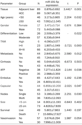
Table 1. Correlations between miR-191-5p expression and clinicopathological features in HCC (Mean ± SD)
Parameter Group N miR-191-5p expression t P Tissue Adjacent liver 89 0.876±0.692 3.441* 0.001
HCC 89 5.067±8.351
Age (years) <50 46 3.173±3.865 2.204 0.032
≥50 43 7.092±11.045
Gender Female 17 3.471±3.714 0.874 0.384 Male 72 5.443±9.088
Differentiation Low 26 2.509±3.379 - 0.022 Moderate 57 6.336±9.944
TNM High 6 4.090±3.077
I+II 23 1.807±1.049 -3.721 0.000 III+IV 66 6.203±9.433
Metastasis No 44 2.840±5.675 2.580 0.012
Yes 45 7.244±9.910
Cirrhosis No 46 5.644±9.625 -6.673 0.503 Yes 43 4.448±6.791
AFP Negative 38 7.277±11.824 -2.144 0.038 Positive 35 2.988±3.359
Embolus No 65 4.427±7.443 1.192 0.236 Yes 24 6.800±10.411
Capsular No 44 6.856±10.200 -2.021 0.047 Yes 45 3.317±5.611
Nodes Single 53 3.186±3.269 2.251 0.030 Multi 36 7.835±12.093
Size <5 cm 14 6.901±11.193 -0.843 0.402
≥5 cm 73 4.8263±7.839
Survival Live 49 5.801±7.863 1.440 0.197 Death 7 15.689±17.927
Vasoinvasion No 57 3.674±6.294 1.987 0.054 Yes 30 7.996±11.009
SD: standard deviation; *Paired samples t-test was utilized.
with high miR-191-5p levels were more likely to suffer from poorer outcomes when the pooled HR was greater than 1.
Statistical analysis for the clinical implication of miR-191-5p in HCC
The miR-191-5p expression data and clinical characteristics of HCC patients were retrieved from
the qualified included profiles
and in-house patient samples. The expression values of miR-191-5p were presented as the mean values and standard devia-tions (SD), and all statistical cal-culation was dealt with using SPSS 22.0. Student’s paired t- test and independent sample t-test were adopted to calculate the difference of miR-191-5p in the two paired or unpaired gro- ups, respectively. One-way analy-sis of variance was employed to estimate the values of more than two groups. The correlation analy-sis was executed by the Spear- man’s method. The distinguishing potential of miR-191-5p in HCC from non-HCC tissues was evalu-ated by the receiver operator characteristic (ROC) curve.
patients with HCC from individuals without
HCC. Furthermore, the pooled specificity and
sensitivity, as well as the positive and negative likelihood ratios, were calculated. The level of the two-sided p values lower than 0.05 was
determined to be statistically significant.
Acquirement of the predicted miR-191-5p tar-gets for and differentially expressed genes in HCC
We applied miRWalk2.0, a platform mainly used for miRNA-targets prediction, to collect the likely targets for miR-191-5p. Twelve data-bases were involved in the prediction analysis [18]. Only the genes nominated by more than four databases were selected as possible tar-gets for miR-191-5p. The differentially expres-
sed genes (DE-genes) were identified by a GEO
microarray (GSE22790), which recorded the values of gene expression in the HCC cell lines after treating with anti-miR-191-5p (the experi-mental group) and without anti-miR-191-5p (the control group). Then, a part of the DE-genes was recognized as potential miR-191-5p tar-gets when it showed elevated expression levels
in this microarray file (|log2 fold changes (FC)|>=1.0, P<0.05).
Signaling pathway analysis for overlapping genes
Finally, the intersection elements of the partic-ularly selected predicted targets of miR-191-5p and the DEGs recognized by GSE22790 were considered the candidate target genes of miR-191-5p. The key genes were adopted to
deter-mine the underlying mechanism of miR-191-5p in HCC. Originally, FunRich3.0 was used to per-form the GO enrichment and pathway analysis of the target genes [19]. The GO analysis aimed
to find the annotations among the biological
processes, cellular component, and molecular function in HCC. Pathway enrichment showed the signaling pathways involved in HCC. The
detailed files were ultimately downloaded to
display the terms involved in the GO and path-way analyses. Moreover, the String database was adopted to construct a visualized and
com-prehensive PPI network, reflecting the relation
-ship among the total selected genes [20]. The genes commonly involved in the most enriched pathways and the PPI network were considered as miR-191-5p potential targets. As the pooled result of the microarray datasets indicated a high expression trend of miR-191-5p in HCC, its target genes were likely to show down-regulat-ed levels. Thus, we utilizdown-regulat-ed the current microar-ray data in the GEO and Armicroar-rayExpress databas-es to analyze the exprdatabas-ession levels of the candi-date targets and the details described above.
Results
Characteristics of the eligible GEO and ArrayExpress profiles
In total, 13 eligible profiles associated with
miR-191-5p and HCC, as well as 40 datasets related to mRNA and HCC, were selected for later analysis. The samples used for the
experi-ments of each profile were obtained from the
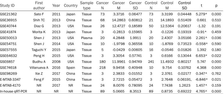
[image:4.612.92.523.86.268.2]HCC and non-HCC tissues, except one from the plasma specimens (GSE50013). The main characteristics are presented in Table 2. The
Table 2. Summary characteristics of the 13 eligible microarray datasets and institute qRT-PCR data Study ID authorFirst Year Country Sample type Cancer N Cancer M Cancer SD Control N Control M Control SD t p
GSE21362 Sato F 2011 Japan Tissue 73 3.3716 0.06477 73 3.3199 0.04448 5.279* 0.000
GSE36915 Shih TC 2013 China Tissue 68 14.2863 0.60812 21 14.1893 0.51409 0.661 0.510
GSE40744 Diaz G 2013 USA Tissue 26 12.4727 0.18589 50 12.5364 0.20617 -1.32 0.191
GSE41874 Morita K 2013 Japan Tissue 3 -0.2813 0.15965 3 -0.1226 0.19319 -0.91* 0.459
GSE50013 Shen J 2013 USA Plasma 20 4.2848 1.9911 20 2.4307 3.05166 2.261* 0.036
GSE54751 Shen J 2014 USA Tissue 10 -1.9798 0.36558 10 -1.8769 0.73523 -0.558* 0.590
GSE57555 Taguchi Y 2015 Japan Tissue 5 -0.0429 0.00605 16 -0.0546 0.01826 1.392 0.180
GSE64632 Peng H 2015 USA Tissue 3 3.3054 0.57741 3 0.6166 0.13044 6.653* 0.022
GSE6857 Budhu A 2008 USA Tissue 180 11.9961 0.94749 241 11.4932 0.80217 5.747 0.000
GSE74618 Villanueva A 2016 Spain Tissue 218 9.9458 0.40948 10 9.754 0.10762 4.368 0.000
GSE98269 Xie Z 2017 China Tissue 3 2.3833 0.01552 3 2.3761 0.02177 0.347* 0.762
E-MTAB-3347 Feng F 2015 China Tissue 3 2.7215 0.05472 3 2.7648 0.06191, -6.846* 0.021
E-MTAB-4170 NR 2017 NR Tissue 24 8.0076 0.78095 24 7.7438 1.2623 1.457* 0.159
In-house qRT-PCR NR NR NR Tissue 89 5.0665 8.3513 89 0.8735 0.69222 4.765* 0.000
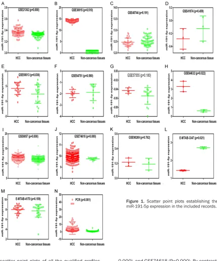
Elevation of MiR-191-5p level in HCC
scatter point plots of all the qualified profiles
are illustrated in Figure 1. Nine records exhib-ited a potential trend of increased miR-191- 5p expression in HCC tissues (GSE21362, GSE36915, GSE50013, GSE57555, GSE64- 632, GSE6857, GSE74618, GSE98269, and
E-MTAB-4170). Specifically, the miR-191-5p
ex-pression was markedly higher in the HCC
tis-sues than in the control group in five records,
namely, GSE21362 (P=0.000), GSE50013 (P= 0.036), GSE64632 (P=0.022), GSE6857 (P=
0.000), and GSE74618 (P=0.000). By contrast, the miR-191-5p levels were lower in the tissue samples from HCC patients than in the control samples in E-MTAB-3347 (P=0.021).
MiR-191-5p expression in clinical HCC patients
We obtained 178 clinical tissue samples from 89 HCC patients, including cancer and their
paired non-cancerous tissues. A significantly higher miR-191-5p expression was identified in
[image:5.612.90.520.72.589.2]corresponding non-cancerous hepatic samples (0.876±0.692; P=0.001, Table 2; Figure 1). The remarkably elevated miR-191-5p expres-sion was detected in the clinically advanced stages (TNM III and IV, 6.203±9.433) rather than in the early stages (TNM I and II, 1.807± 1.049, P=0.000). Similarly, the miR-191-5p lev-els were up-regulated in the HCC tissues with metastasis (7.244±9.91) but not in the non-metastasis tissues (2.84±5.675, P=0.012). A
higher miR-191-5p expression was
identifi-ed in the groups of higher pathological grade
[image:6.612.89.528.76.562.2](4.090±3.077, P=0.022), multi-nodes (7.835± 12.093, P=0.030), non-encapsulated HCC (6.856±10.2, P=0.047), and elderly individuals (7.092±11.045, P=0.032) than in their corre-sponding control groups. By contrast, with regard to AFP examination, the miR-191-5p lev-els were down-regulated in the positive AFP groups (2.988±3.359) in comparison with the negative AFP groups (7.277±11.824, P=0.038). However, no obvious correlation was found between the miR-191-5p expression and the rest of the clinical parameters.
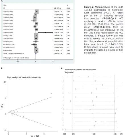
Figure 2. Meta-analysis of the miR-191-5p expression in hepatocel-lular carcinoma (HCC). A. Forest plot of the 14 included records that detected miR-191-5p in HCC applying a random effects model (I2=63.80%, P=0.001). The pooled
result (SMD=0.400>0, 95% CI:
0.139-0.661) was indicative of the miR-191-5p up-regulation in the HCC samples. B. Begg’s funnel plot was used to assess the potential publica-tion bias, and no obvious publicapublica-tion
bias was found (P=0.443>0.05).
Elevation of MiR-191-5p level in HCC
Clinical significance of the miR-191-5p expres-sion in HCC
In total, 1,291 samples were included in a meta-analysis, among which 725 were includ-ed in the experimental groups and 566 in the control groups. The pooled results of all includ-ed records suggestinclud-ed that the miR-191-5p
levels were significantly different among the
samples between patients with HCC and the non-cancerous controls (SMD=0.400, 95% CI=0.139-0.663, P=0.003). The random effect
model was used because of the obvious het-erogeneity (P=0.001, I2=63.8%), as shown in Figure 2. No publication bias was evaluated in this study, as the p value of Begg’s (P=0.443) test was greater than 0.05 (Figure 2). The sen-sitivity analysis is illustrated in Figure 2.
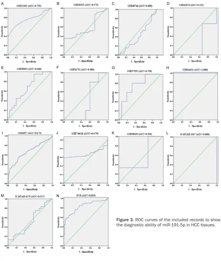
Likely diagnosis ability of miR-191-5p in HCC
The diagnostic value of each eligible record was
varied, as identified by the ROC curves (Figure
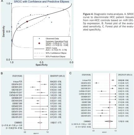
[image:7.612.91.523.70.581.2]0.740 (95% CI=0.700-0.780), suggesting that miR-191-5p could function as an effective bio-marker to diagnose patients with HCC at an earlier stage (Figure 4). The merged diagnostic sensitivity (0.678, 95% CI: 0.567-0.772) and
specificity (0.741, 95% CI: 0.522-0.882) are
shown in Figure 4. The pooled results of the diagnostic odds ratio (6.009, 95% CI: 2.975-12.137), positive likelihood ratio (2.614, 95% CI: 1.418-4.819, I2=78.84%), and negative
likelihood ratio (0.435, 95% CI: 0.348-0.544, I2=42.40%) were also calculated. These results
proved the diagnostic ability of miR-191-5p in HCC. The p value of the publication bias analy-sis was 0.539, and thus this study could be considered to have no publication bias.
Prognostic significance of miR-191-5p in HCC
We acquired three GEO profiles, namely,
GSE10694 (HR=1.280, 95% CI: 0.880-1.850, P=0.197), GSE31384 (HR=0.694, 95% CI: 0.433-1.11, P=0.131), and GSE6857 (HR= 1.204, 95% CI: 0.769-1.887, P=0.413), record-ing the prognostic data of miR-191-5p for the HCC and non-HCC patients. However, none of the three chips, as well as the pooled result (HR=1.042, 95% CI: 0.721-1.504, P=0.828),
had statistical significance. However, there was
[image:8.612.91.523.78.513.2]a tendency in GSE10694, GSE6857, and the pooled result that a higher miR-191-5p level was correlated with poorer outcomes for HCC patients (Figure 5).
Figure 4. Diagnostic meta-analysis. A. SROC curve to discriminate HCC patient tissues from non-HCC controls based on miR-191-5p expression. B. Forest plot of the ated sensitivity. C. Forest plot of the
Elevation of MiR-191-5p level in HCC
Figure 5. Kaplan-Meier survival curve to show the potential miR-191-5p prognostic significance in HCC. A. Forest plots of the three eligible GEO profiles. B and C. A higher miR-191-5p expression implied likely poorer outcomes in
GSE10694 (HR=1.280, 95% CI: 0.880-1.850, P=0.197). D and E. A higher miR-191-5p expression implied likely poorer outcomes in GSE6857 (HR=1.204, 95% CI: 0.769-1.887, P=0.413).
Potential hub genes
In total, 19,034 gene records were predicted by 12 online databases after deleting the repeat-ed genes. To ensure accuracy, the sum of 2,032
genes identified by at least four databases was
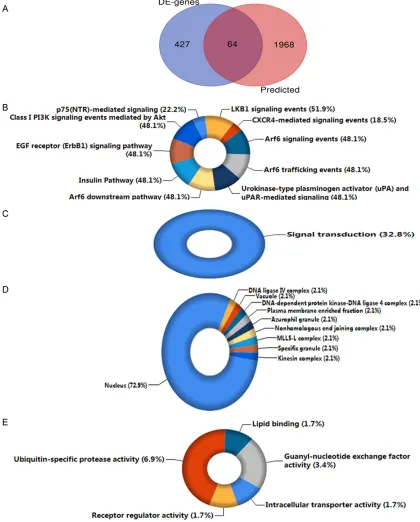
derived as the most likely target genes. By ana-lyzing the gene expression data of GSE22790, we obtained 491 up-regulated DE-genes to integrate with the selected predicted target genes in HCC. Finally, we collected 64
overlap-ping genes, which could be considered as the likely miR-191-5p targets in the regulation of HCC. The overlapping outcome is illustrated in a Venn diagram in Figure 6.
Underlying mechanism of miR-191-5p in HCC by bioinformatics analysis
The candidate targets were then inputted into FunRich 3.1.3 for the subsequent
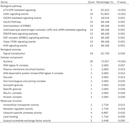
GO terms (P<0.05) are listed in Table 3. Accor- ding to the results, the target genes were highly enriched in several pathways, including p75-neurotrophin receptor (p75-NTR)-mediat-ed signaling, liver kinase B1 (LKB1) signaling events, and CXCR4-mediated signaling events;
all were closely related to the growth of HCC cells (Figure 6). Additionally, the candidate tar-gets were involved in the biological process of signal transduction and related to activities of certain cellular components, such as the nucle-us, DNA ligase IV complex, plasma membrane-Figure 6. Exploration results for the underlying mechanisms of miR-191-5p in HCC. A. Venn diagram of the crucial
[image:10.612.93.513.77.599.2]Elevation of MiR-191-5p level in HCC
Table 3. The most significantly enriched pathways and gene ontology terms of miR-191-5p
Terms Count Percentage (%) P-value
Biological pathway
p75-NTR-mediated signaling 6 22.222 <0.001
LKB1 signaling events 14 51.852 <0.001
CXCR4-mediated signaling events 5 18.519 0.001
Insulin Pathway 13 48.148 0.001
Internalization of ERBB1 13 48.148 0.001
Urokinase-type plasminogen activator (uPA) and uPAR-mediated signaling 13 48.148 0.001
PDGFR-beta signaling pathway 13 48.148 0.001
EGF receptor (ERBB1) signaling pathway 13 48.148 0.001
Class I PI3K signaling events 13 48.148 0.001
Arf6 signaling events 13 48.148 0.001
Biological process
Signal transduction 19 32.759 0.034
Cellular component
Nucleus 35 72.917 <0.001
DNA ligase IV complex 1 2.083 0.007
Plasma membrane enriched fraction 1 2.083 0.013
DNA-dependent protein kinase-DNA ligase 4 complex 1 2.083 0.013
Vacuole 1 2.083 0.013
Non-homologous end joining complex 1 2.083 0.020
Azurophil granule 1 2.083 0.020
Specific granule 1 2.083 0.026
MLL5-L complex 1 2.083 0.026
Kinesin complex 1 2.083 0.029
Molecular function
Intracellular transporter activity 1 1.724 0.013
Receptor regulator activity 1 1.724 0.019
Ubiquitin-specific protease activity 4 6.897 0.032
Lipid binding 1 1.724 0.035
Guanyl-nucleotide exchange factor activity 2 3.448 0.050
enriched fraction, and DNA-dependent protein kinase-DNA ligase 4 complex (Figure 6). Mo- reover, by targeting the likely targets, miR-191-5p could participate in the regulation of the intracellular transporter activity, receptor
regu-lator activity, ubiquitin-specific protease activi -ty, and other molecular functions (Figure 6).
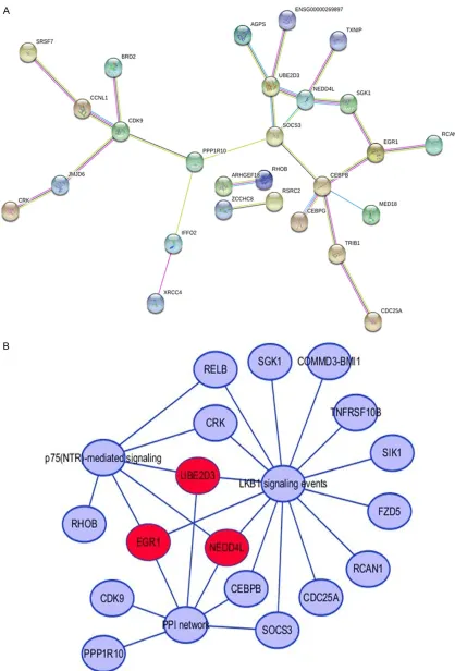
Figure 7 shows the PPI network. The three genes that were greatly involved in the top two enriched pathways and the PPI network could be the vital target genes of miR-191-5p [ear- ly growth response 1 (EGR1), NEDD4L, and UBE2D3], as shown in Figure 7. According to
the pooled result of the profiles and qRT-PCR
data, miR-191-5p showed a high expression in HCC. Thus, the genes with decreased expres-sion levels in HCC were recognized as the likely
targets of miR-191-5p. Particularly, the expres-sion of EGR1 and UBE2D3 showed a lower level trend in HCC (Figure 8). Thus, we hypothesize that EGR1 and UBE2D3 have great potential to be miR-191-5p targets in HCC.
Discussion
In this study, the up-regulation of miR-191-5p was observed in HCC tissues according to the pooled result of our meta-analysis by
microar-ray profiles and qRT-PCR data. The elevated
Figure 7. A. Protein-protein interactions of the 64 included genes. B. The likely target genes of miR-191-5p in HCC;
we regarded the genes as the miR-191-5p targets that were involved in the top two significant pathways and that
Elevation of MiR-191-5p level in HCC
cellular metabolism. EGR1 and UBE2D3 were
identified as the key genes involved in the mod
-ulation network of miR-191-5p in HCC.
Previous studies showed that miR-191-5p was used as an endogenous reference gene based on its suitability for the qRT-PCR analysis of dif-ferent samples from diverse cancer types, including tissues, serum, or plasma samples, obtained from HCC patients [21, 22]. However, growing evidence reveals that miR-191-5p is differentially expressed in certain cancers.
MiR-191-5p has been reported to be signifi -cantly elevated in HCC tissues as a carcinogen-ic factor, thus leading to poor outcomes for patients [23, 24]. He et al. particularly men-tioned that the up-regulation of miR-191-5p in HCC patients was potentially correlated with the hypomethylation occurring in the miR-191-5p locus [23]. Similarly, an increased expres-sion of miR-191-5p was previously found in cholangiocarcinoma (CCA). A recent study utiliz-ing qRT-PCR to examine the miRNA levels in
CCA confirmed that miR-191-5p was overex
-pressed and associated with the deterioration of CCA [25]. Another qRT-PCR analysis deter-mined that miR-191-5p was up-regulated in CCA [26]. Consistent with the aforementioned reports, we observed that miR-191-5p showed a high expression in HCC tissues in our analysis of 1,291 clinical samples and that the overex-pression of miR-191-5p could lead to an unfa-vorable outcome for HCC patients.
MiR-191-5p has been applied as a diagnostic biomarker in several cancers. A recent report showed that miR-191-5p was over-expressed in patients diagnosed with pancreatic cancer
[27]. Wang et al. found the significant diagnos
-tic capacity of miR-191-5p in osteosarcomas by detecting its expression in the serum of patients [28]. A clinical research also men-tioned the early diagnostic value of miR-191-5p in gastric cancer (GC) because it was remark-ably up-regulated in the tissues and serum of GC patients in comparison with the normal con-trols [13]. To date, studies on the diagnostic
significance of miR-191-5p for HCC are scarce.
Li et al. revealed that miR-191-5p could be
used as a specific biomarker combined with
other miRNAs, such as miR-221 and miR-let-7a, to diagnose HCC rapidly [29]. Similarly, one of
our GEO profiles (GSE50013) showed a higher
miR-191-5p level in the HCC plasma sam- ples than in the non-HCC samples. This result
implies that miR-191-5p could have a certain diagnostic capacity in HCC through the meth-ods for examining its expression level in the serum or plasma samples of patients and that
it is greatly beneficial in the early diagnosis of
HCC.
Several studies revealed the underlying mecha-nism of miR-191-5p in HCC. In 2010, Elyakim et al. initially proposed that the over-expression of
miR-191-5p could definitely produce positive
effects on HCC growth; by contrast, its suppres-sion induced HCC cell death in an animal exper-iment, thus implying the potential of miR-191-5p in a new HCC therapy for humans [29].
Afterward, He et al. confirmed the previous
results in their report [23]. A new study discov-ered that an up-regulated miR-191-5p expres-sion was correlated with the induction of epi-thelial mesenchymal transition, which was pos-sibly related to the tumorigenesis of HCC [30].
Additionally, Huang et al. verified the carcino -genic role of miR-191-5p in HepG2 cells in vitro [31].
In our study, we found that p75-NTR-mediated signaling and LKB1 signaling pathways were greatly enriched through a series of bioinfor-matics analyses. p75-NTR, a multifunctional receptor that belongs to both the nerve growth factor receptor family and the tumor necro- sis factor receptor superfamily, demonstrates several biological activities related to neuronal cells, prominently the meditation of neural cell death [32, 33]. p75-NTR was speculated to function as an inhibitor in the growth and prolif-eration of tumor cells and as an inducer in can-cer cell apoptosis [34]. To date, only He et al.
mentioned that a significant decrease in
p75-NTR expression was found in clinical liver can-cer tissues and that increased p75-NTR levels in vitro could drastically attenuate the growth of HCC cells, mainly by inducing a cell cycle arrest [35]. Thus, they hypothesized that p75-NTR could potentially act as a tumor suppres-sor in HCC.
epithelial-Elevation of MiR-191-5p level in HCC
mesenchymal transition in HCC, suggesting the protective role of LKB1 in liver cancer [39]. Wang et al. established that chemokine ligand 17 is a promoter in HCC progression by block-ing the nucleo-cytoplasmic translocation of LKB1 to reinforce the malignant migration and invasion, thus implying the protective role of LKB1 in HCC [40]. Several studies also
identi-fied the similar HCC suppressive function of
LKB1 [41-43]. Thus, we assumed that miR-191-5p could be a vital participant in promoting the occurrence and development of liver malignant tumors by governing the above two pathways as an oncogene. However, further experiments
are required to confirm this speculation.
We observed that EGR1 and UBE2D3 were both involved in the above-described pathways and frequently interacted with other genes in the PPI network. Moreover, the expression lev-els of the two genes were down-regulated in HCC tissues based on the data mining of the current microarray information, thus implying that miR-191-5p could be implicated in HCC carcinogenesis by targeting the two genes. Early growth response 1 (EGR1) encodes a nuclear protein that manifests as a vital regula-tor involved in transcriptional processes, and it activates its target genes to produce proteins associated with differentiation and mitogene-sis [44, 45]. Recent studies showed that EGR1 had a suppressive role in certain cancers (e.g., breast cancer and esophageal squamous cell carcinoma) and that miR-191-5p could induce cancer initiation and progression by negatively regulating EGR1 expression [46, 47]. However, research showing the relationship between EGR1 and miR-191-5p in HCC is scarce, and previous studies failed to evaluate the exact function of EGR1 in HCC. Hao et al. found that EGR1 showed a slight or even no expression in HCC cell lines and tissues and that it could be considered a type II tumor inhibitor [48]. They and Wang et al. [49] validated the inhibitory effect of EGR1 in an in vitro experiment. By con-trast, the tumorigenic role of EGR1 was also reported [50, 51]. Magee et al. summarized in a review that EGR1 could be an activator or a
repressor in liver cancer [52]. We identified a
low expression of EGR1 in HCC tissues based
on the GEO and ArrayExpress profiles. This low
expression could be inversely regulated by miR-191-5p, thus suggesting its potential HCC sup-pressive role. Therefore, further experiments remain to be conducted.
UBE2D3 encodes the ubiquitin conjugating enzymes E2D3 and is implicated in ubiquitina-tion pathways to modulate a series of cellular activities, including cell growth, apoptosis, DNA damage repair, and carcinogenesis [53, 54]. Accumulating evidence shows that the over-expression of UBE2D3 will decrease cell motili-ty and invasiveness likely by inactivating the human telomerase reverse transcriptase, a risk factor in diverse cancers [55, 56]. Additionally, an elevated UBE2D3 expression was proved to increase the radiosensitivity of breast cancer and esophageal carcinoma cells as a promising molecular target [55, 57, 58]. However, the cor-relation between UBE2D3 and miR-191-5p in HCC has not been reported yet. Interestingly, we found that miR-191-5p was potentially
implied in the regulation of ubiquitin-specific
protease activity in our study, thus indicating UBE2D3 to be an inhibitor in HCC through the negative regulation of miR-191-5p. Further studies are required to elucidate our hypo- thesis.
Conclusion
In sum, our comprehensive analysis of in-house qRT-PCR and microarray data could recognize the over-expressed level of miR-191-5p in HCC. The bioinformatics analysis was suggestive of the oncogenic role of miR-191-5p likely by inversely governing two cancer-related path-ways, namely, p75-NTR and LKB1-mediated signaling events, and its underlying target
genes, namely, EGR1 and UBE2D3. These find
-ings strengthen our insight into the cancero-genic effects of miR-191-5p on HCC and pro-vide new perspectives for HCC treatment.
Acknowledgements
The authors are thankful for the use of the GEO and ArrayExpress databases and the software employed in the study. The study was support-ed by the Promoting Project of Basic Capacity for Young and Middle-aged University Teachers in Guangxi (2017KY0111, 2018KY0123), In- novation Project of Guangxi Graduate Edu- cation (YCBZ2017045), the National Natural Science Foundation of China (31760319).
Disclosure of conflict of interest
None.
Medicine, Guangxi Medical University, 22 Shuang- yong Road, Nanning 530021, Guangxi Zhuang Auto- nomous Region, P. R. China. E-mail: slpan@gxmu. edu.cn; Hui-Ping Lu, Department of Pathology, First
Affiliated Hospital of Guangxi Medical University, 6
Shuangyong Road, Nanning 530021, Guangxi Zhu- ang Autonomous Region, P. R. China. E-mail: [email protected]
References
[1] Siegel RL, Miller KD and Jemal A. Cancer sta-tistics, 2019. CA Cancer J Clin 2019; 69: 7-34. [2] Daher S, Massarwa M, Benson AA and Khoury
T. Current and future treatment of hepatocel-lular carcinoma: an updated comprehensive review. J Clin Transl Hepatol 2018; 6: 69-78. [3] Erstad DJ, Fuchs BC and Tanabe KK. Molecular
signatures in hepatocellular carcinoma: a step toward rationally designed cancer therapy. Cancer 2018; 124: 3084-3104.
[4] Zhao H, Kuang L, Wang L, Ping P, Xuan Z, Pei T and Wu Z. Prediction of microRNA-disease as-sociations based on distance correlation set. BMC Bioinformatics2018; 19: 141.
[5] Alfonsi R, Grassi L, Signore M and Bonci D. The double face of exosome-carried microRNAs in cancer immunomodulation. Int J Mol Sci 2018; 19.
[6] Meng X, Dai W, Zhang K, Dong H and Zhang X. Imaging multiple microRNAs in living cells us-ing ATP self-powered strand-displacement
cas-cade amplification. Chem Sci 2018; 9:
1184-1190.
[7] Testa U, Pelosi E, Castelli G and Labbaye C. miR-146 and miR-155: two key modulators of immune response and tumor development. Noncoding RNA 2017; 3.
[8] Pordzik J, Pisarz K, De Rosa S, Jones AD,
Ey-ileten C, Indolfi C, Malek L and Postula M. The
potential role of platelet-related microRNAs in the development of cardiovascular events in high-risk populations, including diabetic pa-tients: a review. Front Endocrinol (Lausanne) 2018; 9: 74.
[9] Varendi K, Kumar A, Harma MA and Andressoo JO. miR-1, miR-10b, miR-155, and miR-191 are novel regulators of BDNF. Cell Mol Life Sci 2014; 71: 4443-4456.
[10] Elyakim E, Sitbon E, Faerman A, Tabak S, Mon-tia E, Belanis L, Dov A, Marcusson EG, Bennett CF, Chajut A, Cohen D, Yerushalmi N. hsa-miR-191 is a candidate oncogene target for hepatocellular carcinoma therapy. Cancer Res 2010; 70: 8077-8087.
[11] Zhang XF, Li KK, Gao L, Li SZ, Chen K, Zhang JB, Wang D, Tu RF, Zhang JX, Tao KX, Wang G, Zhang XD. miR-191 promotes tumorigenesis of human colorectal cancer through targeting C/ EBPbeta. Oncotarget 2015; 6: 4144-4158.
[12] Nagpal N, Ahmad HM, Chameettachal S, Sun-dar D, Ghosh S and Kulshreshtha R. HIF-induc-ible miR-191 promotes migration in breast cancer through complex regulation of TGFbeta-signaling in hypoxic microenvironment. Sci Rep 2015; 5: 9650.
[13] Peng WZ, Ma R, Wang F, Yu J and Liu ZB. Role of miR-191/425 cluster in tumorigenesis and diagnosis of gastric cancer. Int J Mol Sci2014; 15: 4031-4048.
[14] Rong M, Chen G and Dang Y. Increased miR-221 expression in hepatocellular carcinoma tissues and its role in enhancing cell growth and inhibiting apoptosis in vitro. BMC Cancer 2013; 13: 21.
[15] Dang Y, Luo D, Rong M and Chen G. Underex-pression of miR-34a in hepatocellular carcino-ma and its contribution towards enhancement of proliferating inhibitory effects of agents tar-geting c-MET. PLoS One 2013; 8: e61054. [16] Chen WJ, Gan TQ, Qin H, Huang SN, Yang LH,
Fang YY, Li ZY, Pan LJ and Chen G. Implication of downregulation and prospective pathway signaling of microRNA-375 in lung squamous cell carcinoma. Pathol Res Pract 2017; 213: 364-372.
[17] Aguirre-Gamboa R and Trevino V. SurvMicro: assessment of miRNA-based prognostic signa-tures for cancer clinical outcomes by multivari-ate survival analysis. Bioinformatics2014; 30: 1630-1632.
[18] Dweep H and Gretz N. miRWalk2.0: a compre-hensive atlas of microRNA-target interactions. Nat Methods2015; 12: 697.
[19] Pathan M, Keerthikumar S, Chisanga D, Ales-sandro R, Ang CS, Askenase P, Batagov AO, Benito-Martin A, Camussi G, Clayton A, Collino F, Di Vizio D, Falcon-Perez JM, Fonseca P, Fon-seka P, Fontana S, Gho YS, Hendrix A, Hoen EN, Iraci N, Kastaniegaard K, Kislinger T, Kow-al J, Kurochkin IV, Leonardi T, Liang Y, Llorente A, Lunavat TR, Maji S, Monteleone F, Øverbye A, Panaretakis T, Patel T, Peinado H, Pluchino S, Principe S, Ronquist G, Royo F, Sahoo S, Spi-nelli C, Stensballe A, Théry C, van Herwijnen MJC, Wauben M, Welton JL, Zhao K, Mathiva-nan S. A novel community driven software for functional enrichment analysis of extracellular vesicles data. J Extracell Vesicles 2017; 6: 1321455.
[20] Szklarczyk D, Morris JH, Cook H, Kuhn M, Wy-der S, Simonovic M, Santos A, Doncheva NT, Roth A, Bork P, Jensen LJ, von Mering C. The STRING database in 2017: quality-controlled protein-protein association networks, made broadly accessible. Nucleic Acids Res 2017; 45: D362-D368.
Elevation of MiR-191-5p level in HCC
expression analysis in liver carcinoma resec-tion studies. Mol Med Rep 2015; 12: 4683-4691.
[22] Wang Y, Shi J, Wu Y, Xu W, Wang Q, Zhang J, Jiang M and Gu G. Use of Luminex xMAP bead-based suspension array for detecting microR-NA in NSCLC tissues and its clinical applica-tion. Tumori 2012; 98: 792-799.
[23] He Y, Cui Y, Wang W, Gu J, Guo S, Ma K and Luo X. Hypomethylation of the hsa-miR-191 locus causes high expression of hsa-mir-191 and promotes the epithelial-to-mesenchymal tran-sition in hepatocellular carcinoma. Neoplasia 2011; 13: 841-853.
[24] Chen YJ, Thang MW, Chan YT, Huang YF, Ma N, Yu AL, Wu CY, Hu ML and Chiu KP. Global as-sessment of Antrodia cinnamomea-induced microRNA alterations in hepatocarcinoma cells. PLoS One 2013; 8: e82751.
[25] Li H, Zhou ZQ, Yang ZR, Tong DN, Guan J, Shi BJ, Nie J, Ding XT, Li B, Zhou GW and Zhang ZY. MicroRNA-191 acts as a tumor promoter by modulating the TET1-p53 pathway in intrahe-patic cholangiocarcinoma. Hepatology 2017; 66: 136-151.
[26] Kang PC, Leng KM, Liu YP, Liu Y, Xu Y, Qin W, Gao JJ, Wang ZD, Tai S, Zhong XY and Cui YF. miR-191 inhibition induces apoptosis through reactivating secreted frizzled-related protein-1 in cholangiocarcinoma. Cell Physiol Biochem 2018; 49: 1933-1942.
[27] Goto T, Fujiya M, Konishi H, Sasajima J, Fu-jibayashi S, Hayashi A, Utsumi T, Sato H, Iwama T, Ijiri M, Sakatani A, Tanaka K, Nomura Y, Ueno N, Kashima S, Moriichi K, Mizukami Y, Kohgo Y, Okumura T. An elevated expression of serum exosomal microRNA-191, -21, -451a of
pancreatic neoplasm is considered to be effi -cient diagnostic marker. BMC Cancer 2018; 18: 116.
[28] Wang T, Ji F, Dai Z, Xie Y and Yuan D. Increased expression of microRNA-191 as a potential se-rum biomarker for diagnosis and prognosis in human osteosarcoma. Cancer Biomark 2015; 15: 543-550.
[29] Li Y, Zhang L, Liu F, Xiang G, Jiang D and Pu X.
Identification of endogenous controls for ana -lyzing serum exosomal miRNA in patients with hepatitis B or hepatocellular carcinoma. Dis Markers2015; 2015: 893594.
[30] Chen C, Yang Q, Wang D, Luo F, Liu X, Xue J, Yang P, Xu H, Lu J, Zhang A and Liu Q. MicroR-NA-191, regulated by HIF-2alpha, is involved in EMT and acquisition of a stem cell-like pheno-type in arsenite-transformed human liver epi-thelial cells. Toxicol In Vitro 2018; 48: 128-136.
[31] Huang D, Bi C, Zhao Q, Ding X, Bian C, Wang H, Wang T and Liu H. Knockdown long non-coding
RNA ANRIL inhibits proliferation, migration and invasion of HepG2 cells by down-regulation of miR-191. BMC Cancer 2018; 18: 919.
[32] Meeker R and Williams K. Dynamic nature of the p75 neurotrophin receptor in response to injury and disease. J Neuroimmune Pharmacol 2014; 9: 615-628.
[33] Nadezhdin KD, Garcia-Carpio I, Goncharuk SA, Mineev KS, Arseniev AS and Vilar M. Structural basis of p75 transmembrane domain dimer-ization. J Biol Chem 2016; 291: 12346-12357. [34] Wang W, Chen J and Guo X. The role of nerve
growth factor and its receptors in tumorigene-sis and cancer pain. Biosci Trends 2014; 8: 68-74.
[35] Yuanlong H, Haifeng J, Xiaoyin Z, Jialin S, Jie L, Li Y, Huahong X, Jiugang S, Yanglin P, Kaichun W, Jie D, Daiming F. The inhibitory effect of p75 neurotrophin receptor on growth of human he-patocellular carcinoma cells. Cancer Lett 2008; 268: 110-119.
[36] Faubert B, Vincent EE, Griss T, Samborska B, Izreig S, Svensson RU, Mamer OA, Avizonis D, Shackelford DB, Shaw RJ and Jones RG. Loss of the tumor suppressor LKB1 promotes meta-bolic reprogramming of cancer cells via HIF-1alpha. Proc Natl Acad Sci U S A2014; 111: 2554-2559.
[37] Gan RY and Li HB. Recent progress on liver ki-nase B1 (LKB1): expression, regulation, down-stream signaling and cancer suppressive func-tion. Int J Mol Sci 2014; 15: 16698-16718. [38] Kottakis F, Nicolay BN, Roumane A, Karnik R,
Gu H, Nagle JM, Boukhali M, Hayward MC, Li YY, Chen T, Liesa M, Hammerman PS, Wong KK, Hayes DN, Shirihai OS, Dyson NJ, Haas W, Meissner A, Bardeesy N. LKB1 loss links serine metabolism to DNA methylation and tumori-genesis. Nature 2016; 539: 390-395. [39] Qiu B, Wei W, Zhu J, Fu G and Lu D. EMT
in-duced by loss of LKB1 promotes migration and invasion of liver cancer cells through ZEB1-induced YAP signaling. Oncol Lett 2018; 16: 6465-6471.
[40] Wang L, Li H, Zhen Z, Ma X, Yu W, Zeng H and Li L. CXCL17 promotes cell metastasis and in-hibits autophagy via the LKB1-AMPK pathway in hepatocellular carcinoma. Gene 2018; 690: 129-136.
[41] Wu CC, Wu DW, Lin YY, Lin PL and Lee H. Hepatitis B virus X protein represses LKB1 ex-pression to promote tumor progression and poor postoperative outcome in hepatocellular carcinoma. Surgery2018; 163: 1040-1046. [42] Wang YS, Du L, Liang X, Meng P, Bi L, Wang YL,
[43] Chen H, Zhang T, Sheng Y, Zhang C, Peng Y,
Wang X and Zhang C. Methylation profiling of
multiple tumor suppressor genes in hepatocel-lular carcinoma and the epigenetic mecha-nism of 3OST2 regulation. J Cancer 2015; 6: 740-749.
[44] Riffo-Campos AL, Castillo J, Tur G, Gonzalez-Figueroa P, Georgieva EI, Rodriguez JL, Lopez-Rodas G, Rodrigo MI and Franco L.
Nucleo-some-specific, time-dependent changes in histone modifications during activation of the
early growth response 1 (Egr1) gene. J Biol Chem2015; 290: 197-208.
[45] Sekiya T, Kato K, Kawaguchi A and Nagata K. Involvement of CTCF in transcription regula-tion of EGR1 at early G1 phase as an architec-ture factor. Sci Rep 2019; 9: 329.
[46] Di Leva G, Piovan C, Gasparini P, Ngankeu A, Taccioli C, Briskin D, Cheung DG, Bolon B, Anderlucci L, Alder H, Nuovo G, Li M, Iorio MV, Galasso M, Santhanam R, Marcucci G, Perrotti D, Powell KA, Bratasz A, Garofalo M, Nephew KP, Croce CM. Estrogen mediated-activation of miR-191/425 cluster modulates tumorigenici-ty of breast cancer cells depending on estro-gen receptor status. PLoS Genet 2013; 9: e1003311.
[47] Gao X, Xie Z, Wang Z, Cheng K, Liang K and Song Z. Overexpression of miR-191 predicts poor prognosis and promotes proliferation and invasion in esophageal squamous cell carci-noma. Yonsei Med J 2017; 58: 1101-1110. [48] Hao MW, Liang YR, Liu YF, Liu L, Wu MY and
Yang HX. Transcription factor EGR-1 inhi- bits growth of hepatocellular carcinoma and esophageal carcinoma cell lines. World J Gas-troenterol2002; 8: 203-207.
[49] Wang L, Sun H, Wang X, Hou N, Zhao L, Tong D, He K, Yang Y, Song T, Yang J and Huang C. EGR1 mediates miR-203a suppress the hepa-tocellular carcinoma cells progression by tar-geting HOXD3 through EGFR signaling path-way. Oncotarget 2016; 7: 45302-45316.
[50] Li H, Li J, Jia S, Wu M, An J, Zheng Q, Zhang W and Lu D. miR675 upregulates long noncoding RNA H19 through activating EGR1 in human liver cancer. Oncotarget 2015; 6: 31958-31984.
[51] Peng WX, Xiong EM, Ge L, Wan YY, Zhang CL, Du FY, Xu M, Bhat RA, Jin J and Gong AH. Egr-1 promotes hypoxia-induced autophagy to en-hance chemo-resistance of hepatocellular car-cinoma cells. Exp Cell Res2016; 340: 62-70. [52] Magee N and Zhang Y. Role of early growth
re-sponse 1 in liver metabolism and liver cancer. Hepatoma Res 2017; 3: 268-277.
[53] Stewart MD, Ritterhoff T, Klevit RE and Brzovic PS. E2 enzymes: more than just middle men. Cell Res 2016; 26: 423-440.
[54] Middleton AJ, Wright JD and Day CL. Regulation of E2s: a role for additional ubiquitin binding sites? J Mol Biol2017; 429: 3430-3440. [55] Wang W, Yang L, Hu L, Li F, Ren L, Yu H, Liu Y,
Xia L, Lei H, Liao Z, Zhou F, Xie C, Zhou Y. Inhibition of UBE2D3 expression attenuates radiosensitivity of MCF-7 human breast cancer cells by increasing hTERT expression and activ-ity. PLoS One 2013; 8: e64660.
[56] Guan GG, Wang WB, Lei BX, Wang QL, Wu L, Fu ZM, Zhou FX and Zhou YF. UBE2D3 is a posi-tive prognostic factor and is negaposi-tively corre-lated with hTERT expression in esophageal cancer. Oncol Lett2015; 9: 1567-1574. [57] Gao X, Wang W, Yang H, Wu L, He Z, Zhou S,
Zhao H, Fu Z, Zhou F and Zhou Y. UBE2D3 gene overexpression increases radiosensitivity of EC109 esophageal cancer cells in vitro and in vivo. Oncotarget 2016; 7: 32543-32553. [58] Yang H, Wu L, Ke S, Wang W, Yang L, Gao X,