Development of novel fluorescent oligonucleotide probes for use in nucleic acid sequence analysis
216
0
0
Full text
(2) UNIVERSITY OF SOUTHAMPTON. Development of Novel Fluorescent Oligonucleotide Probes for Use in Nucleic Acid Sequence Analysis. Lynda Jane Brown. A Thesis submitted for the Degree of Doctor of Philosophy Department of Chemistry January 2000.
(3) UNIVERSITY OF SOUTHAMPTON. ABSTRACT. FACULTY OF SCIENCE. CHEMISTRY. Doctor of Philosophy DEVELOPMENT OF NOVEL FLUORESCENT OLIGONUCLEOTIDE PROBES FOR USE IN NUCLEIC ACID SEQUENCE ANALYSIS by Lynda Jane Brovm. DNA and RNA labelled probes are used for target recognition in complex systems of nucleic acids. The development of two novel probe systems (heterogeneous and homogeneous assay) for use in nucleic acid sequence analysis is described.. The homogeneous assay proposed required two probes, one terminating in an enzyme (3' , ~-D-galactosidase). and the other tenninating in a fluorophore modified with. ~-D. galactosyl groups (enzyme substrates, 5') to remove its fluorescent properties. Synthesis of each probe is outlined. Fluorescence properties of the two probes hybridised adjacent to one another on a target nucleic acid was investigated.. The heterogeneous assay described involved a resin-bound nucleic acid probe synthesised with a stem and loop structure. The stem consisted of two short annealed ann sequences, one terminating in a quencher and the other in a fluorophore. In buffer only, the resinbound probe was' closed', quenching occurred and the clear glass resin was nonfluorescent. Introduction of a target nucleic acid complementary to the loop region causes the probe to 'open' and the glass beads became brightly fluorescent.. 11.
(4) For Richard. III.
(5) Contents Declaration Abstract Contents Abbreviations Acknowledgements. Chapter 1 1.1. 1.2. 1.3. 1.4. 1.5. 11. IV Vl11. XlI. Introduction to DNA and its Detection. 1. Nucleic acid background. 1. 1.1.1. Primary Structure. 1. 1.1.2. Secondary Structure. 2. 1.1.3. The Genetic Code. 3. Detecting DNA. 4. 1.2.1. DNA Diagnostics. 5. 1.2.2. Classical Methods. 6. 1.2.3. Non-Isotopic Labelling. 7. 1.2.3.1 The Signal Emitting Moiety. 8. 1.2.3.2 The Spacer. 9. 1.2.3.3 The Reactive Group. 9. Labelling Strategies. 11. 1.3.1. Indirect Labelling. 11. 1.3.2. Direct Labelling. 12. 1.3.2.1 Radioactive Labels. 13. 1.3.2.2 Enzyme Labels. 13. 1.3.2.3 Chemiluminescent Labels. 13. 1.3.2.4 Fluorescent Labels. 14. 1.3.2.5 Lanthanide Labels. 16. Fundamentals of Fluorescence. 17. 1.4.1. The Nature of Fluorescence. 17. 1.4.2. Fluorescence Quenching. 18. 1.4.3. Fluorescence Resonance Energy Transfer (FRET). 19. 1.4.4. Time Resolved Fluorescence. 20. 1.4.5. Fluorescence Spectrometry. 22. Molecular Biological Methods Utilised in Fluorescence Assay IV. 23.
(6) 1.5.1. 1.6. Polymerase Chain Reaction. 23. 1.5.1.1 Hot Start PCR. 24. 1.5.2 Fluorescence In Situ Hybridisation. 25. 1.53. 26. UV Thermal Melting. Practical Applications of Fluorescent Probe Systems. 27. 1.6.1. 27. Taqman Assay. 1.6.2 Molecular Beacons. 28. 1.6.3 Scorpion Primers. 30. 1.6.4 Invader Assay. 32. 1.6.5 PNA Probes. 33. 1.6.6 SUNRlSE Primers. 33. 1.6.7 DNA Chip Technology. 34. Development of Novel Homogeneous Assay Systems. 35. 2.1. Introduction. 35. 2.2. Alkaline Phosphatase Approach. 37. 2.2.1. Introduction. 37. 2.2.2 Solid Phase Strategy. 38. Chapter 2. 2.2.3. 2.3. 2.2.2.1 4,4 '-Dimethoxytrityl Ether. 38. 2.2.2.2 p-Methoxybenzyl Ether. 40. 2.2.2.3 t-Butyldimethyl silyl Ether. 41. 2.2.2.4 Levulinyl Esters. 41. 2.2.2.5 Disiloxyl Linker. 41. Solution Phase Strategy. 45. 2.2.4 Conclusions. 46. f)-D-Galactosidase Approach. 47. 2.3.1. 47. Introduction. 2.3.2 Synthesis of Signal Probe. 47. 2.3.3 Evaluation of the Signal Probe. 52. 2.3.4. 53. f)-Galactosidase-Oligonucleotide Enzyme Probe 1 2.3.4.1 Synthesis ofMBS Oligonucleotide. 55. 2.3.4.2 Conjugation to f3-Galactosidase. 56. 2.3.5 Enzyme Probe 1 Activity v. 58.
(7) 2.3.6. SDS Page Electrophoresis. 59. 2.3.7 Assay Evaluation. 60. 2.3.8. Enzyme Probe 1 Hybridisation. 63. 2.3.8.1 UV Thermal Melting. 64. 2.3.8.2 Dot Blot Assay. 65. Factors Influencing Formation and Stability of Hybrids. 66. 2.3.9. 2.3.10 p-Galactosidase-Oligonucleotide Enzyme Probe 2. 68. 2.3.11 Enzyme Probe 2 Hybridisation. 68. 2.3.11.1 UV Thermal Melting. 68. 2.3.11.2 Dot Blot Assay. 69. 2.3.11.3 Molecular Beacon Approach. 69. 2.3.12 Assay Evaluation. 71. Conclusions. 74. Heterogeneous Assay. 76. 3.1. Introduction. 76. 3.2. Molecular Beacons on Beads. 76. 3.2.1. Introduction. 76. 3.2.2. Resin Derivatisation. 78. 3.2.3. Heterogeneous Hybridisation. 79. 2.4. Chapter 3. 3.2.4 Molecular Beacons on Controlled Pore Glass. 81. 3.2.5. Molecular Beacons Immobilised on Alternative Supports. 84. 3.2.5.1 Polystyrene Beads. 85. 3.2.5.2 TentagefT M Beads. 86. 3.2.5.3 Macroporous Beads. 86. Conclusions and Future Work. 87. 3.2.6. Heterogeneous Capture Assay. 89. 3.3.1. Principle. 89. 3.3.2. Results. 89. 3.3.3. Conclusions. 91. Enzyme Labile Linkers. 92. 4.1. Introduction. 92. 4.2. p-Galactose Linker. 94. 3.3. Chapter 4. VI.
(8) 4.3. 4.2.1. Principle and Preparation. 94. 4.2.2. Oligonucleotide Synthesis and Linker Cleavage. 97. 4.2.3. Conclusions. 97. Peptoid Linkers. 98. 4.3.1. Phenylalanine Linker 1. 98. 4.3.1.1 Principle and Preparation. 98. 4.3.1.2 Oligonucleotide Assembly and Linker Testing. 99. 4.3.1.3 Conclusions. 102. Phenylalanine Linker 2. 102. 4.3.2.1 Principle and Preparation. 102. 4.3.2.2 Linker Cleavage. 104. 4.3.2.3 Conclusions. 106. Lysine Linker. 107. 4.3.3.1 Principle and Preparation. 107. 4.3.3.2 Linker Cleavage. 109. 4.3.3.3 Conclusions. 111. 4.3.2. 4.3.3. Chapter 5 5.1. 5.2. 5.3. 5.4. Experimental. 112. Preparation of Compounds. 112. 5. 1. 1 General Methods. 112. 5.1.2. List of Compounds. 114. 5.1.3. Experimental. 117. Preparation of Resins. 166. 5.2.1. General Methods. 166. 5.2.2. Resin Derivatisation. 166. Preparation of Synthetic Oligonucleotides. 169. 5.3.1. General Methods. 169. 5.3.2. Oligonucleotide Sequences. 170. Molecular Biology. 174. 5.4. 1 General Methods. 174. 5.4.2. 175. Experimental. Chapter 6. Publication. Chapter 7. References. 185 Vll.
(9) Abbreviations A. Angstrom unit (=10- 10 m). A. adenine. a. attomole. ABI. Applied Biosystems. Abs. absorbance. Ac. acetyl. AMPS. ammonium persulphate. Aq. aqueous. BCIP. 5-bromo-4-chloro-3-indolyl phosphate. BSA. bovine serum albumin. tBu. tertiary buty I. C. cytosine. BC. carbon [NMR]. CDCb. deuteriated chloroform. CPG. controlled pore glass. CPG-LCAA. controlled pore glass-long chain alkyl amino. Cy. cyanine (dye). o. chemical shift (parts per million). DAB. diaminobenzimide tetrahydrochloride. DABCYL. 4-(4' -dimethylaminophenylazo )benzoic acid. DBU. 1,8-diazabicyclo( 5.4.0 )undec-7 -ene. DCC. dicyclohexylcarbodiimide. Dde. 4,4-dimethyl-2,6-dioxocyclohex-l-y(idene )ethane. DDQ. 2,3-dichloro-5 ,6-dicyano-l ,4-benzoquinone. DIPEA. diisopropy lethy lamine. DIC. disopropy lcarbodimide. DIG. digoxigenin. DMAP. 4-dimethylaminopyridine. DMF. N,N-dimethylfonnamide. DMSO. dimethylsulphoxide. Vlll.
(10) DMT. 4,4' -dimethoxytrityl. DNA. deoxyribonucleic acid. DNP. 2, 4-dinitropheny1. DVB. diviny lbenzene. dNTP. 2' -deoxynucleoside triphosphate molar extinction coefficient. EDANS. 5-(2' -aminoethyl)aminonapthalene-l-sulfonic acid. EDC. dimethylaminopropyl-3-ethylcarbodiimide. EDTA. ethylenediamine tetracetic acid. ES. electrospray (mass spectrometry). EtOH. ethanol. EtOAc. ethy 1 acetate. F. fluorophore. FAM. 5( 6)carboxyfluorescein. FAB. fast atom bombardment. FISH. fluorescence in situ hybridisation. Fmoc. fluorenylmethyloxycarbonyl. ~gal. ~-galactosidase. G. guanme. lH. proton [NMR]. Hepes. 4-(2-hydroxyethy I)-I-piperazine ethanesulfonic acid. HEX. hexachlorinated fluorescein. hr. hour. HRP. horseradish peroxidase. HPLC. high perfonnance liquid chromatography. Hz. Hertz. mer. nucleotides in length. MBS. maleimidobenzoy1-N- hy droxysuccinimide ester. MeCN. acetonitrile. MeOH. methanol. mp. melting point. NBT. nitroblue tetrazolium chloride IX.
(11) NBS. N-hydroxysuccinimide ester. NMP. N-methyl pyrrolidone. NMR. nuclear magnetic resonance. OD. optical density. PAGE. polyacrylamide gel electrophoresis. PBS. phosphate buffered saline. PC. personal computer. PCR. polymerase chain reaction. PEG. polyethylene glycol. Ph. phenyl. Phe. phenylalanine. PNA. peptide nucleic acid. ppm. parts per million. PS. polystyrene. Piv. pivaloyl. Q. quencher. RNA. ribonucleic acid. RP. reverse phase. SDS. sodium dodecyl sulphate. SSC. trisodium citrate. T. thymidine. TBAF. tetrabutylammonium fluoride. TBS. t-butyldimethylsilyl. TCA. trichloroacetic acid. Temed. N,N,N',N',-tetramethylethylenediamine. TET. tetrachlorinated fluorescein. THF. tetrahydrofuran. TG. tentagel. tIc. thin layer chromatography. Tm. melting temperature. Tris. tris(hydroxymethyl)aminomethane. U. uri dine x.
(12) UV. ultraviolet. Vis. visible. Xgal. 5-bromo-4-chloro-3-indolyl-f3-D-galactopyranoside. Xl.
(13) Acknowledgements To my supervisor, Prof. Torn Brown, thank you for the ideas, the support and the encouragement over the years, without which the research would not have been possible.. My gratitude also to Nycomed Amersham, not only for their generous financial support of my research, but for the continued advice and inspiration of my two industrial supervisors Dr. Alan Hamilton and Dr. Jon Cummins. The remainder of the project funding was provided by the BBSRC.. A special thanks to Dr. Sarah Allinson and Dr. Richard Brown for their time spent proofreading this entire document, and to all the past and present members of the Brown group I have had the pleasure of working alongside over the years.. Thank you to Dr. John Langley and Miss Julie Herniman for all their mass spectrometry expertise and assistance and to Mrs Joan Street for providing excellent NMR resources. Thank you to Mr. Jamie Nicol for preparation of the Methyl Red quencher, to Miss Cathy Richards for synthetic assistance, to Dr. Andrew Stuart (Oswel Research Products) for guidance in protein purification and to Dr. Catherine McKeen (Oswel Research Products) for the early experimental investigations.. To my family for their encouragement through the good times and the bad, an especial thanks to you for always being there.. And finally, to Richard for his continued love and support.. XlI.
(14) Chapter 1 Introduction to DNA and its Detection.
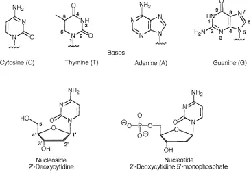
(15) ~H,aLJL''''''. 1 - Introduction. 1.0 Introduction to DNA and its Detection Nucleic Acid Backgroun.d 1.1.1 Primary Structure In all organisms there are only two forms of nucleic acid, deoxyribonucleic acid (DNA) and ribonucleic acid (RNA) possessing the genetic infonnation of life. Each of these high molecular weight acids is composed of individual monomeric structural units called nucleotides. Nucleotides are phosphate esters ofnucleosides and are the building blocks of polymeric DNA. A nucleotide consists of a pentose sugar, a nitrogen-containing heterocyclic base and covalently bound phosphate groups. The bases of DNA and RNA are monocyclic pyrimidines; cytosine (C), thymine (T) and uracil (U) or bicyclic purines; adenine (A) and guanine (G). Nucleosides are assembled by the att.achment of one of the heterocyclic bases to the l' position of a sugar ring fonning a glycosidic linkage. In RNA the sugar is ribose whereas in DNA the sugar is 2'-deoxyribose.. c: r IN~O I. I. 6. NH2. 0. eY~ ~ Y-/'. ~H. l~ N 2 0 1/. ~N. H2N~N. I. N. ~N7 I ~6 3. '4. 75. ....,."..,.. """"'". ~. Bases Cytosine (C). Thymine (T). Adenine (A). Guanine (G). NH2. N~. HO. 8. ~ O~NjJ o-rr-°-Vo-J 8 )---/. 0. OH Nucleoside 2'-Deoxycytidine. Figure 1.1. Nucleotide 2'-Deoxycytidine 5'-nlonophospllate. Structure of four heterocyclic bases, a nucleoside and nucleotide. 1.
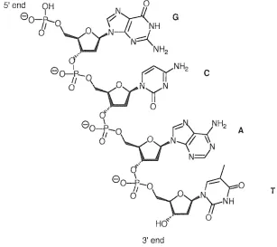
(16) 1 - Introduction The addition of one or more phosphate groups to the 5', 3' or 2' position of the pentose ring gives rise to nucleoside mono-, di- or triphosphates. In DNA and RNA, deoxyribonucleotides or ribonucleotides respectively are joined into a polymer by the covalent linkage of phosphate groups of the 5' hydroxyl of one ribose and the 3' hydroxyl of the next. This linkage is kl10v.m as the phosphodiester bond and it is this that gives the nucleic acid direction. At physiological pH each phosphate group is deprotonated (hence the tenn 'acid') causing nucleic acids to be highly charged molecules.. 1.1.2 Secondary Structure The characteristic secondary structure of DNA is known as the "double helix". James Watson and Francis Crick first deduced the structure of this highly orga.TJ.ised duplex in 1953 1,2 making use of the X-ray fibre diffraction data of Rosemary Franklin3,4 and Maurice Wilkins 5,6 and the infonnation contained in Chargaffs rules (see later). The structure is described as 1:\vo separate, antiparallel chains of DNA. wound around each other following a helical pathway. The coiling produces a double helix that is right-handed; the negatively charged sugar-phosphate chain forms the external backbone. 5' end. 9H. N. 0. eO--!Jv-v{:Ct W. NH2. cJ. eo--k~. 6'. G. --V. ~NH2. /0, .... C. NyN. ". 0. (J. 80~_ II q. ~~NH2. I. ~OVN. o. \.J. r \\. Nd. -L r-Yo. C{. 8. 0. -f-o o. A. T. "(yN/rNH HO":-<. 0. 3'end. Figure 1.2 - Phosphodiester bonds and covalent structure of a DNA strand 2.
(17) 1 - Introduction The planar heterocyclic bases form the core of the helix by stacking one above the other providing considerable stability to the helix. This conformation leads to the formation of major and minor grooves that follow the coiled path of the molecule. The two strands are held together by "base-pairs" formed by hydrogen bonds between individual bases on facing strands and around 10 base pairs make up one tum of the helix.. Under the influence of early work by Avery, MacLeod and McCarty7, Erwin Chargaffin the early 1950's published rules indicating the amount of adenine always equalled the amount of thymine and the same was true for guanine and cytosine in DNA 8. This feature is explained, as the two strands are complementary in terms of sequence. The base pairs are always purine-pyrimidine pairs, where A only pairs with T and G only pairs with C, therefore the sequence of one strand directly defines the sequence of the other.. G:C. A:T. Figure 1.3 - Hydrogen bonding between the DNA base pairs. 1.1.3 The Genetic Code 9 The human genome contains approximately 10 5 genes, encoded by about 3% to 5% of the total 3 x 10 9 base pairs in human DNA. The DNA sequence is found on 23 chromosomes; 22 pairs of autochromosomes and 1 pair of sex chromosomes. A gene is simply a segment of a DNA molecule that codes for the synthesis of a protein, and in eukaryotes is divided into coding regions (exons) and non-coding regions (introns). Each gene can consist of up to 2 million base pairs.. 3.
(18) 1 - Introduction Replication. n DNA. TranscriPt/, RNA. .. PROTEIN. Translation. Figure 1.4. Central dogma of molecular biology. The central dogma of molecular biology describes the flow of the genetic information from DNA through RNA to a final protein product. The sequence in which amino acids in a protein are joined together during protein synthesis is ultimately determined by the sequence of nucleotide bases in a gene encoding that protein. Each amino acid is coded by a codon (groups of three nucleotides). There are 64 non-overlapping codons which make up the" genetic code", 61 coding for amino acids and 3 stop codons. As there are twenty amino acids, the code is degenerate, some amino acids being coded by more than one triplet. The code is universal and the basis of the heredity information of nucleic acids.. In order for the sequence of bases to be deciphered during protein synthesis the doublestranded DNA is transcribed into single-stranded mRNA. The information carried by the mRNA is then used as a template for the synthesis of protein molecules during translation in which other RNA molecules, principally tRNA and rRNA, act as key intermediates 10, 11.. 1.2. Detecting DNA. The fundamentals of using DNA diagnostically involves: (i) defining the nucleic acid sequences that are essential in the expression of proteins associated with disease; (ii) determination of the presence of these sequences in an individual. Nucleic acid hybridisation is the most powerful method for revealing and quantifYing specific DNA or RNA. The association of complementary strands can be exploited in the detection of nucleic acids. A probe is a short piece of DNA or RNA that is labelled to allow its detection, subsequent to hybridisation. A suitable probe system should be highly specific, have low detection limits and be simple to prepare and purifj 12-15.. 4.
(19) 1 - Introduction. 1.2.1 DNA Diagnostics Applications of routine nucleic acid screening depend upon the development of sensitive, rapid, accurate and economical procedures, \vhere ideally PCR amplification is used in conjunction with appropriate probe technology.. Single nucleotide substitutions and insertions and deletion mutations can lead to mutant alleles or gene variants that are associated with genetic disease e.g. sickle cell anaemia 16 , cystic fibrosis, phenylketonuria 17 and Huntington'S disease. These defects can be screened for directly or by probing for linked genetic markers coinherited with the mutant gene 18 . Analysis is important in pre~natal diagnosis and in genetic counselling in affected families for subsequent pregnancies. Genetic information can be also be used to determine susceptibility in individuals to exogenous risks such as diet or environmental factors.. Infectious disease e.g. measles, rubella, HIV19 Hepatitis A and B, salmonella and candida can be clinically diagnosed by the detection of pathogenic organisms by means of DNA probes. Selection of DNA sequence allows tailoring of a test to a particular disease, for example HIV screening of donated blood, or for differentiation between closely related organisms e.g. Herpes Simplex Virus types 1 and 2. Genes related to resistance to antibiotics for treatment of disease can also be screened.. Applications in forensic science based upon the identification of individuals at a genetic level are more accurate than traditional identification by blood types, fingerprints or physical characteristics. DNA can be extracted from body fluids and be used to deduce if individuals are related in paternity testing, or for compatibility in bone marrow transplants. Criminal investigation employs the technique ofDN.A fingerprinting which exploits the highly variable micro satellite sequences of the human genome to yield a "bar code" of an individuapo .. 5.
(20) ,-,U<U.IJ' ..d. 1 - Introduction. 1.2.2 Classical Methods The first DNA probes to be implemented were labelled radioactively. The labels are generally radioactive deoxyribonucleoside triphosphates incorporated directly into nucleic acid molecules by enzymatic methods. These methods employ include nick translation21, random priming22 ,23, polymerase chain reaction 24 ,25 and the 3' end modification by terminal transferase 26,27.. Labelling at the 5'end is carried out by phosphorylation using l2P-ATP and T4 polynucleotide kinase. The choice of labelling strategy is detennined by the nature of the probe, DNA or RNA, double-stranded or single-stranded. The choice of isotope depends on the nature of the probe, the method of incorporation and the sensitivity and stability required. Isotopes such as 3H ,. 1251 and 14C. are often used to label nucleoside triphosphates.. The phosphodiester bond can be modified to contain 32p or 35 S in the case of phosphothioates28 ,29.. 60.2 days. 87.9 days. 12.3 years. 5730 years. Figure 1.5 - The half-lives of commonly employed radioisotopes. An early example of the use of a radiolabelled DNA probe was described by Southern in 19752 8. The technique he describes, known as Southern Blotting, allows the mapping of DNA fragments relative to restriction endonuclease sites. DNA is cut up into fragments by restriction enzymes; the fragments are separated electrophoretically and immobilised on to a nitro-cellulose membrane by blotting.. 32p. labelled probes specific to sites on the target. nucleic acid sequence are introduced and after stringent washing, any bands containing complementary sequences can be visualised by autoradiography.. Quantification of the amount of a specific DNA or RNA present in a sample can be deduced by a similar technique known as dot or slot blotting30 . Here the nucleic acid is hybridised to a membrane as a dot (or slot) carefully washed, and then the intensity of. 6.
(21) ,-,nULlL,",'. 1 - Introduction. probe concentration remaining on the membrane is measured. The reading directly represents the amount of target sequence present in the sample.. Radiolabelling has the advantages of very high sensitivity. For example. 1251 has. a lower. detection limit of lOamol, and radiolabels are easily incorporated. However, there are numerous conflicting disadvantages associated with this technique: ... hazardous handing; regulated by the Home Office short half-life; short shelf-life expensIve limited signal emissions time consuming methodology waste disposal. 1.2.3 Non-isotopic Labelling In order to overcome the disadvantages associated with radioactive labels the search began for safe and convenient alternatives. The most common methods to emerge employed colourimetric31 ,32, chemiluminescent33 , bioluminescent34 or fluorescent 35 ,36 reporter groups. The labels allow the incorporation of more than one label molecule per oligonucleotide 37 and also permit the simultaneous detection of several DNA targets within one experiment. This has also enabled the development of automated DNA sequence analysis as a different coloured fluorophore can be used for each reaction specific for each of the four bases 38 . Combinatorial labelling of probes has been used to increase the number of target sequences that can be detected at the same time 39 .. A label is a molecule that can be attached to a protein or nucleic acid and is capable of releasing a signal. Ideally a label for a DNi\. probe would possess the following properties:. ... attached to DNA or RNA under mild conditions with a simple, cheap and reproducible protocol. ... attachment should not significantly alter the signal generating properties of the label or effect hybridisation properties of oligonucleotide. 7.
(22) ,,",U,CLIJ"'-". 1 - Introduction. detectable at low concentrations using simple instrumentation produce a hybridisation signal which is distinguishable from that of an unreacted signal stable to hybridisation conditions e.g. elevated temperatures, detergents and solvents40 the corresponding reagent is highly specific for its target chemically cheap and variable to permit synthetic modification of functionality (changes to physical properties such as solubility, emission characteristics and charge) co. allow simultaneous detection of several labels in one experiment. co. multiple addition of the label to a single system. co. stable to long term storage. co. easily disposed of. The label essentially consists of three parts, the signal emitting moiety, a spacer and a reactive group.. ,,~__. -. Y. __~). Reactive group. Figure 1.6. \._ _ _ _-.,. .. v. \....._ _...... ,.... _ _~J. Spacer. Signal emitting moiety. ~_--"J. v. Components of a label. 1.2.3.1 The Signal Emitting Moiety In direct labelling strategies this is generally a luminescent 1abel, but can be an enzyme for example alkaline phosphatase directly conjugated to an oligonucleotide probe41 . The label molecule has to either exhibit a satisfactory quantum yield in aqueous solution or turnover a signal-generating substrate. Indirect methods employ either enzymes or haptens (small foreign molecules that can be attached to a macromolecule to which antibodies can be raised) as the label, and the signal is generated sequentially. Enzymes include horseradish peroxidase conjugated to an antibody42 and a common hapten system is based on the highly specific binding between biotin and strepavidin43 .. 8.
(23) '---'UCl.LJL'-l. 1 - Introduction. 1.2.3.2 The Spacer Many simple aliphatic and aromatic molecules have been used to separate the luminescent moiety from the labelled substance44 . An important prerequisite of the spacer is that it does not interfere with the luminescent properties of the label. This bridging group can be used to change the hydrophobicity or hydrophilicty of the molecule, and alter the flexibility of the label relative to the labelled substance thereby assisting hybridisation.. 1.2.3.3 The Reactive Group general terms, this can be any group capable of reacting with a nucleic acid, under mild conditions, to fonn a covalent linkage. There are a few examples of non-covalent labelling of nucleic acids such as the intercalation of ethidium bromide45 in double-stranded DNA, however covalent coupling has proved to be more popular and versatile. Examples of some significant labelling reactions are described:. (i) Free amino groups on DNA or nucleosides can couple with N-hydroxysuccinimide esters, isothiocyanates or activated carboxyl groups of luminescent dyes yielding stable thiourea or amide bonds. The amino groups are either those present on the heterocyclic bases or generated by the derivatisation of 5' or 3' end of DNA. These reactions are usually mild and can be carried out in aqueous solution (a and b, figure 1.7)46-50.. (ii) The nucleic acid is synthesised with a free thiol at one terminus. This undergoes Michael addition with a,j3-unsaturated ketones attached to the luminescent label (c, figure. 1.7)51.. (iii) Labels are functionalised with azido groups which upon activation form highly reactive nitrene intermediates that react with many different types of chemical bonds in the nucleic acids. However this results in non-specific labelling of the nucleic acid52 ,53.. (iv) Labelling of nucleoside triphosphates by the reaction of activated sites on the label and free amino groups attached to the purine or pyrimidine bases at convenient sites. The triphosphates are enzymatically incorporated into the DNA probe by methods such as nick translation or peR Cd, figure 1.7)54. 9.
(24) 1 - Introduction (v) Labels are synthesised as phosphoramidites that can be incorporated into DNA during solid phase oligonucleotide synthesis. One or more of these monomers can be placed internally or at the 3' or 5' terminus of the oligonucleotide (e, figure 1.7)32,35,55 (a). o. H2N~. +. 8-spacer-{ ) O-N. P --,.... 8-spacer---{. HN~. ". (b). 8-spacer-N=C=S. 8-spacer-NH N H - 0. [( S. (c). (0-s. r-y. 8-spacer-~s/~. ~. pace. + HS-@J. -. o. o. o. (d). NH2 I N:::;.-' OH. 0. o. I. \. 18. o. CL ) l NH U Spacer. 80 --,.... 0 3. -. ~:::;.-'. \ ~-. 0. 08. I. 3. OH. OH. (e). -0-1--<. fl'\--spacer---. V. 0. (. 01.,,"000>'". + HO----1'NAl. ~. ~. s~ 0-spacer-0-p-0~. ~. 68. CN. 8. ~ = nucleic acid. = luminescent molecule. Figure 1.7- Popular methods of label incorporation into nucleic acids. 10.
(25) 1 - Introduction. 1.3. Labelling Strategies. 1.3.1 Indirect Labelling. Indirect labelling requires a DNA probe labelled with a small molecule that is detectable by a highly specific binding protein. A classic case is the incorporation of a hapten into a probe that is recognised by an antibody. The antibody is either covalently linked to a luminescent molecule or conjugated to an enzyme (alkaline phosphatase, horseradish peroxidase). The antibody will bind tightly to the hapten and its presence is detected directly, or in the case of an enzyme case by the turnover of a substrate that results in a colourimetric, chemiluminescent or fluorescent product. A more complicated, yet more sensitive method involves the binding of a primary antibody to the hapten of the probe, followed by binding of a secondary antibody that recognises the first antibody. In this case it is the secondary antibody which is labelled for detection. Antibodies that recognise specific haptens are produced in mammals by exposure to the hapten coupled to a protein carrier for immunisation. Substrate \. .,ace Enzyme. \. D'. Y. 0. (E')'----. Q .. Colour or light. ~ Hap!e, ""T """" ""III "'"'' ""'" '""" "'"" "'"'" Antibody. Hybridised DNA probe '''''''' ""'". Target ON. A. Membrane. Figure 1.8 - Indirect labelling strategy. The most commonly studied hapten is biotin56 . Linking to DNA occurs by the reaction of reagents such as N-biotinyl-6-aminocaproic acid N-hydroxysuccinimide ester or biotin hydrazide 57 (figure 1.9) with an amino group on DNA, or by enzymatic incorporation of biotinylated nucleotides. Reporter groups are attached to strepavidin and avidin, proteins (not antibodies) that bind very tightly to biotin, and hybridisation can be detected. Biotin 1 1. ~1.
(26) ~u.aIJL',",J.. 1 Introduction. has the disadvantage that it is present at high levels in certain tissues, leading to high background signals. However, amplification of signal occurs in the presence of extra biotin as there are four possible binding sites on avidin only one of which is occupied by the biotin attached to the probe. Other widely applied indirect detection systems include digoxigenin (DIG) in combination with anti-DIG antibodies, and dinitrophenyl group (DNP) with anti-DNP antibodies 58 ,59.. (i). (ii). HO. H. Figure 1.9 - i, N-biotinyl-6-aminocaproic acidN-hydroxysuccinimide ester, ii, digoxigenin. 1.3.2 Direct Labelling Direct labelling occurs when a reporter molecule is bound immediately to a nucleic acid probe. The specific molecular recognition of a nucleic acid probe annealing to its complementary target sequence enables rapid direct detection60 . The label must not interfere with the hybridisation process and must be stable to hybridisation conditions. The nucleic acid probe can be a long polynucleotide or a short synthetic strand. Synthetic probes are advantageous as they can be easily prepared in large quantities, and the probe length can be used to control the melting temperature of the probe-target duplex. Alteration of the hybridisation temperature can be used to favour formation of the desired duplex over mismatch hybrids, and can allow the assay to proceed at lower temperatures and with shorter annealing times.. \ I /"" 0=J -'". Colour or light. Label Hybridised DNA probell'""' IlIHII!. "liT. "11'" Jill III niH U. In Iill lillill IInll: H1Hili. Target DNA. Membrane. Figure 1.10 - Direct labelling strategy. 12.
(27) ,--,wc<uv'-". 1 - Introduction. 1.3.2.1 Radioactive Labels These are commonly employed and exhibit high signal sensitivities especially when the probe is multiply labelled. However radio labels are associated with numerous disadvantages (section 1.2.2).. 1.3.2.2 Enzyme Labels E!lzymes are covalently attached to nucleic acids. The enzymes can be detected by the turnover of a substrate to produce colour or light. Typical systems use alkaline phosphatase41 ,61 with 5-bromo-4-chloro-3-indolyl phosphate/nitroblue tetrazolium (BCIP/~TBT) or horseradish peroxidase 62 with diaminobenzimide tetrahydrochloride. (DAB). The conditions of the assay must not cause the enzyme to be denatured; therefore the activity of the enzyme must be monitored. These labels offer the highest sensitivities with low background signals and visualisation is rapid.. 1.3.2.3 Chemiluminescent Labels Chemilumescence is the emission of light as a result of a chemical reaction. The ent"IJalpy of the reaction causes an atom to be promoted to a vibronically excited state, when it decays a photon is emitted.. C + Reactant. I>'. Pr* ---_)I> Pr+. tru. Catalyst. The emitter is chemically different from the original molecule therefore one molecule can only produce one photon. However in some cases the product is capable of fluorescing but may only be transiently stable. Chemiluminescent molecules have been used to detect nucleic acids, for example luminol is a chemilwninescent substrate for horseradish peroxidase, and has been used in conjugation with a biotin-strepavidin system63 . Fluorescamine is intrinsically non-fluorescent but reacts in milliseconds with primary aliphatic amines to yield a fluorescent derivative (figure 1.11).. 13.
(28) 1 - Introduction. ---... RNH2. Luminal. CY.. ~ I I O~.. H r.OOH. N. I ~. ~. i. h. Fluorescamine. Figure 1. 11 - Chemiluminescent molecules. 1.3.2.4 Fluorescent Labels Fluorescent molecules absorb light of a specific wavelength and emit light of Imver energy and longer wavelength. Attachment ofthese molecules to nucleic acids pennits direct detection if monitored at the correct emission wavelength. In principle, fluorescence measurements offer extremely high sensitivities, however in practice the sensitivity can be limited by light scattering, background fluorescence and quenching effects. Many fluorescent dyes are commercially available and their excitation and emission wavelengths are represented in figure 1. 12. F----JJ>. F* _ _ _ _JJ>. F + hv Light. The most commonly employed fluorescent moieties include 6-carboxyfluorescein (F AM)35,60,64,65, its tetra and hexachlorinated analogues (TET and HEX) and carboxy-Xrhodamine (ROX)66. These have proved popular due to their high absorption and emission wavelengths and the range of dyes will allow the detection of several targets within one experiment67 . On the negative side, the broad nature of their emission peaks complicates the simultaneous detection of multiple sequences. They are also sensitive to pH68 and susceptible to photobleaching. The inadequacies of these dyes has led to the development of alternatives such as the CyDyes ™and the BODIPY™ spectral range dyes.. 14.
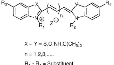
(29) '-..-uuvv,,<. 1 - Introduction. o. CIIl!!I. o. o •. •O •. 0. •. 0. 0. 0. •. •. O. 0. BODIPYTR BODiPY FL. DIll. DIIIII. Texas Red. •. u. Rhodamine Casade Blue Lucifer Yellow. •. r---r---,.---,r---r--r--r-0--r----r---,-,.--...,.-iIII-,--r---r---r-.--.,..--.--r--,. 300. I I I I 400I I I I I 500I I I I I 600I I I I I 700I Wavelength (nm). o. Excitation. •. Emission. Figure 1.12 - Excitation and emission maxima of a range of fluorescent dyes. Cyanine Dyes (CyDye™) These covalent labelling agents, introduced by Amersham Life Science 69 , emit in the farred region of the visible spectrum and have the general structure shown in figure 1.13. These dyes have many of the properties of ideal fluorescent probes including high extinction coefficients, high quantum yields, and excellent photostability. The spectral properties (excitation and emission maxima 500-750nm) are selected by the appropriate choice of heterocyclic nuclei. eX and Y) and the length of the polymethine chain (n). The. groups Rl - ~ are variable, providing the desired functionality, charge, reactivity and solubility. Cyanine dyes are popular, as they are commercially available and pH insensitive. They can be used to detect proteins 70,71, antibodies, peptides and nucleic acids 72, 73.. 15.
(30) ,->U,CUJV'-'i. 1 - Introduction. x + Y = S,O,NR,C(CH3l2 n = 1,2,3, ...... Rl - R4 = Substituent. Figure 1.13 - Generic structure of a cyanine dye. BODIPY™ dyes These versatile dyes which have been patented by Molecular Probes Inc. span the visible spectrum by alteration of the substituents RJ and R2 which can either increase or decrease the level of conjugation74 . The substituents can be used to alter the solubility and the dyes are pH insensitive (figure 1.14). The improved spectral characteristics of these dyes have been exploited in automated DNA sequencing. It is reported that the improved sensitivity allows a reduction in reagent consumption by 33%, compared to conventional dye primers 75 .. Rr R2 = Substituent R3 = Linker. Figure 1.14. Generic structure of a BODIPY dye. 1.3.2.5 Lanthanide Labels Lanthanides form chelates that are highly fluorescent with large Stokes shifts and extremely long lifetimes 76 -78 . DNA has been directly labelled with lanthanide chelates by incorporation of labelled deoxynuc1eoside triphosphates by nick translation, random priming, peR and ruthenium phosphoramidites. Hurskainen et ai. describe a method using the amino groups on cytidine, which undergo a transamination reaction in the presence of sodium bisulfite diamine. The free primary aliphatic amine groups react with an 16.
(31) '"-'uuvv'"',. 1 - Introduction. isothiocyanate derivative of an europium chelate. This process causes multiple labelling of the DNA, which can affect the efficiency of hybridisation by significantly altering the melting temperature of the probe-target duplex. It has been reported that the optimum system contains four to eight europium chelates per hundred bases. The sensitivity of these chelates is comparable to the detection limits achieved with radioisotopes. NH2. ~Jp. ~N~ I. . H2N~. NH 2. NaHS03 , pH 6. Figure 1.15. 1.4. Labelling of cytosine with a europium chelate. Fundamentals of Fluorescence. 1.4.1 The Nature of Fluorescence 79 ,80 Fluorescence is the property of some atoms and molecules to absorb photons of incident light of a particular wavelength (excitation wavelength) and after a short period (fluorescence lifetime) re-emit a lower energy photon (emission wavelength). The process begins when a fluorophore absorbs a photon from an external light source, which causes the promotion of an electron from its ground state (So) to the first excited singlet state (Sl). The excited molecule then undergoes a conformational change and is subject to collisions with surrounding molecules. These interactions cause discharge of thennal energy from the molecule to its environment (internal conversion). After a short fluorescence lifetime the electron falls back to the ground state and a photon of lower energy, longer wavelength is emitted (fluorescence). Each fluorophore can repeat the cyclic process many times before. 17.
(32) 1 - Introduction photobleaching prevails. The 'shift' in energy or wavelength from excitation to emission is called the Stokes shift and this is fundamental to the sensitivity of fluorescence measurements, a large Stokes shift leads to low background signals.. The fluorescence quantum yield is the ratio of the number of fluorescence photons emitted to the number of photons absorbed. It is a measure of the emission efficiency of a fluorophore. Physical measurements can be complicated by background signals that may originate from endogenous sample constituents.. 2---------------S2. 1. o. ;. ,. i, Internal conversion(10· 2s) 1. ; J. Absorbtion (10'1 5S ). Fluoresence (1 O,9S ). 2 So. 1. o. Figure 1.16 - Jablonski energy level diagram. 1.4.2 Fluorescence Quenching 81 ,82 The fluorescence energy of an excited fluorophore can be transferred non-radiative1y to a molecule in close proximity resulting in the loss of fluorescence emission. No photons are emitted and the phenomenon is known as quenching. There are two main quenching processes both requiring molecular contact between fluorophore and quencher:. (i) Collisional (or dynamic); quencher molecules collide with the excited fluorophore and energy is dissipated as heat, significantly reducing the fluorescence lifetime of the fluorophore. Collisional quenchers include molecular oxygen, acrylamide, nitromethane, purines, pyrimidines and some olefins.. (ii) Static (or complex); a complex is formed between the fluorophore and quencher which is non-fluorescent. Alternatively resonance energy transfer (section 1.4.3) quenches the fluorophore. 18.
(33) '--'~'lUIJ.'-'~. 1 - Introduction. Collisional quenching only affects the excited states of the fluorophore and therefore no changes in the absorption spectra are observed. In contrast, ground state complex formation will result in perturbation of the absorption spectrum of the fluorophore.. Ex. II. ;v·'-I. I"',~. F+a Collisional quenching. no emission. 'V. 'V. '#. Fluorescence emission. (F. 0)". F*. F*. ---"'ks. Fluorescence emission. F.O Static quenching. Figure 1.17 Principles of collisional and static quenching. 1.4.3 Fluorescence Resonance Energy Transfer (FRET) An excited fluorophore (donor, D) may transfer its energy to a neighbouring chromophore or fluorophore (acceptor A) non-radiatively through induced dipole-induced dipole interactions. This occurs when the dipoles are in an approximately parallel orientation. The criterion for occurrence is the overlap of the emission spectra of the donor and the excitation spectmm of the acceptor. Forster in the late 1940' s first proposed a theof'j 83 , which was later con finned by Stryer and co-workers 84 , describing long-range molecular interactions by resonance energy transfer. His theory related the interchromophore distance (r) and the spectroscopic properties of the chromophore. The rate of energy transfer between the donor and the acceptor is inversely proportional to the sixth power of the distance (1/r6 ). The optimum distance (r) for non-radiative transfer of energy is between 10-. IooA and it is this distance that governs the efficiency of the process. The extent of energy transfer can be measured because the fluorescence of the donor (both intensity and lifetime) is significantly reduced, and the acceptor, if it is fluorescent, increases in emission (sensitised emission).. FRET has been used in many biochemical and structural biological applications as a qualitative or quantitative tool. The subject has been extensively reviewed with references to applications for peptide and protein interactions and the use of FRET as a spectroscopic. 19.
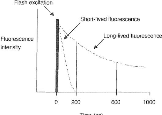
(34) Chapter 1 - Introduction ruler84 -87 . FRET is an extremely powerful tool for probing nucleic acid structure 88 , sequence 89 and hybridisation90 . Examples include examining triple helical DNA91 and the structure of a four-way DNA junction at varying salt concentrations 92 .. .",, .·:: -..... . ···· .... Donor Absorbance Normal Donor Emission. ~. Quenched Donor Emission Acceptor Absorbance Acceptor Emission Sensitised Emission. Figure 1.18 - Illustration of quenched donor emission and sensitised acceptor emission. 1.4.4 Time Resolved Fluorescence 93 ,94 Fluorescent measurements are plagued by background signals arising from Raman and Rayleigh scattering or the emission of organic fluorophores within biological samples (350600nm). This can limit the sensitivities of the commonly employed short-lived fluorescent dyes (10-9 to lO-1O S) . However, the use offluorophores with long fluorescent lifetimes permits selective detection of signals, by delaying measurements until all other species have decayed. For an interference-free measurement of a signal, the label lifetime must be at least ten times longer (> lOOns) than the decay of background signals (::::; IOns). It was the advent of rare-earth metal ion complexes used as labels that transformed the. technology of time-resolved fluorescence. The chelates ofEu (III)77, Tb (III)78, Sm (III) and Ru (III) with aromatic chromophores, all have long fluorescent lifetimes (IllS to 1ms), large Stokes shifts (>200nm), narrow emission bandwidths (10nm) and long emission wavelengths.. Luminescence of these chelates is not strictly fluorescence or phosphorescence. The organic ligand absorbs energy and is promoted from So to the excited SI state, where it rapidly loses energy (intersystem crossing) and falls to the excited Tl state. From this level. 20.
(35) 1 - Introduction the molecule can either return to So ground state (phosphorescence) or transfer energy intramolecularly to the 4f energy level of the central metal ion. If this phenomenon occurs the metal ion moves into its own Sl state and can decay to So emitting radiation (ion fluorescence). When a sample is excited by short light pulses short-lived fluorophores will quickly dissipate to zero. Time-resolved fluorescent measurements are taken after this period eliminating unwanted background signals.. DELFIA® (dissociation enhanced lanthanide fluorescence immunoassay, Wallac Oy, Turku, Finland) is a commercially available assay format that uses a non-luminescent lanthanide chelate (e.g. diazaphenyl-ethylenediamine tetraacetic acid-Eu (III)) as a label for an antibody or nucleic acid. The final step of the assay involves lowering the pH to induce dissociation of the lanthanide complex and capture of the lanthanide in a solubilising micelle. The micelle contains reagents capable of forming a luminescent chelate with the ion (e.g. fluorinated aromatic ~-diketones) enabling detection95 . Flash excitation. I~ Fluorescence intensity. II. Short-lived fluorescence. ''-'/ . '. ,. ". ". Long-lived fluorescence. / '". ..... I. o. 200. 600. 1000. Time (ns). Figure 1.19 - Principle of time-resolved fluorescence. Enzymes as either labels or analytes can be detected by the turnover of a substrate to release a product that can chelate witl) lanthanide ion and fonn a luminescent complex. An example of this is the glucose oxidase substrate 1,10 phenanthroline-2,9-dicarboxylic acid dihydrazide that gives the detectable chelate Eu (III)-phenanthroline-dicarboxylic acid96 . 21.
(36) Chapter 1 - Introduction. 1.4.5 The Fluorescence Spectrometer The basic task performed by a fluorimeter is delivery of excitation energy to the fluorescencing species, and separation of the weaker emitted light from the brighter excitation light97 . This ensures only emitted light is detected and a sensitive and defined image is generated. Most modem spectrometers consist of four main elements; an excitation source, wavelength selection devices, the sample cell, and a detector. The basic construction can be seen in figure 1.20. Light emerges from the source (e.g. xenon or mercury lamps) and enters the excitation monochromator. In modem spectrometers, monochromators are based on diffraction gratings, not prisms, allowing the selection of a narrow band of light by rotation of the concave grating. Typical monochromators have entrance and exit slits, which are variable. Large slit widths produce increased signal levels but with higher signal: noise ratios, smaller slit widths give higher resolution but at the expense of light intensity. The chosen wavelength of light travels to the sample, the emitted light generated then passes through the emission monochromator to a photomultiplier. Here, incident photons hitting the photocathode are accelerated through a series of dynodes towards an anode, this causes the signal to be amplified and the final output is proportional to the emitted light intensity (figure 1.20).. --s. L =lens S = variable slit C = sample cell Mex =excitation monochromator Mem = emission monochromator P = photomultiplier. -s. P. PC. Figure 1.20 - Typical set-up of a fluorescence spectrometer. 22.
(37) '-'HUIJL'.... '. 1 - Introduction. To measure the excitation spectrum of a fluorophore, the emission monochromator is set at a desired wavelength (usually the emission maximum) and the excitation monochromator is scanned through the absorption bands. To measure the emission spectrum the appropriate excitation wavelength is chosen and wavelength scanned with the emission monochromator to detennine the distribution of light emitted by the sample.. Fluorescence spectroscopy is a highly sensitive technique; signals can originate from sources other than those of the fluorophore of interest. Interference can arise due to Rayleigh and Raman scattering, background fluorescence of solvents, light leaks in the instrumentation or stray scatter due to particulates. Acquisition of spectra is complicated by the wavelength-dependent nature of the source, monochromators and photomultipler tubes all playing an important part in instrument design 98 .. 1.5. Molecular Biological Methods Utilised in Fluorescence Assay. 1.5.1 Polymerase Chain Reaction The polymerase chain reaction (peR) is a technique used widely in molecular biology 99 to amplify a section of target DNA that is flanked by two kno'vvn genetic sequences. Two short oligonucleotides are prepared (usually synthetically) and are designed such that each is complementary to one end of the target strand. These oligonucleotides are known as primers, and are typically 18-30 bases in length, with similar (%G+C) content to ensure similar annealing temperatures. The region of the template bound by the primers is amplified by a series of cycles. In the first cycle the duplex target is dissociated into two single stands by heating to 95°C, subsequent cooling to approximately 55°C allows the primers to anneal with their 3' ends pointing towards each other. The temperature is now increased to 72°C, the optimum temperature for activity of the thermostable Taq polymerase. This polymerase is isolated from the thermophilic bacterium Thermus aquaticus and has a half-life over two hours at 95°C so is suited well to the experiment 100. The polymerase, in the presence ofMg2+, uses nucleotide triphosphates to extend the primers along the length of the target producing two new double-stranded sections of DNA.. 23.
(38) Chapter 1 - Introduction The second cycle begins again by heating to 95°C effecting the denaturation of the newly synthesised molecules. Each single strand now acts as a template for primer annealing and extension. During the second cycle primers annealing to newly synthesised molecules can only be extended as far as the first primer, affording molecules of the correct length. All of the following cycles amplify the correct length products, which soon outnumber the original target molecules. In practice n cycles will amplify a particular sequence 2n times 24 , 101 .. ~: Target DNA. 3' 5'. (i). 3h==. y. 3'. 5'. !:$. ... -. ,../ 5'. /. -. .(ii). 5'. ~. ---- .............. 5'. -. Cycle 1 : (i) denaturation (ii) primer annealing (iii) polymerisation. ,../. 5'. c. 3'. '---. .. 3' .,---. .............. --. .,--c. Cycle 2: (i) denaturation (ii) primer annealing (iii) polymerisation. .. .... 5'. 3'. 5'. 3'. "". -............... •. '--- 3'===-. (iii). 5'. -. .,---. -. ... 3'. c. -I>. C. .-. .... Cycle 3: (i) denaturation (ii) primer annealing (iii) polymerisation. - - DNA bracketed by the primers -. DNA outside primer region. -~. Primer 1. -. Primer2. Figure 1.21 - The first three cycles of a polymerase chain reaction. 1.5.1.1 Hot Start peR Hot start PCR is designed to alleviate some of the problems associated with background signals in PCR such as primer-dimer formation and non-specific priming. This technique is carried out manually. Initially the reaction mixture lacks a crucial component such as magnesium ions or Taq polymerase. Heating during the first cycle ensures there is no binding, at this point the tube can be opened and the missing component added. PCR now. 24.
(39) 1 - Introduction continues as nonnal. The major disadvantage of this technique is the possibility of contamination during the addition ofthe missing component to the open reaction tube. Alternatives include adding a reagent that can bind to the polymerase and inhibit its action (e.g. an antibody) which is denatured in the first heating cycle102 , or enclosing an essential component in wax, again released upon heating.. 1.5.2 Fluorescence In Situ Hybridisation (FISH)103 Fluorescence in situ hybridisation is an essential tool in genetic analysis as it allows the identification of the presence and location of cellular DN.A or RNA within morphological preserved chromosome preparations with sensitivity and ease. The principle lies in the annealing of a labelled probe to its complementary strand in fixed cells or tissues followed by detection of the label (radioactive or lwninescent). FISH is different to other probe systems as the target is embedded in a complex matrix that can hinder probe access and destabilise probe: target hybrids formed. The probe (DNA or RNA) is usually prepared by one of four methods: nick-translation; random priming; peR or chemical synthesis and is labelled during each process. The length of the probe is dependent on the application, but the typical length is between 200 bp and lkb. Longer probes increase non-specific background but short probes can be difficult to detect due to insufficient hybridisation, high probe concentrations decrease the signal: noise ratio. It is important that the target is accessible to the probe and must be retained in situ, not degraded by nucleases. Visualisation limits span from an entire chromosome to a 40kb chromosomal section. FISH employs both indirect and direct labelling strategies, however methods generating signal amplification have proved most popular.. FISH has been used in toxicological studies to monitor the effect of exposure to radiation on chromosomal aberrations (structtl.ral and numerical) 104. Chromosome painting 105 ,106 by combinatorial or ratio labelling of specific probes has led to the painting of all twenty four different human chromosomes being labelled with distinct colours which can provide a general screening test for chromosome abnormalities. Other applications include monitoring changes in aneuploidy in sperm (a major cause of birth defects) and genetic mapping by the use of probes labelled with multiple colours allows gene order along a. 25.
(40) Chapter 1 - Introduction chromosome to be determined l07 . Complex and unusual structural rearrangements in genes that lead to tumours can be assessed by comparative genomic hybridisation (CGH). Two probes are generated one specific for normal DNA and the other for tumour DNA, each are labelled differently. Equimolar amounts of the probes are introduced into the test cells; the nature of the signal is a measure of the extent of hybridisation of one probe over another 108.. Figure 1.22 - Human metaphase chromosomes stained with DAPI (Image from Violette Paragas and Jeff Pollack, Molecular Probes, Inc.). 1.5.3 UV Thennal Melting. The aromatic bases of nucleic acids absorb UV light with a maximum absorption at around 260nm. This absorption is constant for the bases, however the extinction coefficient depends on the environment. Individual nucleotides absorb the greatest quantity ofUV light, followed by single-stranded DNA and least of all double stranded DNA. The reduction in absorption from single to double stranded DNA is caused by the fixing of the bases in a hydrophobic environment by stacking, the change is known as hypochromicity.. Heating double stranded nucleic acids causes denaturation by disrupting the ordered stacking of the bases through breakdown of hydrogen bonds. The process can be conveniently monitored by an increase in UV absorbance as the double strands unwind to. 26.
(41) 1 - Introduction single strands. The thermal denaturation of dsDNA is co-operative indicating that the ends and the AT-rich internal regions de stabilise adjacent regions of the helix. This leads to a progressive and concerted melting of the whole structure at a well-defined temperature, corresponding to the mid-point of a smooth transition. This temperature is known as the melting temperature (Tm). UV melting experiments can provide both quantitative and qualitative data about the nature, purity and degree of hybridisation of a sample.. 1.6. Practical Examples of Fluorescent Probe Systems. 1.6.1 Taqrnan™ Assay The Taqman™ assay is a widely used format for the detection of accumulation ofPCRspecific products 109, 110. A linear probe is assembled consisting of an oligonucleotide labelled with a fluorogenic donor (5' FAM) and acceptor (internal or 3' TAMRA). Irradiation of the intact probe causes excited emission by the donor, which is quenched by the process ofFRPT (section 1.4.3). The probe can hybridise to the complementary template and during polymerisation the probe will be cleaved due to the inherent 5' ~ 3' nucleolytic activity ofTaq DNA polymerase. Cleavage causes separation of the donor and acceptor that resulting in an increase in fluorescent intensity, due to loss of quenching. Measurement of the rise in fluorescent intensity directly indicates the generation of probespecific amplicons.. The design and performance of the probe requires three main considerations:. (i) Degree of quenching; dependent on choice and separation of the fluorophore and quencher pair, the conformational flexibility and purity of the probe.. (ii) Hybridisation of the probe; internal quenchers can disrupt base pairing and reduce Tm, a 2~3°C reduction in Tm has been reported for tvvo internal TAMRA molecules 109 .. (iii) Efficiency of probe cleavage by Taq polymerase; dependant on the accessibility of the probe to the enzyme and the complementarity between the probe and template.. 27.
(42) Chapter 1 - Introduction 1. Polymersation Forward 5' primer. Q Probe ,.. 3"==~"~:::::~---. 5'. •. Quencher. o. Fluorophore (quenched) Fluorophore. 2. Probe Displacement. 5,~~===f~~~~3~'___ 3~. 5'. 3. Cleavage of Probe. ,, '\.."..... 5' 3,======~"::----I'l==~3r.' _ _ _ 5' 4. Completion of Polymerisation. -. , ... 5'. '-'". -". 3'. ...... 3"~~============~·~5'. Figure 1.23 - The Taqman™ assay. The Taqman fonnat has applied to be rapid and accurate detection of hepatitis C virus in 48 patient samples 111 and detection of salmonella in food samples in less than 20 hours producing no false negatives or false positive results 112. The assay has also been used to develop a test for parental DNA for a deletion sequence linked to the genetic syndrome cutaneous malignant melanoma 113.. 1.6.2 Molecular Beacons Tyagi and Kramer have described the recent discovery of a novel probe technology for the detection of specific nucleic acids in homogeneous solution l14 . These probes, named 'Molecular Beacons', fluoresce only upon hybridisation to their target. They produce sensitive, real-time signals that indicate the degree of hybridis ation of the probe to a target nucleic acid. The essential feature of the probe is their stem and loop structure. The loop portion is an oligodeoxynucleotide sequence that probes for its complementary target nucleic acid in solution. The stem is constructed of two short oligodeoxynucleotide arms. 28.
(43) Chapter 1 - Introduction either side of the loop, one ann is tenninally labelled with a fluorophore and the other with a quencher. Ideally the quencher should be non-fluorescent. Annealing of the anns causes intennolecular energy transfer from the fluorophore to the quencher and this energy is dissipated as heat. In this confonnation the probe is non-fluorescent. Hybridisation of the loop to its target causes a confonnational change, the stem anns dissociate, and the fluorophore and quencher are no longer in close proximity; fluorescence is restored.. Molecular Beacon Probe Non-fluorescent. +. Target Nucleic Acid. Probe-Target Hybrid Fluorescent. Figure 1.24 - Principles of a molecular beacon assay. The original molecular beacons synthesised by Kramer' s group utilised 5-(2'aminoethyl)aminonapthalene-1-sulfonic acid (EDANS) as a fluorophore and 4-(4 ' dimethylaminophenylazo) benzoic acid (DABCYL) as the quencher. DABCYL was later shown to be an efficient quencher (98%) ofa series of spectral dyes (emission maxima 475615nm) due to Van der Waal contact of the fluorophore and quencher pennitting direct energy transfer 115.. H. ~~NH2. ~ 0=1=0 08. Na@. EDANS. Figure 1.25 - Structure of a commonly employed fluorophore and quencher pair 29.
(44) '-../H'U.IJV...... 1 - Introduction. Molecular beacons must be designed so that the arm sequences are unrelated to the target and are long enough to form a stem (>4 bp) but not so long that dissociation is difficult «12 bp). In addition, hybridisation of the arm sequences in the hairpin loop must produce a weaker interaction than target-to-probe annealing.. Molecular beacons have been used to detect and quantify PCR ampIicon concentration by the intensity of fluorescence at the mmealing stage in each cycle. They are pa.rticularly suited to this application, exhibiting fast hybridisation kinetics and allowing sealed tube experiments. The probes exhibit allelic discrimination, only binding to their perfectly complementa.ry targets and have the ability to recognise single base mutations. These principles have allowed the use of multicoloured probes in a single experiment 115,116.. Practical examples of their application to real systems include detection of the single point mutation of cytosine to thymidine in the methylenetetrahydrofolate reductase (MTHFR) gene. This mutation has been related to an increased risk of cardiovascular disease and neural tube defects. Molecular beacons specific for the wild type or mutant sequence demonstrated high levels of specificity for their target 117. it mutant form arising from a conservative substitution (V641) in the coding region of the CCR2 genotype has a protective effect against HIV-l disease progression. A molecular beacon assay that accurately discriminated between the wild type labelled with fluorescein, and mutant form labelled with hexachlorinated fluorescein has been described 118 .. In addition the probes have been used for the detection of mRNA119, 120, in surfaceimmobilised hybridisation studies 121 , as PNA-DNA hybrid probes 122 , sequence analysis of amplified segments of Mtuberculosis DNA123 and in the spectral genotyping of the alleles of the ~-chemokine receptor S (CCRS)19.. 1.6.3 Scorpion Primers A recently described technology combines a highly specific probing region with a primer for a peR reaction65 . The assay is unimolecular and therefore kinetically and. 30.
(45) Chapter 1 - Introduction thermodynamically favoured. The probe region is chemically attached to the 5' end of one of the PCR primers and is blocked against copying by a hexaethylene glycol spacer. The primer binds to the target and is extended by polymerisation. The newly synthesised strand contains a sequence complementary to the probe sequence, annealing occurs and fluorescence is detected. The principal superiority of the assay is its unimolecular nature, inferring zero-order kinetics increasing the efficiency of the probing process (the probe is held in close proximity to its target) and reducing the time-scale of hybridisat ion. Intermolecular systems (TaqmanTM109 and molecular beacons 124) are limited by the formation of alternative intra-strand secondary structures at the assay temperature and the competition of re-annealing complementary PCR amplicons.. Scorpions have shown high specificity in single tube genotyping experiments of the two main variants of the Hereditary Haemochromatosis gene: C282Y, the major disease causing mutation and H63D, a prevalent polymorphism of uncertain clinical relevance. Scorpions have also been shown to accurately distinguish between matched and mismatched allelic polymorphs of ex on 10 of the BRCA2 gene.. Scorpion in Dark State •. o. Quencher Fluorophore (quenched) Fluorophore. I. Blocker. ~PCRprimer. Primer Extension. Probe-Target Hybridisation. Figure 1.26 - Stages of a Scorpion detection assay. 31.
(46) Chapter 1 - Introduction. 1.6.4 Invader Assay. Third wave technologies have described an isothermal, quantitative assay that requires no PCR and generates amplified signals upon probe-target hybridisation 125 . Termed "Invader Assay" the system is comprised of two probes that hybridise adjacent on a target nucleic acid. The probes are designed so that the 3' end of the upstream Invader probe overlaps the 5' end of the dO\VIlstream, labelled, signal probe. This invasion of the signal probe-target duplex region causes the displacement (by at least one base) of a portion of the 5' signal probe, creating a branched structure with a single stranded flap. The junction between the flap and the partially invaded duplex is recognised and cleaved by the Cleavase ™ enzyme 126.. Hybridisation of probes. 111;m 1111111 11. Target DNA Invader probe. J. Cleavase binding and cleavage. ",;W IIIII IIII .......... Signal probe Cleavase enzyme. ! :t. 1111110. ........... J. ~~. ". Dissociation of cleaved probe. ! 1117irrn 11111 11. Amplification. Figure 1.27 - Third wave Invader assay format. Detection can be effected by direct gel analysis, FRET or mass spectrometry. The relatively high temperature of the assay causes the cleaved signal probe to dissociate and another intact signal probe to anneal, whilst the invader probe remains bound to the target. Providing the signal probe is in excess there will be an amplification of signal. It has been shown, using Human cytomegalovirus genome as a target, that cleavage generates a signal. 32.
(47) 1 - Introduction. that is proportional to the number of copies of target DNA present. Carry-over contamination, false-positives and background signals are all alleviated as signal is only procured in the presence of a target 127 .. l.6.5 PNA Probes. PNA oligomers are not substrates for DNA, RNA or protein modifying enzymes, and will hybridise to DNA and RNA by normal \Vatson and Crick base pairing128. They bind strongly under conditions in which normal nucleic acids hybrids are disfavoured, for example at low salt concentrations. PNA probes are assembled by standard FMOC or BOC chemistry and C~'1 be labelled directly with fluorescent dyes 122 , enzymes, or visualised indirectly with haptens such as biotin and dinitrophenol. PNA probes have high specificity, affinity and stability at low salt concentrations in comparison with DNA probes. DNA probes often fail to distinguish certain regions of highly structured rR..NA. However PNA probes detect these highly structured rRNA regions with significantly higher fluorescent signals, exhibiting improved performance at high temperatures and low salt concentrations.. l.6.6 SUNRISE Primers. An amplification system has been developed called SUNRlSETNI The basis is the incorporation of fluorescence energy-transfer labelled primers into PCR products. These primers have specific target sequences at the 3' end and hairpin structures at the 5' end that are non-fluorescent prior to PCR. Incorporation of the primer during PCR causes the hairpin to be disrupted a,'ld fluorescence is detectable. The fluorescent signal intensifies with increasing number ofPCR cycles as more primers are incorporated I 29.. Following this a Universal SUNRISE Primer was reported to allow the incorporation of an ~d"'ntl·cal ~~l·me" i \..~ hal·rp~n 1 "1-1-1 JJI 1.. 1 .1. ;nto ".lJ an'\! tArget nnAl",;,., "a .1.1. U.\o..IL'-'J.\.I. "c~d130 u .1 . •. An ;nl·tl·al PI'R st"'p l·ncorpo~"'+"'s " 1u(,.\.,.o U l.l .1 1.. ~1-. \,.;.L. primer that carries an oligonucleotide tail (15 bases) that is complementary to the sequence of the universal hairpin primer. Following incorporation of the tail, the hairpin primer takes over and is unwound in the same manner as before. 33.
(48) ,,--,u,~ ..n~i. 1 - Introduction. The primers have been successfully applied to a closed tube amplification detection fonnat to distinguish between the normal (\VG4) and mutant (RG4) alleles ofthe. ~(3)-adrenergic. receptor gene 131.. 1.6.7 DNA Chip Technology DNA chip technology utilises high-density microscopic arrays of nucleic acids immobilised on solid surfaces for biochemical analysis. DNA or RNA samples from biological sources are labelled enzymatically by the incorporation of labelled primers or nucleoside triphosphates during peR amplification. Hybridisation of the targets to the microarrays and subsequent detection can be used for polymorphism detection, DNA sequencing and genotyping. For example analysis of the CFTR gene was carried out by the preparation ofa microplate loaded with 428 features to identifY mutations in exon 11, and a microarray containing 96,600 20mers was used to identifY mutations over the entire 3.45 kb of exon 11132.. The oligonucleotide arrays are often prepared on glass due to its inert chemical properties, low intrinsic fluorescence a.lld the ability to derivitise its surface. The strategies for the preparation of these libraries using photolithography and micro-spotting, has been excellently reviewed by B.Lemieux et al. 133 and E.M. Southern 134. 34.
(49) Chapter 2 Development of Novel Homogeneous Assay Systems.
(50) Chapter 2 - Homogeneous Assay. 2.0 Development of Novel Homogeneous Assay Systems. The preceding chapter introduced the reagents, techniques and strategies employed in the design of systems to probe genetic sequences. The following chapter aims to record the work carried out in the design, synthesis and testing of a novel two-probe assay.. 2.1. Introduction. The aim of this project was to further develop the use of fluorescence as a detection method for DNA sequences. When designing a novel assay the incorporation of the following features was considered to be beneficial:. (i) Homogeneous assay (all components in solution) is advantageous over its heterogeneous counterpart 135 (solid and solution phase components) as it does not rely on the hybridisation of a probe to an insoluble support and therefore displays faster hybridisation kinetics. In homogenous assay formats there is no requirement for washing steps as the signal is generated only when the labelled probe is hybridised to the target.. (ii) Enzyme-linked probes are highly specific for their substrates and provide high detection sensitivities, similar to those attained using radioisotopes 61 .. (iii) Isothermal signal generation simplifies an assay by reducing the number of steps perfonned.. (iv) In certain cases, the use of more than one probe in an assay increases the specificity for a particular target as two or more adjacent nucleic acid sequences can be probed.. An assay was proposed (figure 2. 1) that required a probe labelled at the 5' end that is initially non-fluorescent and complementary to a known sequence on a target nucleic acid (signal probe). This probe is 'dark', as the fluorescent label it bears has been chemically modified with masking groups to remove its fluorescent characteristics. The masking. 35.
(51) Chapter 2 - Homogeneous Assay groups are substrates that can be cleaved from the label by an enzyme-catalysed reaction. A second probe consists of the conjugate formed when an enzyme is linked to the 3' end of an oligonucleotide (enzyme-probe). The enzyme-probe is complementary to a sequence downstream (further along the target DNA) of the upstream signal probe sequence on the target DNA. There will be little or no fluorescence from either the signal probe or enzymeprobe individually in solution. When the two probes are mixed there may be a small amount of background fluorescence detected due to the enzymatic hydrolysis occurring when the two probes meet in solution. However, in the presence of a target nucleic acid the two probes can hybridise adjacent to each other, bringing the 3' enzyme and the 5' substrate molecules into close contact. Cleavage of the masking groups by the enzyme will release a strong fluorescent signal, which can be detected. (i). 1. (ii). ~. J (iii). ~. J. o. Nonfluorescent label. •. Fluorescent label Enzyme. Q. Substrate protecting groups. -. Probe DNA. -. Target DNA. (iv). Figure 2. 1- (i) An oligonucleotide is labelled with a fluorophore (5') which has been chemically modified to remove its fluorescent properties (signal probe), the probe is introduced to the target nucleic acid. (ii) The probe hybridises to its complementary sequence on the target nucleic acid. (iii) An oligonucleotide labelled with a enzyme (3 ' ) is introduced (enzyme-probe). (iv) The oligonucleotide-enzyme conjugate hybridises adjacent to the signal probe, the enzyme cleaves the substrate groups attached to the fluorophore and fluorescence is visualised.. 36.
(52) Chapter 2 - Homogeneous Assay The assay is desirable as is homogeneous, isothermal, requires two probes and exploits enzyme detection. The close proximity of the substrate and enzyme is caused only by hybridisation of the two probes adjacent to one another on a target; consequently fluorescence is only generated in the presence of a target nucleic acid. The assay format is sensitive, specific and the fluorescent signal generated \vill be directly proportional to the number of enzyme-probes hybridised.. Fluorescein is a well-known dye that will only fluoresce when its phenolic hydroxyl groups are unsubstituted. This allows the lone pairs on the oxygen to participate in the highly conjugated delocalised system and stabilise the lowest excited state. When the phenolic hydroxyl groups are unprotected the wavelength of emission is in the visible region and fluorescence is observed. However, protection of the hydroxyl groups masks this fluorescence by shifting the wavelength of emission into the ultraviolet region. A nonfluorescent probe can be created by attachment of a phenol-protected fluorescein monomer to the 5'end of an oligonucleotide.. 2.2. Alkaline Phosphatase Approach. 2.2.1 Introduction. Alkaline phosphatase catalyses the hydrolysis of phosphomonoesters to produce inorganic phosphate and the corresponding alcohol. It is a relatively small enzyme with a molecular weight of about 89 kDa and is dimeric in structure. Alkaline phosphatase has been applied to indirect 136 and direct labelling strategies61 ,62 in nucleic acid hybridisation probe technology with detection reported at the level of one attomole 137 . Indirect procedures described in the literature involve attachment to a hapten or to an antibody specific for a hapten, in both cases detection is achieved by enzymatic turnover of a substrate releasing light or colour. Probe assembly in direct methodologies involves either covalently attaching the enzyme or the substrate to an oligonucleotide. Hybridisation is measured by the increase in colour or light caused by turnover of the substrate such as 5-bromo-4-chloro-3indolyl phosphate I nitroblue tetrazoliuIn (BCIP/NBT). The chemistry involved in. 37.
(53) Chapter 2 - Homogeneous Assay conjugation of alkaline phosphatase to oligonucleotides is routine 138 presenting a convenient starting point for assay development.. Preparation of a signal probe required the attachment of a label modified with substrates for alkaline phosphatase (phosphate esters). Fluorescein diphosphate was chosen as a suitable label modified with a reactive spacer for attachment to an oligonucleotide (figure 2.2). Several synthetic approaches to prepare the signal probe are described in the following sections.. eQ. 0,. I. oe. p, e d 0. '/. eif'\\o. H. N. o. R. o-p.-o~. ~. 6e. oligonucleotide. Figure 2.2 - Fluorescein diphosphate signal probe. 2.2.2 Solid Phase Strategy This approach involved synthesising a fully protected phosphoramidite monomer of fluorescein that was compatible with standard solid phase DNA synthesis 139. The monomer could be coupled to the 5' end of a resin bound oligonucleotide on a DNA synthesiser, and following selective removal of the fluorescein protecting groups, on-resin phosphorylation of the phenolic hydroxyls could be attempted. The choice of protecting groups for tl}e phenolic hydroxyls of fluorescein was imperative, as their selective removal required orthogonality between these protecting groups and those of the heterocyclic bases. In addition any deprotection reagents applied had to leave the oligonucleotide-resin linkage intact allowing subsequent on-resin phosphorylation.. 2.2.2.14,4'-Dimethoxytrityl Ether 4,4' -Dimethoxytrityl ethers are acid labile and repeatedly removed from the 5' hydroxy! of nucleosides during solid phase DNA synthesis, inferring compatibility for protection of the. 38.
Figure


+7


Related documents