Int J Clin Exp Pathol 2017;10(3):2461-2477 www.ijcep.com /ISSN:1936-2625/IJCEP0039841
Original
Article
Identification and function analysis of macrophages
subsets in chronically inflamed human epididymis
Wenzhong Zheng3*, Yan Wang1*, Jianbo Chen1, Yuhong Zhu1, Minghua Li1, Longlong Fan1, Xianxin Li1,2 1Department of Urology, Peking University Shenzhen Hospital, Shenzhen PKU-HKUST Medical Center, Shenzhen, P. R. China; 2Department of Surgical, Shenzhen Sun Yat-sen Cardiovascular Hospital, Shenzhen, P. R. China; 3Department of Anesthesiology, The Third Affiliated Hospital of Guangzhou Medical University, Guangzhou, P. R. China. *Equal contributors.
Received September 11, 2016; Accepted September 28, 2016; Epub March 1, 2017; Published March 15, 2017
Abstract: Chronic epididymitis is amongst the leading causes of male infertility. The main clinical manifestations of chronic epididymitis included oligozoospermia and asthenospermia. Our previous studies showed that over-ex-pression of dendritic cells (DCs) and secreting cytokines were found in chronically inflamed human epididymis. They were thought to play a key role in the immune response of chronic epididymitis. The object of this study was to inves-tigate the expression and characteristic of macrophage subsets in chronic epididymitis and its controls. Our study demonstrated that both inducible nitric oxide synthase (iNOS) +CD68+ M1 and CD163+CD206+ M2 macrophages were significantly increased in the chronic inflammation of human epididymis compared with its controls. Moreover, CD68+iNOS+ M1 and CD163+CD206+ M2 macrophages were observed to capture spermatozoa in the lumen of cauda epididymidis. The CD68+ M1 macrophages in chronically epididymitis expressed cytokines IL-6 and IL-23p19; meanwhile, CD163+ M2 macrophages expressed transforming growth factor-β (TGF-β), vascular endothelial growth factor (VEGF) and IL-23p19. In addition, Th1 (CD3+IFN-γ+) cells were predominantly distributed in the chronically epididymitis. Taken together, our results indicate that epididymal macrophages may play an important role in the development of chronic epididymitis and induce oligozoospermia and asthenospermia of male infertility patients.
Keywords: Chronic epididymitis, male infertility, M1/M2 macrophages, oligozoospermia and asthenospermia
Introduction
Chronic infection and inflammatory conditions
of the male reproductive tract is one main pathogeny of male sterility. Etiologies of chronic
inflammation in male reproductive tract are
numerous and include chronic orchitis, epidi- dymitis, prostatitis, seminal vesiculitis and ure-thritis [1, 2]. Compared to the testis or seminal vesicles, the epididymis is not only considered to be the most common site of intrascrotal
inflammation but also a significant cause of
male infertility [3]. The incidence of chronic epi-didymitis is more prevalent than orchitis and which very often accompanied with epididymo-orchitis, as the elaborate blood-testis barrier (BTB) appears to be more restrictive compa- red to the blood-epididymis barrier (BEB) [4]. Although earlier studies have showed that T cells, dendritic cells and macrophages were detected in the interstitial compartment and
epithelium area of epididymis, but their
immu-nological characters remain to be clarified especially under inflammatory conditions of
human epididymitis [5-7].
Macrophages are a heterogeneous population of antigen presenting cells (APCs) that are cru-cial regulators of innate immune and adap-
tive immune system, which can be classified
into two major types: M1 macrophages and M2 macrophages [8]. The classically activa- ted macrophages (M1 macrophages) are
pro-inflammatory and play a pivotal role in host
ses to anti-inflammatory reactions as well as
tissue remodeling which are linked with
trans-forming growth factor-β (TGF-β) and vascular endothelial growth factor (VEGF) production,
and cell surface expressed CD163 or CD206 that stimulate Th2 (antibody-mediated) type
responses and promote further amplification
of M2-type responses [10].
The post-testicular and, especially, the epidi- dymis environment contain an intricate net-work of macrophages and DCs; but the imm- unophysiology of this highly specialized and intriguing mucosal system remains poorly
defined [11]. The identification and removal of
potentially defective sperm or the possible presence of spermatozoa “mass control” mec- hanisms is one of the most controversial as- pects of epididymal physiology [12-15]. Prev- ious studies have showed that the obstruc- tion of male genital tract is linked with macro-phages undergoing spermatophagy in the lumen of human epididymis [16-18]. Although “spermiophagy” or “spermatophagy” has been previously reported [13, 16], but there has no convictive evidence presented, until nowa- days, that macrophage, DCs and other white
blood cells (WBCs) type play a significant role in the identification or destruction of defective
sperm in the normally functioning epididymis of human.
Therefore, unraveling the characterization and function of macrophage subsets are helpful for understanding the mechanisms of immune tolerance to mature sperm or spermatozoa “mass control” in the normal environment of epididymis and illustrating the pathological
mechanisms of chronic inflamed epididymis.
The purpose of this study was to identify the distribution and intensity of macrophage of
var-ious phenotypes in the chronically inflamed
human epididymis and its control to elucidate the contribution of macrophages to the sper-matozoa “mass control”, immune tolerance and autoimmunity of human epididymis. To
address this question, we firstly characterized
the subtype of the macrophages in chronic epididymitis and normal epididymis; Secondly, we detected the macrophages, DCs and T lym-phocyte in the lumen of cauda epididymidis and analyzed the relationship between those immune cells and spermatozoa; Subsequently,
we investigated the cytokine profile of macro
-phages under chronic inflammation of human
epididymis; Finally, we assessed the balance
between Th1 (CD3+IFN-γ) and Th2 (CD3+IL4+)
which associated with M1/M2 paradigm. Materials and methods
Antibodies for immunohistochemistry and im-munofluorescence staining
For morphology (immunohistochemistry and
immunofluorescence) staining, the following
antibodies were used as primary antibodies: polyclonal rabbit anti-human CD3 (ab5690,
IgG; Abcam, Cambridge, UK), monoclonal mo-use anti-human CD4 (ab67480, IgG; Abcam),
monoclonal mouse anti-human CD68
(66231-1-Ig, IgG1; ProteinTech, Wuhan, CHN), poly -clonal rabbit anti-human CD163 (ab87099,
IgG; Abcam), monoclonal mouse anti-human CD163 (MAB-0206, IgG; MaiXin-Bio; CHN),
mo-noclonal mouse anti-human HLA-DR (ab8085,
IgG2a; Abcam), polyclonal rabbit anti-human IL-23p19 (ab45420, IgG; Abcam), polyclonal rabbit anti-human TGF-β (ab66043, IgG;
Abc-am), polyclonal rabbit anti-human iNOS (ab-
15326, IgG; Abcam), polyclonal rabbit anti-human IL-4 (ab9622, IgG; Abcam), polyclonal rabbit anti-human IFN-γ (ab56070, IgG;
Ab-cam), polyclonal rabbit anti-human IL-6
(21865-1-AP, IgG; ProteinTech), polyclonal rabbit anti-human CD206 (ab64693, IgG; Abcam), poly
-clonal rabbit anti-human VEGF (ab46154, IgG;
Abcam), monoclonal mouse anti-human CD11c
(60258-1-Ig, IgG2a; ProteinTech), polyclonal rabbit anti-human DC-SIGN (LS-B479, IgG; Life Span, WA Seattle, USA), monoclonal mouse anti-human CD123 (LS-C311924, IgG1; Life
Span), polyclonal rabbit anti-human IL-10
(20850-1-AP, IgG; ProteinTech), Mouse anti-human IgG (A57H, IgM; Gene Tex, California, USA), rabbit anti-human IgG (GTX22410; Gene Tex), mouse anti-human IgG1 (GTX76251, IgG2a; Gene Tex) and mouse anti-human IgG2a (code X0943; DaKo, Copenhagen, DEN) were
served as negative control. Mouse anti-human
Vimentin (ab8978, IgG1; Abcam) were severed
as positive control.
Epididymal tissue specimens
Chronic epididymitis and matched specimens were obtained from patients undergoing uro-logical work-up between 2005 and 2015 at the
Macrophages subsets in chronic epididymitis
Written informed consent was obtained from all study subjects and received institutional
review board approval from Peking University Shenzhen Hospital and Shenzhen PKU-HKUST
Medical Center Research Ethics Committee according to the principles which expressed in the declaration of Helsinki.
Immunohistochemistry
The immunohistochemical staining was per-formed by using a standard three-step
tech-nique. 4-μm sections of the epididymal biop
-sies were deparaffinized in dimethylbenzene
and rehydrated in alcohol gradient. Antigen retrieval was performed with EDTA (pH 9.0). Pho-phate-buffered saline (PBS, pH 7.45) was severed as wash buffer. Endogenous peroxi-dase activity of biopsies was blocked by 3% H2O2 solution at room temperature for 12
min-utes and non-specific binding of the
secon-dary antibody was decreased by 7.5% normal bovine serum at 37°C incubator for 30 min-utes. Tissue sections were treated with the primary antibodies at the optimal dilutions (Tables 1, 2) and overnight at 4°C refriger-
ator in a humidified chamber. After washing
steps, the slides were treated with the secon- dary antibodies and streptavidin-peroxidase solutions at 37°C incubator for 30 minutes. The staining reactions were developed brown
by using DAB Kit (DAB-0031, MaiXin-Bio; CHN)
and nuclear counterstaining was performed
with Mayer’s hematoxylin. The sections of
ly-mph node and psoriatic skin were severed as the positive control. In addition, the primary antibodies were replaced with anti-vimentin antibodies. The negative controls were obtai-
embedded in paraffin for immunofluorescence
and immunohistochemistry staining studies. Inclusion criteria for chronic epididymitis were previous history of bacterial epididymitis, clini-cal symptoms of continuous epididymal pain (over 6 weeks), increased numbers of dendritic cells and/or macrophages in semen, diagnosis of epididymitis by scrotal Doppler ultrasono- graphy, and the patients has strongly willing-ness to receive epididymectomy. On the other hand, the exclusion criteria were the genitouri-nary tract infection, prostatitis, previous vas- ectomy or scrotal surgery, chronic pelvic pain syndrome, and concurrent diseases such as a granuloma or an epididymal cyst both of them can have scrotal pain symptom. According to the histopathological evaluation, ten patients
with both signs of inflammation and
impai-red epididymal structure were included in this study (Figure 1A, 1B). For comparison, ten
[image:3.612.90.524.72.195.2]non-inflamed epididymal samples (Figure 1C) were collected from patients who underwent testec-tomy for castration therapy of prostate cancer.
Figure 1. Microphotographs of haematoxylin and eosin stained in inflamed and control epididymis. Both the caput (A) and cauda (B) of inflamed epididymis, the histopathological changes were characterized by mononuclear cell infiltrates in the interstitial area. Furthermore, peritubular fibrosis and the disordered morphology of the epithelial epithelium were formed. Control epididymis without mononuclear cell infiltrates (C). *represent the lumen of epi -didymis. Bar =100 μm.
Table 1. Primary antibodies for
immunohisto-chemistry and immunofluorescence staining
Against Dilution Specificity Isotype Source
DC-SIGN 1:800 DC/Macrophage IgG Rabbit
CD4 1:200 Th cell IgG Mouse
CD68 1:350 Macrophage IgG1 Mouse
CD163 1:500 M2 Macrophage IgG Rabbit CD163 1:200 M2 Macrophage IgG Mouse HLA-DR 1:800 DC/Macrophage IgG2a Mouse iNOS 1:500 M1 Macrophage IgG Rabbit CD206 1:800 M2 Macrophage IgG Rabbit
[image:3.612.91.302.301.421.2]ed to represent the expression intensity of immune-positive cells. Data are expressed as mean ± SD and were analyzed by SPSS
soft-ware (version 16.0). Statistical significance was
assessed using Independent Samples t-test and a p-value less than 0.05 was considered
to indicate a statistically significant difference.
Results
Increased numbers of
iNOS+HLA-DR+CD68+DC-SIGN+ M1 macrophages and CD163+CD206+ M2 macrophages in inflamed epididymis
As little know about the expression of M1 and
M2 macrophages in normal or inflamed epi-didymis, we, therefore, firstly characterized the
subtype of macrophages in chronic epididy- mitis and its controls. The results of our study showed that mostly of CD68+, iNOS+ macro-phages and HLA-DR+ cells were found close and under the epithelium area of the control epididymis (Figure 2A, 2D, 2G), meanwhile,
DC-SIGN+ cells were localized in the
inters-titial compartment (Figure 2J). As expected, CD68+, iNOS+ macrophages and HLA-DR+,
DC-SIGN+ cells were detected within both the
interstitial area and the lumen of the epididy-
mitis and the number were significantly higher
compared with control epididymis (Figure 2B, 2C, 2E, 2F, 2H, 2I, 2K-N). In addition, our
dou-ble staining of immunofluorescence studies
showed that iNOS was co-localized with CD68,
HLA-DR and DC-SIGN respectively (Figure
3A-L). In this study, CD68 was co-localized
with DC-SIGN (Figure 3M-P). Therefore, we
speculate that M1 macrophages in the infla-med epididymis were specificity identified as iNOS+HLA-DR+CD68+DC-SIGN+ macrophages
and play an important role in the pathological
process of chronic inflamed epididymis.
Unlike M1 macrophages, M2 were specificity identified as CD163+ and CD206+ cells. Mostly
of CD163+ macrophages were found in the ned by substitution of the primary antibodies
with isotype control IgG, IgG1 or IgG2 respec -tively in the procedure of staining. The slides were visualized and photographed with Leica Application Suite (Version 4.2.0, Oberkochen,
Germany) under a Leica DM 4000B
micro-scope.
Immunofluorescence (double staining)
Briefly, 4-μm sections of the epididymal biop
-sies were deparaffinized in dimethylbenzene
and rehydrated in alcohol gradient. Antigen retrieval was performed with EDTA (pH 9.0). PBS was severed as wash buffer. Endogenous peroxidase activity of biopsies was blocked by 3% H2O2 solution at room temperature for 20
minutes and non-specific binding of the sec -ondary antibody was decreased by 7.5% nor- mal bovine serum at 37°C incubator for 30 minutes. Tissue sections were incubated respectively with the mixture of two primary
antibodies overnight at 4°C in a humidified
chamber. After washing with PBS three times, the slides were incubated with the mixture of two secondary antibodies (CY2 conjugated
goat anti-rabbit IgG and CY3 conjugated goat anti-mouse IgG; Jackson Immunoresearch)
for 45 min at 37°C incubator. The nuclear of tissues were mounted with Hoechst 33342
(H3570, Life Technologies; OR, USA). The tis -sues of lymph node and psoriatic skin were severed as positive control. The negative con-trols were obtained by substitution of the pri-mary antibodies with isotype in the procedure of staining. The slides were visualized and photographed with Leica Application Suite
(Version 4.2.0, Oberkochen, Germany) under a
Leica DM 4000B microscope. Quantitative analysis
The images data of immunohistochemical were
analyzed and quantified by Image-Pro Plus
[image:4.612.87.373.96.162.2](Version 6.0) using manually handwork and segmentation for color image. Ten random
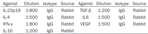
Table 2. Dilution of antibodies used for immune-detection of cytokines
Against Dilution Isotype Source Against Dilution Isotype Source IL-23p19 1:800 IgG Rabbit TGF-β 1:200 IgG Rabbit
IL-4 1:500 IgG Rabbit IL6 1:500 IgG Rabbit
IFN-γ 1:800 IgG Rabbit VEGF 1:500 IgG Rabbit
IL-10 1:200 IgG Rabbit
fields of microscopic were
sel-ected from each slide (×200)
and quantified by two blinded
Macrophages subsets in chronic epididymitis
results showed that the alternatively activ- ated CD163+CD206+ M2 macrophages were
the predominant component of the inflamma
-tory epididymis and abundantly infiltrate into the epididymal lumen under chronic inflamed
conditions.
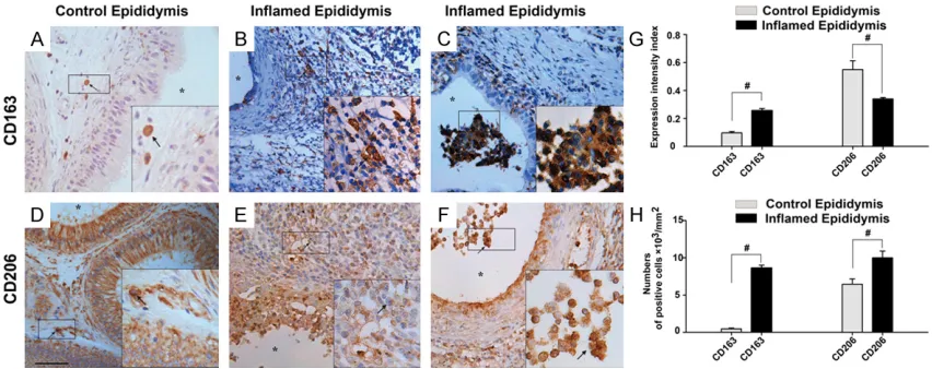
iNOS+CD68+ M1 and CD163+CD206+ M2 macrophages capturing spermatozoa in the lumen of cauda epididymidis
As M1 and M2 macrophages were abundantly
infiltrate into the epididymal lumen under chr-onic inflamed conditions, we, therefore, investi -gated the relationship between those macrop- hages and spermatozoa. Interestingly, we obs- interstitial compartment of the control epi-
didymis (Figure 4A), meanwhile, CD206 were detected both within the interstitial and epit- helium area of the control epididymis (Figure 4D). Interesting, epithelial cell of control epi-didymis strongly expressed CD206 (Figure 4D), which has not previously been reported.
Compared with the controls, significantly
inc-reased numbers of CD163+, CD206+ cells were localized both the interstitial
compart-ment and the lumen of the inflammatory epi -didymis (Figure 4B, 4C, 4E-H). Furthermore,
immunofluorescence studies of double
stain-ing show that CD163 was co-localized with CD206, meanwhile, did not co-localized with
[image:6.612.90.526.68.447.2]CD68, HLA-DR and DC-SIGN (Figure 5A-P). The
Macrophages subsets in chronic epididymitis
cells (APCs) which could induce an immuno- deviation of T helper (Th cell) cell towards a Th17 immune response associated with tes- ticular damage were detected in azoospe-
rmic testis with chronic inflammation [21].
Moreover, our recent studies demonstrated that DCs through secreting cytokine IL-23p19 induce an increased recruitment of Th17 cells
under chronic inflammation of human epidid
ymis [22]. The results of immunohistochemi-
cal and immunofluorescence (double staining)
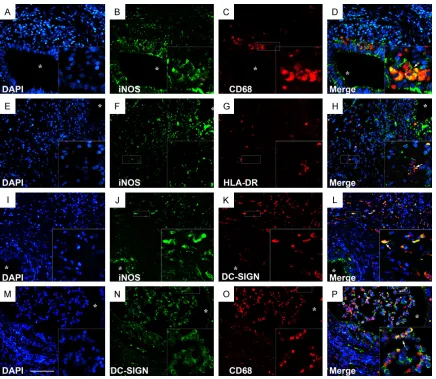
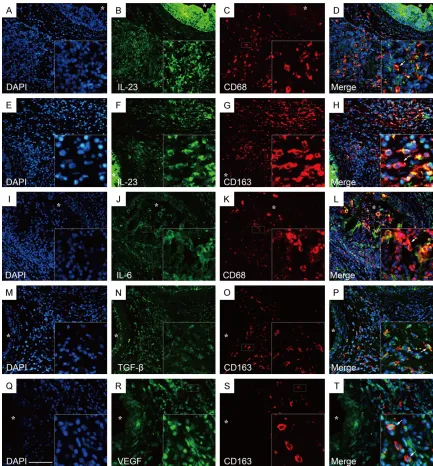
studies revealed that an increased number of IL-23p19+ cells could be detected in epididy- mitis tissue (Figure 9A, 9L) and its expression could be localized to epididymal CD68+ and CD163+ macrophages (Figure 10A-H).
In this study, we also test the expression
pro-files of other cytokines such as VEGF, TGF-β1, IL-10 and IL-6 in the inflamed epididymis and
its controls. IL-6+ cells were detected mostly in the epididymal epithelium of the controls (Figure 9B), in addition, the subepithelial IL-6+ cells formed a closely network in the epithe- lium area as well as CD1a+ DCs with a satel- lite morphology and exhibits a dense network in normal human epididymis [22]. Meanwhile,
in the inflamed epididymis, the IL-6+ cells
were detected within both the interstitial and erved large numbers of spermatozoa-attached
cells in the lumen of inflamed cauda
epididy-mis which indicated that sperm were captured and degraded by those cells (Figure 6A, 6B). Previous studies have showed that oligozoo-spermia and asthenooligozoo-spermia are associated macrophages undergoing spermatophagy in the semen of infertility patients [19, 20]. Accor- dingly, we detected M1 and M2 macrophages
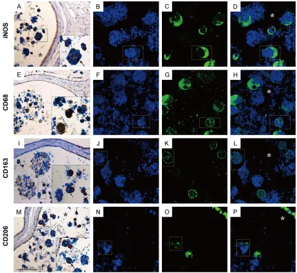
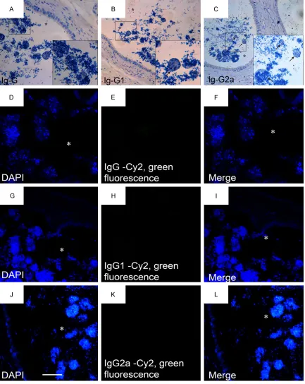
in the lumen of inflamed cauda epididymis by using immunofluorescence and immunohisto -chemistry staining. Both the M1 and M2 macro-phages were found capturing spermatozoa in the lumen of cauda epididymidis (Figure 7A-P). In this study, the negative controls were obt- ained by substitution of the primary antibo-
dies with isotype control IgG, IgG1 or IgG2
respectively in the procedure of
immunohisto-chemistry and immunofluorescence staining
(Figure 8A-L).
The inflammatory infiltrate is characterized by the recruitment of IL-6, IL23p19 producing M1 macrophages and TGF-β, VEGF, IL-23p19 producing M2 macrophages in inflamed epi-didymis
[image:7.612.94.520.74.243.2]Previous study indicated that IL-23p19 prod- ucing CD11c+ and CD68+ antigen presenting
Figure 5. Immunofluorescence double staining of CD163+CD206+, CD163+CD68-, CD163+DC-SIGN- and CD163+HLA-DR- M2 macrophages in the inflamed epididymis. Two color immunofluorescence was used to de -termine CD163+CD206+, CD163+CD68-, CD163+DC-SIGN- and CD163+HLA-DR- M2 macrophages expression: CD163, CY3, red fluorescence (C, G, K, O); CD206, CD68, DC-SIGN and HLA-DR, CY2, green fluorescence (B, F, J, N). CD163+CD206+ M2 macrophages, merged (D); CD163+CD68- M2 macrophages, merged (H); CD163+DC-SIGN- M2 macrophages, merged (L); CD163+HLA-DR- M2 macrophages, merged (P); Nuclei were labeled with DAPI (A, E, I, M); respectively. Arrows represent positive cells. *represent the lumen of epididymis. Bar =100 µm. (Original magnification, ×200; Inset: ×400).
Figure 6. Spermatozoa-attached cells in the lumen of inflamed cauda epididymis. Magnification: ×200 (A), ×600 (B). HE represent the head of
spermatozoa. N&T represent the neck and tail of spermatozoa. *rep-resent the lumen of epididymis. Bar =100 µm.
epithelium area of inflamed
epididymis (Figure 9G) and its expression could be localized to epididymal CD68+ (Figure 10
I-L) but not CD163+ macro-phages (data not show).
[image:8.612.91.524.68.447.2] [image:8.612.90.367.557.699.2]Macrophages subsets in chronic epididymitis
anti-inflammatory reactions and tissue remo-deling is associated with TGF-β, IL-10 and VEGF under chronic inflamed environment [10, 23-25]. In this study, TGF-β+ cells were
found mostly in the epithelium and sparsely distributed in the interstitial compartment of control epididymis (Figure 9C). In addition, the
subepithelial TGF-β+ cells formed a closely
network in the epididymal epithelium of con-trols which consisted with IL-6+ and CD1a+ cells in normal epididymis. On the other hand,
the numbers of TGF-β+ cells were significantly
elevated in inflamed epididymis (Figure 9H, 9L) and its expression could be localized to epididymal CD163+ M2 macrophages (Figure 10M-P). Unlike TGF-β, the expression of VEGF
was specially found in the intraepithelium area of control epididymis (Figure 9D). Meanwhile,
the expression of VEGF was greatly increased in the inflamed epididymis (Figure 9I, 9K, 9L)
and double staining of immunofluorescent studies showed that VEGF could be
[image:9.612.93.524.70.466.2]ted in the principal cells of epididymal epit- helium rather than immune cells of normal control epididymis (Figure 9E). On the other
hand, IL-10+ cells were sparsely distributed in the epididymal interstitial compartment of
[image:10.612.91.524.72.616.2]inflamed epididymis (Figure 9J).
Macrophages subsets in chronic epididymitis
epididymis (K, L). Quantitative enumeration was performed at the single cell level in immunohistochemically stained sections and mean optical density summary was used to represent the expression intensity of immune-positive cells by in situ analysis using Image-Pro Plus software. The results are expressed as the means ± SEM. Arrows represent the positive cells. *represent the lumen of epididymis. #represent P<0.05. NS represent no significance. Bar =100
[image:12.612.88.521.141.607.2]µm. (Original magnification, ×200; Inset: ×400).
Macrophages subsets in chronic epididymitis
Th1 (CD4+IFN-γ+) cells were predominantly chronically epididymitis
It is well known that M1 and M2 macropha-
ges can stimulate antigen-specific T cells and
induce Th1/Th2 type responses respectively through surface molecules and secreting cyto-kines [25-27]. CD3+ and CD4+ T cells have been found in the epididymis for a very long time [28], However, Th1 and Th2 cells were characterized recently as a distinct lineage of
IFN-γ and IL-4-producing CD4+ T cells respec -tively remain to be elucidated.
The populations of CD4+ T cells were low in control epididymis, and most of them were detected in the interstitial compartment (Figure 11A). For comparison, the numbers of CD4+ T
cells were significantly increased in peritubular areas of inflamed epididymis and also concen
-trated in the inflammatory infiltrate (Figure 11B, 11H).
Our immunohistochemical study suggests that
the cytokines IFN-γ is expressed in controls
at low levels (Figure 11E). On the other hand,
the expression of IFN-γ was significantly inc-reased in inflamed epididymis (Figure 11F-H) and its expression could be localized to epi- didymal CD4+ T lymphocytes (Figure 12E-H). From this part of experiments, we specul- ate that M1 macrophages could induce an increased recruitment of Th1 cells under chr-
onic inflammation of human epididymis.
Discussion
Macrophages are versatile controller between innate and adaptive immunity system which
[image:13.612.94.519.78.344.2]play a critical role in non-specific defense, and
also promote initiate specific defense mecha -nisms by recruiting other immune cells such as T lymphocytes [29]. Along with DCs specialized APCs, macrophages are foremost among the cells which present antigens, exerting a pivo- tal role in initiating an immune response, but the distribution, characteristics and func-tion of macrophages subsets in the normal
and inflamed epididymis have not been well defined in the previous study. Recently, we
observed that a high population of CD11c+, CD123+, CD209+ DC subsets and Th17 (CD4+
IL-17A) cells in the inflamed epididymis [22]. In this study, we demonstrated for the first time
that: (i) iNOS+, HLA-DR+, CD68+, CD163+ and CD206+ cells were found in the normal epi- didymis of human. (ii) The number of both
iNOS+HLA-DR+CD68+DC-SIGN+ M1 and CD-163+CD206+ M2 macrophages was signifi
-cantly increased in inflamed epididymis. (iii)
iNOS+, CD68+ M1 macrophages and CD163+, CD206+ M2 macrophages capturing sper- matozoa in the lumen of cauda epididymidis. (iV) IL-6, IL-23p19 producing M1 macroph-
ages and TGF-β, VEGF, IL-23p19 producing M2 macrophages were significantly elevated in inflamed epididymis. (V) Th1 (CD4+ IFN-γ+)
cells were predominantly distributed in the
inflamed epididymis.
A majority of macrophages which are known as the mononuclear phagocyte system are sta-tioned at strategic sites where accumulation of
foreign particles or microbial invasion is likely
to occur. Under a certain condition, the BEB
and immune cells in the epithelium of normal epididymis may cooperate to prevent auto antigens from entering the circulation and initi-ating a pathological immune reaction against sperm [30]. Hence, the strategic localization of
CD68+, iNOS+, HLA-DR+, DC-SIGN+, CD163+,
CD206+ macrophages/DCs indicate that they may play a pivotal role in the afferent arm of the adaptive immune reaction in normal epi-didymis, by taking up luminal antigens from pathogens and apoptotic cells.
Our studies showed that abnormal large am- ount of CD68+ macrophages were found in
the inflammatory infiltrates as well as con-centrated in the lumen of inflamed epididymis, which were significantly correlated with the
population of iNOS+ cells. Furthermore, imm-
unofluorescence studies of double staining
show that CD68 was co-localized with iNOS, meanwhile, did not co-localized with CD163
which specificity expressed in M2 macro -phages. It is well known that “killer” M1
macro-phages are activated by IFN-γ and LPS, and secrete high levels of inflammatory cytokines
such as IL23p19, IL-6 and low levels of
anti-inflammatory cytokines like IL-10 [31, 32].
Acc-ordingly, our observations showed that epidi- dymal CD68+iNOS+ M1 macrophages express high level of MHC class II molecular (HL-DR)
and represent inflammatory cytokines IL-23
[image:14.612.91.522.70.257.2]Macrophages subsets in chronic epididymitis
and IL-6 under chronic inflammation of human
epididymis. Collectively, these results indicate
that a pro-inflammatory response involving M1
macrophages might play an important role in the cascade of events which leading to epididy-mis rupture.
Interestingly, we found that CD163+CD206+
M2 macrophages were significantly increased and expressed tolerance cytokines VEGF and TGF-β in the inflamed epididymis, which sug
-gest that the anti-inflammatory activities of
M2 may have a role the constructive proces- ses such as tissue repair, and further turn off damaging immune activation by secreting
anti-inflammatory cytokines like VEGF and TGF-β. On the other hand, our study showed
that both CD68+ M1 and CD163+ M2
macro-phages in inflamed epididymis represent pro-inflammatory cytokines IL-23. It is relevant to
know that IL-23 produced by antigen presen- ting cells was thought to play an important in role in terminal differentiation of Th17 cells which associated with the development of inf- lammatory response and autoimmunity [33]. Our previous study indicated that IL-23 pro- ducing CD11c+ DCs could induce an immu- nodeviation of T helper (Th cell) cell towards a Th17 immune response associated with
epi-didymis damage were detected in inflamed
epididymis [22]. Therefore, we speculate that both CD11c+ DCs and M1/M2 macrophages represent the main source of IL-23 which ini-
tiating and promoting inflammatory cascade
and survival of Th17 cells. Furthermore, unlike the classical “repair” designation, M2
macro-phages with pro- and anti-inflammatory cyto
-kine profiles may enact both stimulatory and
inhibitory effects on the development of chr-
onic inflamed epididymis.
Our data showed that IFN-γ was represented not only in control but also in inflamed
epidi-dymis with elevated number; meanwhile, the
expression intensity of IL-4 was significantly decreased. IFN-γ secreted by CD4+ T cells is a pro-inflammatory cytokine that acts as a
potent mediator in initiating and promoting
inflammatory cascade of M1 macrophages,
and via positive feedback, M1 macrophages
can stimulate antigen-specific T cells and ind-uce IFN-γ (Th1) type response [34]. IFN-γ also
inhibits the production of cytokines IL-4, an important cytokine associated with the Th2
type response, and thus it also acts to pres- erve its own response [35]. These studies provide credence for indication that macro-phages induce an increased recruitment of
Th1 cells under inflamed condition of chronic
epididymitis and leading to epididymis rupture. The most remarkable of our study is that both M1 and M2 macrophages were found capturing spermatozoa in the lumen of cauda
epididymidis. In accordance with this finding,
earlier studies demonstrated that in the pati- ents with chronic accessory gland infections or occurrence of antibody-coated sperma- tozoa in their semen, the presence of macro-phages “spermatophagy” activity on ejacul-
ated sperm is significant [36]. Previous
stu-dies have showed that the inhibitory power of
seminal plasma, such as PGE1, 19-OH-PEG1 and PGE2, protected spermatozoa from cap -ture by APCs [37]. It is relevant to know that
the inflamed epididymis does not store the seminal plasma and under a pro-inflammatory
conditions. Taken together, we speculate that the excessively activated macrophages under
chronic inflamed epididymis probably make the
spermatozoa “mass control” mechanisms out of control which induce oligozoospermia and asthenospermia of male infertility patients. In summary, our data suggests that the chr-
onic inflamed epididymis is characterized by
the recruitment of different macrophage sub-sets, such as iNOS+, CD68+, HLA-DR+ M1 and CD163+, CD206+ M2 macrophages, and which capture spermatozoa in the chronic inf- lamed epididymis. These cell populations, by means of cell interactions and their secretory products, may play an important role in the initiation of immune reaction and autoimmune response which leading to epididymis rupture. In the future, unraveling the characterization, function and regulation of epididymis
macro-phage subsets in inflamed epididymis is
criti-cal to the design of better strategies for the treatment of immunological male infertility. Acknowledgements
Disclosure of conflict of interest
None.
Address correspondence to: Dr. Xianxin Li, Depart- ment of Urology, Shenzhen PKU-HKUST Medical Center, Peking University Shenzhen Hospital, 1120 Lianhua Road, Futian District, Shenzhen 518036, P. R. China. Tel: (+86)755-83923333-8886; Fax: (+86)755-83929927; E-mail: [email protected]
References
[1] Schuppe HC, Meinhardt A, Allam JP, Bergmann M, Weidner W and Haidl G. Chronic orchitis: a neglected cause of male infertility? Andrologia 2008; 40: 84-91.
[2] Rusz A, Pilatz A, Wagenlehner F, Linn T, Diemer T, Schuppe HC, Lohmeyer J, Hossain H and Weidner W. Influence of urogenital infections and inflammation on semen quality and male fertility. World J Urol 2012; 30: 23-30.
[3] Haidl G, Allam JP and Schuppe HC. Chronic epididymitis: impact on semen parameters and therapeutic options. Andrologia 2008; 40: 92-96.
[4] Hedger MP and Winnall WR. Regulation of ac-tivin and inhibin in the adult testis and the evi-dence for functional roles in spermatogenesis and immunoregulation. Mol Cell Endocrinol 2012; 359: 30-42.
[5] Da Silva N, Cortez-Retamozo V, Reinecker HC, Wildgruber M, Hill E, Brown D, Swirski FK, Pittet MJ and Breton S. A dense network of dendritic cells populates the murine epididy-mis. Reproduction 2011; 141: 653-663. [6] Gibbings DJ, Marcet-Palacios M, Sekar Y, Ng
MC and Befus AD. CD8 alpha is expressed by human monocytes and enhances Fc gamma R-dependent responses. BMC Immunol 2007; 8: 12.
[7] Serre V and Robaire B. Distribution of immune cells in the epididymis of the aging Brown Nor-way rat is segment-specific and related to the luminal content. Biol Reprod 1999; 61: 705-714.
[8] Benoit M, Rodrigues A, Zhang Q, Fourre E, Vi -gier Kde O, Tatibouet JM and Jerome F. Depoly -merization of cellulose assisted by a nonther-mal atmospheric plasma. Angew Chem Int Ed Engl 2011; 50: 8964-8967.
[9] Mills CD. Anatomy of a discovery: m1 and m2 macrophages. Front Immunol 2015; 6: 212. [10] Benoit M, Desnues B and Mege JL.
Macro-phage polarization in bacterial infections. J Im-munol 2008; 181: 3733-3739.
[11] Da Silva N and Barton CR. Macrophages and dendritic cells in the post-testicular environ-ment. Cell Tissue Res 2016; 363: 97-104.
[12] Cooper TG, Yeung CH, Jones R, Orgebin-Crist MC and Robaire B. Rebuttal of a role for the epididymis in sperm quality control by phago-cytosis of defective sperm. J Cell Sci 2002; 115: 5-7.
[13] Jones R. Sperm survival versus degradation in the Mammalian epididymis: a hypothesis. Biol Reprod 2004; 71: 1405-1411.
[14] Sutovsky P. Ubiquitin-dependent proteolysis in mammalian spermatogenesis, fertilization, and sperm quality control: killing three birds with one stone. Microsc Res Tech 2003; 61: 88-102.
[15] Sutovsky P, Moreno R, Ramalho-Santos J, Dominko T, Thompson WE and Schatten G. A putative, ubiquitin-dependent mechanism for the recognition and elimination of defective spermatozoa in the mammalian epididymis. J Cell Sci 2001; 114: 1665-1675.
[16] Holstein AF. Spermatophagy in the seminifer-ous tubules and excurrent ducts of the testis in Rhesus monkey and in man. Andrologia 1978; 10: 331-352.
[17] McGinn JS, Sim I, Bennett NK and McDonald SW. Observations on multiple sperm granulo-mas in the rat epididymis following vasectomy. Clin Anat 2000; 13: 185-194.
[18] McDonald SW. Cellular responses to vasecto-my. Int Rev Cytol 2000; 199: 295-339. [19] Pelliccione F, D’Angeli A, Cordeschi G, Mihalca
R, Ciociola F, Necozione S, Francavilla F and Francavilla S. Seminal macrophages in ejacu-lates from men with couple infertility. Int J An-drol 2009; 32: 623-628.
[20] Oren-Benaroya R, Kipnis J and Eisenbach M. Phagocytosis of human post-capacitated sper-matozoa by macrophages. Hum Reprod 2007; 22: 2947-2955.
[21] Duan YG, Yu CF, Novak N, Bieber T, Zhu CH, Schuppe HC, Haidl G and Allam JP. Immunode -viation towards a Th17 immune response as-sociated with testicular damage in azoosper-mic men. Int J Androl 2011; 34: e536-545. [22] Duan YG, Wang P, Zheng W, Zhang Q, Huang W,
Jin F and Cai Z. Characterisation of dendritic cell subsets in chronically inflamed human epi -didymis. Andrologia 2016; 48: 431-40. [23] Hasita H, Komohara Y, Okabe H, Masuda T, Oh
-nishi K, Lei XF, Beppu T, Baba H and Takeya M. Significance of alternatively activated macro -phages in patients with intrahepatic cholangio-carcinoma. Cancer Sci 2010; 101: 1913-1919. [24] Luo H, Hao Y, Tang B, Zeng D, Shi Y and Yu P.
Mouse forestomach carcinoma cells immuno-suppress macrophages through transforming growth factor-beta1. Mol Med Rep 2012; 5: 988-992.
Macrophages subsets in chronic epididymitis
Welters MJ, van Hall T and van der Burg SH. M2 macrophages induced by prostaglandin E2 and IL-6 from cervical carcinoma are switched to activated M1 macrophages by CD4+ Th1 cells. J Immunol 2011; 187: 1157-1165. [26] Duan W and Croft M. Control of regulatory T
cells and airway tolerance by lung macro-phages and dendritic cells. Ann Am Thorac Soc 2014; 11 Suppl 5: S306-313.
[27] Bain CC and Mowat AM. Macrophages in intes-tinal homeostasis and inflammation. Immunol Rev 2014; 260: 102-117.
[28] Ritchie AW, Hargreave TB, James K and Ch -isholm GD. Intra-epithelial lymphocytes in the normal epididymis. A mechanism for tolerance to sperm auto-antigens? Br J Urol 1984; 56: 79-83.
[29] Garg SK, Delaney C, Shi H and Yung R. Chang -es in adipose tissue macrophag-es and T cells during aging. Crit Rev Immunol 2014; 34: 1-14.
[30] Yeung CH, Wang K and Cooper TG. Why are epididymal tumours so rare? Asian J Androl 2012; 14: 465-475.
[31] Novoselov VV, Sazonova MA, Ivanova EA and Orekhov AN. Study of the activated macro-phage transcriptome. Exp Mol Pathol 2015; 99: 575-580.
[32] Wang N, Liang H and Zen K. Molecular mecha -nisms that influence the macrophage m1-m2 polarization balance. Front Immunol 2014; 5: 614.
[33] Allam JP and Novak N. Local immunological mechanisms of sublingual immunotherapy. Curr Opin Allergy Clin Immunol 2011; 11: 571-578.
[34] Mills CD. M1 and M2 Macrophages: Oracles of Health and Disease. Crit Rev Immunol 2012; 32: 463-488.
[35] Salem ML. Estrogen, a double-edged sword: modulation of TH1- and TH2-mediated inflam -mations by differential regulation of TH1/TH2 cytokine production. Curr Drug Targets In-flamm Allergy 2004; 3: 97-104.
[36] Blanco AM, Palaoro L, Ahedo MI, Palamas M and Zanchetti F. Phagocytosis of ejaculated spermatozoa. Acta Cytol 1992; 36: 251-258. [37] Rennemeier C, Schwab M, Lermann U, Albert