Original Article
Yeast as a model to study the roles of aging regulation
path way Sip2-Sch9 in α-synuclein pathogenesis
Ziyan Fang1*, Caifeng Guo1*, Xiaoming Zhou1, Yi Huang2, Yixuan Zeng2, Zhong Pei2, Yuping Ning1
1Department of Neurology, The Affiliated Brain Hospital of Guangzhou Medical University (Guangzhou Huiai Hospi
-tal), Guangzhou, China; 2Department of Neurology, The First Affiliated Hospital of Sun Yat-sen University, Guang
-zhou, China. *Co-first authors.
Received February 5, 2017; Accepted February 26, 2017; Epub May 1, 2017; Published May 15, 2017
Abstract: A-synuclein is the main component of Lewy body which is main pathological presentation of Parkinson’s disease. This study was aimed at observing the impact of Sip2 and Sch9 gene on α-synuclein (α-syn) toxicity, ag-gregation and reactive oxygen species (ROS) level in yeast. The pRS414-α-syn, pRS416-RFP and pRS416-α-syn-RFP plasmid were constructed, and were transformed into the YL36, and YL36+ΔSip2 or YL36+ΔSch9 yeast. Expres-sion of α-syn was testified using western-blot. The spotting assay and growth curve were adopted to observe α-syn toxicity, the distribution of α-synwas observed with fluorescent microscope, and ROS level were detected with H2D-CFDA. The results show that yeast transformed into α-syn plasmid showed obviously growth retardation induced in galactose while no in glucose. The α-syn protein express on the cytomembrane induced in galactose for 4 h, and the aggregation was found from cytomembrane to cytoplasm for 8 h induction. A higher ROS level induced in galactose was detected than that uninduced in glucose. The yeast displayed obvious growth retardation when the gene for Sip2 is removed, while the yeast showed even better growthwhen the gene for Sch9 is removed. The expression and aggregation of α-syn, and ROS level were more obviously increased when the gene for Sip2 is removed. However, the expression of α-syn and ROS level was also increased when the gene for Sch9 is removed, but the aggregation was decreased. Our study showed that the ΔSch9 in yeast was protective for growth and aggregation formation while ΔSip2 in yeast could aggravate the growth retardation, expression and aggregation of α-syn, and the ROS level.
Keywords: α-synuclein, aggregation, ROS, yeast, Sip2, Sch9
Introduction
A-synuclein (α-syn) is the main component of Lewy body which is main pathological presenta-tion of Parkinson’s disease (PD), Research shows the pathological accumulation of α-syn in the brain is a typical neuropathological fea-ture of PD [1-3]. And there is growing evidence that α-syn can spread from diseased to healthy cells or tissues, and then contributing to dis-ease worsening [4-6]. However, the pathogenic mechanism of α-synas a role in neurodegener-ative diseases is not completely understood. It can form oligomers and aggregation in concen-tration dependence pattern which result in tox-icity to cells. In the formation of the aggrega-tion, ROS plays an important part. With the function of protein quality control system, abnormal protein aggregation is not evident in health neuron, which can also neutralize exces-sive ROS in cell. However, some mutations,
resulting in neurodegenerative diseases, main-ly produce some proteins with evident aggre-gated trend and injure the function of the pro-tein quality control system, or have the declined capacity for antioxidant defense [7-9].
In addition to that, the increased incidence of neurodegenerative diseases is related with the age, which can lead to abnormal protein aggre-gation and declined function of the protein quality control system, and elevated ROS level beyond capacity of antioxidant defense mecha-nisms [10, 11]. Therefore, researches exploring intrinsic factor to prolong life may be more attractive. For instance, the latest research has shown that Sip2 acetylation-Sch9 phosphoryla-tion was an intrinsic aging defense pathway [12, 13].
is a practical model to study neurodegenerative diseases like protein aggregation, protein mis-folding, oxidative stress and apoptosis, et al [14- 16]. Features of diseases like Alzheimer and Parkinson’s disease have been successfully modeled in yeast. The basic cellular machinery for neuronal function is conserved in yeast making it an attractive unicellular, genetically and biochemically tractable model organism to study pathways underlying neurodegeneration diseases. As the extension of cultivation in the stationary phase, the injury in cell is accumu-lated, except for that, the cell also display many features in aged mammal such as decline in protein synthesis, decreased metabolism, and changes of cell morphology. And the research-es on yeast in stationary phase have indicated some genes regulating the age. Hence, the sta-tionary phase yeast (aged yeast) is taken as an ideal model to clarify the interaction between genes in neurodegenerative disease research. In view of these, the purpose of this study is using yeast as model to explore the role of Sip2-Sch9 in a-syn toxicity.
Materials and methods
Plasmids and vector construction
The commercialized pRS416 (H4ARS, CEN3, Ampr, Ori, Ura) and pRS416 (H4ARS, CEN3, Am- pr, Ori, trp) plasmids, which were cloned with the gene of gal1 promoter, c-myc tag, and cycle-terminator, were donated by the School of Life Sciences and Biomedical Center of Sun Yat-sen University. The RFP cDNA was isolated from pTol2HuC-mRFP cDNA via PCR amplification using the primers list in the Table 1 that includ-ed, respectively, a BglII and XboI restriction site for cloning into the pRS416 plasmid. The α-syn cDNA was isolated from HtrA2 cDNA via PCR amplification using the primers list in the Table
1 that included, respectively, the XhaI and BglII restriction site for cloning into the pRS416 plas-mid. PCR kit Q5 and other enzymes were from NEB (#M0491L). PCR conditions were 98°C for 1 min, followed by 30 cycles of 98°C for 10 sec, and 65°C for 20 sec, and then 72°C for 30 sec. To confirm the fidelity of the PCR and cloning steps, all GFP and α-syn-GFP fusion constructs were sequenced.
Yeast strains and cultivation
Yeast strains used in this study were derived from YL36 (matα, ade2, ura3, his3, trp1, leu2, can100), which was donated by the School of Life Sciences and Biomedical Center of Sun Yat-sen University. Deletion of sip2 or sch9 was achieved by replacement of the gene with the PCR product containing a Kan gene which was amplificated using the pFa6a-kanMX6 as the template. Yeast cells were grown in non-selec-tive medium (YPD) at 30°C and transformed as described. Selective synthetic complete medi-um (SCM) contained 2% glucose or 2% galac-tose and lacked the nutrient corresponding to the marker (tryptophan for pRS414 and uracil for pRS416). Yeast were first grown in SCM with glucose and lacking tryptophan/uracil over-night then shifted to SCM containing galactose for 12 hours to induce α-syn expression. After pre-incubation in galactose medium, yeast cells were shifted to SCM with glucose to shut off the promoter for 2 hours, and observed with a fluorescent microscope.
Spot assay and growth profile
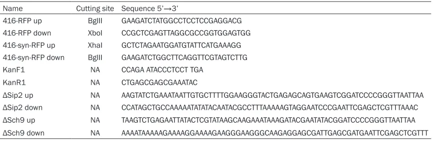
[image:2.612.90.537.83.225.2]For growth assays on solid medium, a serial dilution of yeasts was made in SCM medium ranging from an OD600 of 10-1 to 10-4. Of these dilutions, 5 μl was spotted on SCM agar plates, which were then incubated at 30°C for at least Table 1. Primers and their sequences
Name Cutting site Sequence 5’→3’
416-RFP up BglII GAAGATCTATGGCCTCCTCCGAGGACG 416-RFP down XboI CCGCTCGAGTTAGGCGCCGGTGGAGTGG 416-syn-RFP up XhaI GCTCTAGAATGGATGTATTCATGAAAGG 416-syn-RFP down BglII GAAGATCTGGCTTCAGGTTCGTAGTCTTG KanF1 NA CCAGA ATACCCTCCT TGA
KanR1 NA CTGAGCGAGCGAAATAC
48 h. At least three independent transformants were tested for the survival plating to rule out clonogenic variation of the effects. To deter-mine survival upon a-syn expression, 5 ml yeast from overnight cultures inoculated in tubes on SCM was diluted to OD600 of 10-2, and was grown at 30°C of 250 rpm for 28 h. The OD600 of yeast was tested every 4 h. The growth pro-files were then established by measuring OD600 in a DU 800 Protein nucleic acid spec-trophotometer (Beckman Coulter, California,
bated for 30 min in a Thermo Lab systems Fluoroskan Ascent microplate reader at 37°C. Results
Construction and identification of pRS414-α-syn, pRS416-RFP and pRS416-α-syn-RFP plasmids
[image:3.612.92.522.73.254.2]The pRS414-α-syn was successfully construct-ed. The positive stripes within 400-500 bp in 1-4 holes by PCR were observed in Figure 1A,
Figure 1. Electrophoresis of the PCR products was identified. A. The positive stripes within 400-500 bp shows that α-syn DNA was successfully amplified from pRS414-α-syn plasmid (arrow). B. The positive stripes within 700 bp shows that RFP DNA was successfully amplified from pRS416-RFP plasmid (arrow). C. The positive stripes within 400-500 bp shows that α-syn DNA was successfully amplified from pRS416-α-syn-RFP plasmid (arrow).
Figure 2. The protein expression of α-syn in YL36+pRS414-α-syn yeast. Induced, YL36+pRS414-α-syn yeast cultivated in the galactose medium; uninduced, YL36+pRS414-α-syn yeast cultivated in the glucose medium. Compared with uninduced, *P<0.05.
USA). For all assays, every exper-iment was repeated three times. In order to test growth retarda-tion caused by native α-Syn, the growth assay of the correspond-ing strains were compared to that of a control strain trans-formed with the empty vectors. Measurement of intracellular ROS in yeast
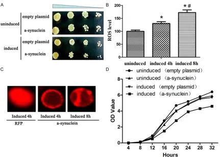
[image:3.612.90.365.327.568.2]incu-Figure 3. Effect of α-syn on physiological status of yeast. A. The growth retardation were observed in YL36+pRS414-α-syn yeast. B. The ROS level were detected in yeast. Compared with uninduced, *P<0.05; compared with induced for 4 h, #P<0.05. C. The expression of α-syn and RFP in yeast. D. The growth profiles were then established by
mea-suring OD600. Experiments were performed in triplicate.
which were in accordance with the stripe size of α-syn. The pRS416-RFP was successfully con-structed. The positive strips approximately 700 bp in 1-7 holes by PCR were observed in Figure 1B, which were in accordance with the stripe size of RFP. The pRS416-α-syn-RFP was suc-cessfully constructed. The positive strips within 400-500 bp in 4-7 holes by PCR were observed in Figure 1C, which were in accordance with the stripe size of α-syn. The α-syn-RFP fusion con-structs were sequenced to be with the right direction, no bass error and missing in genes. Construction and identification of
YL36+pRS414-α-syn yeast
YL36+PRS414-α-syn yeast was cultivated both in the glucose and galactose medium. The expression of α-syn cultivated in the galactose medium was significantly higher than in the glu-cose medium analyzed by western blot (Figure 2).
α-syn toxicity, aggregation and ROS level in yeast
[image:4.612.92.523.69.378.2]When cultured in the galactose medium for 4 h, the α-syn-RFP was emerged to be the small granular distributed in the cell membrane. When cultivated for 8 h, the α-syn-RFP incre- ased and was emerged to be the large granular aggregations in both the cell membrane and cytoplasm. While the YL36+pRS416-RFP yeast induced in the galactose medium, the RFP was uniformly distributed in the cytoplasm (Figure 3C).
The value of OD600 was certified to increase as the time gone (0-32 h). And the growth curve was present the S shape. At the same time point, the OD600 value of YL36+pRS414-α-syn yeast was much lower than the YL36+pRS414 yeast. And when cultivated in the galactose medium, the OD600 value of YL36+pRS414-α-syn yeast was significantly higher in the glucose medium (Figure 3D).
Identification and toxicity of YL36+ΔSip2 or YL36+ΔSch9 yeast
The wild yeast without the positive stripe within 700-800 bp as the negative control and the ΔOmi yeast with the positive stripe as the posi-tive control, there were posiposi-tive stripes in 1-5
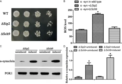
α-syn-RFP yeast (P<0.05). And the ROS level in the YL36+ΔSch9+pRS416-α-syn-RFP yeast was higher than in the YL36+pRS416-α-syn-RFP yeast (P<0.05, Figure 5A, 5B). When com-pared to cultivated in the glucose medium, the expression of α-syn was significantly higher in YL36+ΔSip2+pRS414-α-syn and YL36+ΔSch9+ pRS414-α-syn yeast cultivated in galactose medium (Figure 5C, 5D).
α-syn toxicity, aggregation and ROS level in ΔSip2 and ΔSch9 yeast
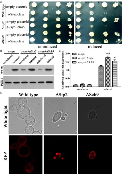
[image:5.612.91.394.71.319.2]All these 6 groups (YL36+pRS416, YL36+pRS- 416-α-syn-RFP, YL36+ΔSip2+pRS416, YL36+ ΔSip2+pRS416-α-syn-RFP, YL36+ΔSch9+pRS- 416, and YL36+ΔSch9+pRS416-α-syn-RFP) yeasts had no obvious differences in growth cultivated in glucose medium. But when culti-vated in galactose medium, the YL36+ΔSip2+ pRS416-α-syn-RFP yeast displayed an obvious-ly growth retardation even when the OD600 value was 10-2 point. The growth of YL36+ ΔSch9+pRS416-α-syn-RFP yeast was better than the YL36+pRS416-α-syn-RFP yeast, or even as good as the YL36+pRS416 yeast induced in the galactose medium (Figure 6A).
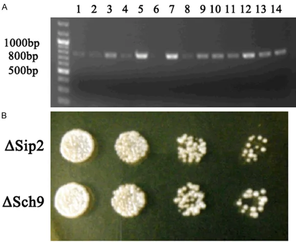
Figure 4. Construction and identification of YL36+ΔSip2 or YL36+ΔSch9 yeast. A. The YL36+ΔSip2 (1-5 holes) and YL36+ΔSch9 (8-14 holes) yeast were success-fully constructed. The wild yeast without the positive stripe within 700-800 bp as the negative control (6 holes) and the ΔOmi yeast with the positive stripe as the positive control (7 holes). B. The YL36+ΔSip2 and YL36+ΔSch9 yeast grow in the G418 plate.
holes and in 8-14 holes which indicated that the YL36+ΔSip2 and YL36+ ΔSch9 yeast were suc-cessfully constructed, re- spectively (Figure 4A). The YL36+ΔSip2 and YL36+ΔSch9 yeast grew well while the wide yeast could not grow in the G418 plate. There were no differences between them in growth after cul-tivating in YPD plate at 30°C for 3 days (Figure 4B).
Identification of YL36+ΔSip2+pRS414-α-syn and
YL36+ΔSch9+pRS414-α-syn yeast
YL36+pRS416-Figure 5. Construction and identification of YL36+ΔSip2+pRS414-α-syn and YL36+ΔSch9+pRS414-α-syn yeast. A, B. The ROS level were detected in yeast. WT, wild type yeast with pRS414-α-syn overexpression; ΔSip2, yeast with pRS414-α-syn overexpression and Sip2 knockout; ΔSch9, yeast with pRS414-α-syn overexpression and Sch9 knock-out. Compared with WT, *P<0.05. C, D. The protein expression of α-syn in yeast.compared with induced, *P<0.05.
The α-syn expression of YL36+ΔSip2+pRS414-α-syn yeast and YL36+ΔSch9+pRS414-YL36+ΔSip2+pRS414-α-syn yeast were both higher than the pRS414-α-syn yeast (P<0.05), and the α-syn expression of YL36+ΔSip2+pRS414-α-syn yeast was even higher than the YL36+ΔSch9+pRS414-α-syn yeast (P<0.05, Figure 6B, 6C).
When cultivated in galactose medium for 12 h, compared to the YL36+pRS416-α-syn-RFP yeast, the α-syn-RFP of YL36+ΔSip2+pRS416-α-syn-RFP yeast was increased to be the large granular aggregations in the cytoplasm, while the α-syn-RFP of YL36+ΔSch9+pRS416-α-syn-RFP yeast was decreased to be distributed only in the cytomembrane with light red, which was even less than the α-syn-RFP of YL36+pRS416-α-syn-RFP yeast (Figure 6D).
Discussion
The quantity of α-syn is the precondition for the formation of toxic. The wild type and A53T mutation α-syn were expressed in yeast through
the way of secretion, the distribution of α-syn gathered on the cell membrane lead to the for-mation of inclusions [17-19]. The forfor-mation pro-cess of inclusion was started from the small nuclear formation in [20-22]. The study adopt-ed the transformadopt-ed pRS plasmid series with gal promoter, in which the α-syn could be induced in galactose while not in glucose. Yeast with α-syn expression induced in galactose had the growth retardation but the control had not the growth retardation, α-syn expression was in- creased as the extension of time in galactose medium by western blot, suggesting that α-syn caused growth retardation was positive corre-lated with the α-syn expression. Study clarified that α-syn level polyploidization were more like-ly to lead to the conclusion of the earlier onset of Parkinson’s disease.
[image:6.612.93.518.74.367.2]forming inclusions, also detected the positive phase change with the content of ROS. Although α-syn was observed the concentration depen-dence of aggregation and toxicity to the cells, but the research was not specific the intermedi-ate products lead to aggregation for the cyto-toxicity. Although studies proposed that the early stage of oligomers formation had cytotox-ic effect. The formation of intracytoplasmcytotox-ic inclusion was observed in yeast with wild type α-syn expression which was speculated that the formation of inclusion was a major factor for toxicity to die, while the rise of ROS level was both the stimulating factor and the product for formation of inclusions. When α-syn folding abnormally, the molecular chaperone regulato-ry proteins were the first to regroup which were degraded by the ubiquitin protein or CMA path-way. But when more α-syn folded abnormally to produce the aggregation, autophagy pathway was temporarily activated to keep the steady of proteins when these aggregation proteins were too much to be degraded by the pathway reminded above. With the further increased α-syn aggregation, the autophagy-lysosome pathway was damaged which lead to cytotoxic-ity, and the excessive α-syn diffused between cells that further aggravated the toxicity to cells.
The study was first aimed to construct the Sip2 and Sch9 single gene deletion which were certi-fied to be harmless for the growth of yeast, then the yeast was transferred with α-syn to induce its expression. The two strains had a distinct difference on yeast growth, the α-syn toxicity in Sip2 deletion strain was more aggra-vated than the wild yeast while in Sch9 strain, the α-syn toxicity was relieved than the wild strain.
Sch9, Serine/threonine protein kinase, was found in transposon mutation screening for modulating the yeast age that the deletion of Sch9 gene lead to great life extension in yeast [26-28]. The level of Sch9 phosphorylation was found in aged yeast, and the upregulated phos-phorylation level of Sch9 by TOR in TOR-Sch9 pathway could decline the life in yeast. This study was designed to reduce the phosphoryla-tion level by deleting the Sch9 gene, which turned out that both α-syn toxicity and aggrega-tion could be relieved. In conclusion, it was speculated that the decline of Sch9 phosphory-lation level had the function against the α-syn toxicity that was the possible mechanism of life
extension, and at the same time to state that toxicity protein was an important factor of age. Though sch9 deletion yeast had more α-syn expression and increased ROS level than the wild group, there was no obvious α-syn aggre-gation, which could be explained that the over-expressed α-syn was not enough to form the aggregation (or might be the upregulated deg-radation of α-syn). The TOR pathway involved in autophagy, but Sch9 as the substrate of TOR, the deletion of Sch9 was speculated to increase the activated function of autophagy to clear the α-syn in cell that the light red protein was dis-tributed in the cell membrane as observed in this study. However, there was a need to detect the autophagy in the study to certify the pro-tected function of phosphorylation deficiency in yeast.
Sch9 is TOR the substrate, the TOR pathway involved in cell autophagy [29], speculated that the lack of Sch9 can increase the activation of autophagy to clear the alpha syn, results in a decrease of alpha syn in the yeast cell, so the distribution of the membrane as the observed pink ring light circle. So you need under the lack of alpha syn expression of Sch9 detect the changes of autophagy further clear Sch9 phos-phorylation of lack of protection.
Sip2, a subunit of Snf1 of AMPK, was a modera-tor of Snf1. When Sip2 acetylation increased, the interaction between Sip2 and Snf1 was strengthened that downregulated the activity of Snf1 kinase, then declined phosphorylation of downstream Sch9 was participated in the age regulation in yeast, which proved the function of upstream Sip2 acetylation in the α-syn toxic-ity. The α-syn toxicity and aggregation were obvious increased in yeast with reduced acety-lation level by deleting the Sip2 gene. And the toxicity resistant function was verificated in Sip2 in our study.
Acknowledgements
We acknowledge that financial support was provided by the National Natural Science Foundation of China (No. 81571333), and Guangzhou Medical and Health Science and Technology Project (No. 20171A011268). Disclosure of conflict of interest
Address correspondence to: Yuping Ning, Depart- ment of Neurology, The Affiliated Brain Hospital of Guangzhou Medical University (Guangzhou Huiai Hospital), N 36 Mingxin Road, LiwanDistrict, Guang- zhou 510370, Guangdong, China. E-mail: [email protected]; Zhong Pei, Department of Neurology, The First Affiliated Hospital of Sun Yat-sen University, Guangzhou 510080, Guangdong, China. E-mail: [email protected]
References
[1] Al-Hilaly YK, Biasetti L, Blakeman BJ, Pollack SJ, Zibaee S, Abdul-Sada A, Thorpe JR, Xue WF, Serpell LC. The involvement of dityrosine cross-linking in α-synuclein assembly and deposition in Lewy Bodies in Parkinson’s disease. Sci Rep 2016; 6: 39171.
[2] Lulla A, Barnhill L, Bitan G, Ivanova MI, Nguyen B, O’Donnell K, Stahl MC, Yamashiro C, Klärner FG, Schrader T, Sagasti A, Bronstein JM. Neu-rotoxicity of the Parkinson disease-associated pesticideziram is synuclein-dependent in ze-brafish embryos. Environ Health Perspect 2016; 124: 1766-1775.
[3] Hegarty SV, Sullivan AM, O’Keeffe GW. The Epigenome as a therapeutic target for Parkin-son’s disease. Neural Regen Res 2016; 11: 1735-1738.
[4] Breid S, Bernis ME, Babila JT, Garza MC, Wille H, Tamgüney G. Neuroinvasion of α-synuclein- prionoids after intraperitoneal and intraglossal inoculation. J Virol 2016; 90: 9182-93. [5] Flagmeier P, Meisl G, Vendruscolo M, Knowles
TP, Dobson CM, Buell AK, Galvagnion C. Muta-tions associated with familial Parkinson’s dis-ease alter the initiation and amplification steps of α-synuclein aggregation. Proc Natl Acad Sci U S A 2016; 113: 10328-33.
[6] Luo HT, Zhang JP, Miao F. Effects of pramipex-ole treatment on the α-synuclein content in serum exosomes of Parkinson’s disease pa-tients. Exp Ther Med 2016; 12: 1373-1376. [7] Bonow RO, Leon MB, Doshi D, Moat N.
Man-agement strategies and future challenges for aortic valve disease. Lancet 2016; 387:1312-23.
[8] Halliwell B. Proteasomal dysfunction: a com-mon feature of neurodegenerative diseases? Implications for the environmental origins of neurodegeneration. Antioxid Redox Signal 2006; 8: 2007-19.
[9] Halliwell B. Oxidative stress and neurodegen-eration: where are we now? Jneurochem 2006; 97: 1634-58.
[10] Paschen W, Mengesdorf T. Endoplasmic reticu-lum stress response and neurodegeneration. Cell Calcium 2005; 38: 409-15.
[11] Hol EM, Scheper W. Protein quality control in neurodegeneration: walking the tight rope be-tween health and disease. Jmol Neurosci 2008; 34: 23-33.
[12] Koutsilieri E, Scheller C, Grünblatt E, Nara K, Li J, Riederer P. Free radicals in Parkinson’s dis-ease. Jneurol 2002; 249 Suppl 2: II1-5. [13] Lu JY, Lin YY, Sheu JC, Wu JT, Lee FJ, Chen Y, Lin
MI, Chiang FT, Tai TY, Berger SL, Zhao Y, Tsai KS, Zhu H, Chuang LM, Boeke JD. Acetylation of yeast AMPK controls intrinsic aging indepen-dently of caloric restriction. Cell 2011; 146: 969-79.
[14] Mulak A, Bonaz B. Brain-gut-microbiota axis in Parkinson’s disease. World J Gastroenterol 2015; 21: 10609-20.
[15] Cebollero E, Reggiori F. Regulation of autopha-gy in yeast Saccharomyces cerevisiae. Biochim Biophys Acta 2009; 1793: 1413-21.
[16] Haynes CM, Caldwell S, Cooper AA. An HRD/ DER-independent ER quality control mecha-nism involves Rsp5p-dependent ubiquitination and ER-Golgi transport. J Cell Biol 2002; 158: 91-101.
[17] Roy A, Rangasamy SB, Kundu M, Pahan K. BPOZ-2 gene delivery ameliorates Alpha-Synu-cleinopathy in A53T transgenic mouse model of Parkinson’s disease. Sci Rep 2016; 6: 22067.
[18] Yamasaki T, Fujinaga M, Kawamura K, Furut-suka K, Nengaki N, Shimoda Y, Shiomi S, Takei M, Hashimoto H, Yui J, Wakizaka H, Hatori A, Xie L, Kumata K, Zhang MR. Dynamic changes in striatal mGluR1 but not mGluR5 during pathological progression of Parkinson’s dis-ease in human Alpha-Synuclein A53T trans-genic rats: a multi-PET imaging study. J Neuro-sci 2016; 36: 375-84.
[19] Deusser J, Schmidt S, Ettle B, Plötz S, Huber S, Müller CP, Masliah E, Winkler J, Kohl Z. Seroto-nergic dysfunction in the A53T alpha-synuclein mouse model of Parkinson’s disease. J Neuro-chem 2015; 135: 589-97.
[20] Temple MD, Perrone GG, Dawes IW. Complex cellular responses to reactive oxygen species. Trends Cell Biol 2005; 15: 319-26.
[21] Dixon C, Mathias N, Zweig RM, Davis DA, Gross DS. Alpha-synuclein targets the plasma mem-brane via the secretory pathway and induces toxicity in yeast. Genetics 2005; 170: 47-59. [22] Zabrocki P, Pellens K, Vanhelmont T,
Vande-broek T, Griffioen G, Wera S, Van Leuven F, Winderickx J. Characterization of alpha-synu-clein aggregation and synergistic toxicity with protein tau in yeast. FEBS J 2005; 272: 1386-400.
S. The Parkinson’s disease protein alpha-synu-clein disrupts cellular Rab homeostasis. Proc Natl Acad Sci U S A 2008; 105: 145-50. [24] Prusiner SB, Woerman AL, Mordes DA, Watts
JC, Rampersaud R, Berry DB, Patel S, Oehler A, Lowe JK, Kravitz SN, Geschwind DH, Glidden DV, Halliday GM, Middleton LT, Gentleman SM, Grinberg LT, Giles K. Evidence for α-synuclein prions causing multiple system atrophy in hu-mans with parkinsonism. Proc Natl Acad Sci U S A 2015; 112: E5308-17.
[25] Stefanis L. alpha-Synuclein in Parkinson’s dis-ease. Cold Spring Harb Perspect Med 2012; 2: a009399.
[26] Sinclair D, Mills K, Guarente L. Aging in Sac-charomyces cerevisiae. Annu Rev Microbiol 1998; 52: 533-60.
[27] Longo VD. Mutations in signal transduction proteins increase stress resistance and lon-gevity in yeast, nematodes, fruit flies, and mammalian neuronal cells. Neurobiol Aging 1999; 20: 479-86.
[28] Longo VD, Fabrizio P. Regulation of longevity and stress resistance: a molecular strategy conserved from yeast to humans? Cell Mol Life Sci 2002; 59: 903-8.