Original Article
Expression of G protein-coupled estrogen receptor 30
(GPR30) and extracellular signal-regulated kinase
1/2 (p-ERK1/2) in the endometrioid endometrial
cancer and their correlation
Jing Zhao1, Qianyu Wu2, Weijuan Xin3
1Department of Obstetrics and Gynecology, Shanghai First Maternity and Infant Hospital, Tongji University, School
of Medicine, Shanghai, China; 2Department of Obstetrics and Gynecology, East Hospital, Tongji University, School
of Medicine, Shanghai, China; 3Department of Obstetrics and Gynecology, Obstetrics and Gynecology Hospital of
Fudan University, Shanghai, China
Received October 30, 2015; Accepted December 26, 2015; Epub February 1, 2016; Published February 15, 2016
Abstract: Purpose: This study aimed to explore the expressions of G protein-coupled estrogen receptor 30 (GPR30) and extracellular signal-regulated kinase 1/2 (p-ERK1/2) in the endometrioid endometrial cancer (EEC) and evalu-ate their correlation. Methods: Expressions of GPR30 and p-ERK1/2 were measured in 22 cases of EEC, 20 cases of atypical hyperplasia endometrium (EAH) and 20 cases of normal endometrium. Results: The positive rate of GPR30 expression in EEC was significantly higher than in normal endometrium and EAH (P<0.05), and significant difference was also observed between normal endometrium and EAH (P<0.05). Significant difference in GPR30 expression was also found between EECs with different depths of myometrial invasion: the deeper the myometrial invasion, the higher the positive rate was (P<0.05). GPR30 expression had no relationship with other clinicopathological charac-teristics (P>0.05). The positive rate of p-ERK1/2 expression in EAH was significantly higher than in normal endome-trium, but it in EEC was comparable to that in normal endometrium and EAH (P>0.05). p-ERK1/2 expression was not related to clinicopathological characteristics (P>0.05). The positive rate of GPR30 and p-ERK1/2 co-expression was 5.0% in normal endometrium, and there was no relationship between GPR30 expression and p-ERK1/2 expression (P>0.05). The positive rate of GPR30 and p-ERK1/2 co-expression was 35.0% and 45.6% in EAH and EEC, respec-tively, and positive relationship was observed between GPR30 expression and p-ERK1/2 expression in both groups (P<0.05). Conclusion: GPR30 may exert its effects via regulating p-ERK1/2 signaling pathway in EEC.
Keywords: Endometrioid endometrial cancer, G protein-coupled estrogen receptor, extracellular signal regulated kinase1/2, immunohistochemistry
Introduction
Endometrial cancer (EC) is a common malig-nancy in women [1]. About 72% of EC patients are diagnosed at stage I-II, but 28% of EC patients are diagnosed when regional or distal metastasis has been present (about 20% at stage III and about 8% at stage IV) [2]. Of differ-ent types of EC, endometrioid endometrial can-cer (EEC) is a major one and accounts for 80-90%. Long term estrogen stimulation in the absence of antagonism of progesterone is a major cause of EEC. Recent studies identify a 7-transmembrane Gprotein-coupled estrogen
involved in several intracellular processes including proliferation, differentiation and me- tabolism. ERK1/2 can be activated by several mitogens including growth factors, to promote the transcription and expression of oncogenes and genes related to the regulation of cell cycle, leading to the proliferation of cancer cells and the inhibition of their apoptosis [6]. Phosphorylated ERK (p-ERK) is the activated form of ERK. After entering the cells, p-ERK1/2 may initiate the expression of some oncogenes such as c-jun, c-fos and c-myc, which then pro-motes cells to progress from G1 phase to S phase, resulting in malignant proliferation. Acconcia et al [7] used estrogen and tamoxifen (TAM) to induce the cytoskeleton remodeling and migration of EC cells and found E2 and 4-OHT could rapidly activate ERK1/2, C-Srk and FAK signaling pathway to alter the cytoskel-eton of EC cells and promote their migration and invasion. The binding of estrogen to GPR30 may activate ERK via Src, Raf, Ras and Mek, leading to the cell proliferation and prolonga-tion of cell cycle. Filardo et al [8] found that, in GPR30 positive MCF-7 breast cancer cells, estrogen could activate ERK, but estrogen failed to activate ERK in MDA-MB-231 breast cancer cells negative for GRP30. After transfec-tion with GRP30 in MDA-MB-231 breast cancer cells, estrogen was able to activate ERK. These findings suggest that GPR30 plays an impor-tant role in the ERK/MAPK signaling pathway. In the present study, immunohistochemistry was performed to detect the GPR30 and acti-vated p-ERK1/2 in normal endometrium, atypi-cal hyperplasia endometrium (EAH) and endo-metrioid endometrial cancer (EEC), analyze the correlation between GPR30 and p-ERK1/2 and explore the role of GPR30 in the pathogenesis of EEC and the non-nuclear regulation of estro-gen, which may provide theoretical evidence for the clinical therapy of EEC.
Materials and methods
Clinical characteristics
Patients (n=22) who received surgery due to ECC in the Dongfang Hospital of Tongji University between January 2009 and February 2012 were included. The median age was 55.0
years (range: 46-68 years). ECC was staged according to the FIGO staging system. G1 EEC was found in 5 patients, G2 EEC in 12, and G3 EEC in 5. Patients (n=20) with EAH were includ-ed. The median age was 47.5 years (range: 40-56 years). In control group, normal endome-trium was collected from 20 patients who received surgery due to benign uterine diseas-es. The median age was 48.5 years (range: 40-61 years). Chemotherapy, radiotherapy and hormone therapy were not performed in these patients before study. The age was comparable among three groups.
Detection of GPR30 and p-ERK1/2 in the endometrium
SP method was used for immunohistochemis-try for GPR30 and p-ERK1/2 according to man-ufacturer’s instructions. Antigen retrieval was conducted with a microwave oven. There were positive control group (known positive) and negative control group (primary antibody replaced with PBS). Rabbit anti-human GPR30 polyclonal antibody (ab98075; Abcam, USA; 1:50), rabbit anti-human p-ERK1/2 polyclonal antibody (BS4621; Bioworld, USA; 1:50), HRP conjugated goat anti-rabbit secondary antibody (111-035-003; Jackson, USA), immunohisto-chemistry kit (SP9001) and DAB detection kit (Beijing Zhongshan GoldenBridge Biotech Co., Ltd) were used in the present study.
Determination of protein expression
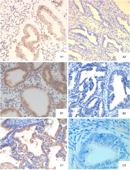
Figure 1. GPR30 protein expression (SP method; ×20). A: normal endometrium; A1: GPR30 positive; A2: GPR30 negative; B: EAH; B1: GPR30 positive; B2: GPR30 negative; C: EEC; C1: GPR30 positive; C2: GPR30 negative
suggests positive. Scoring was conducted by 2
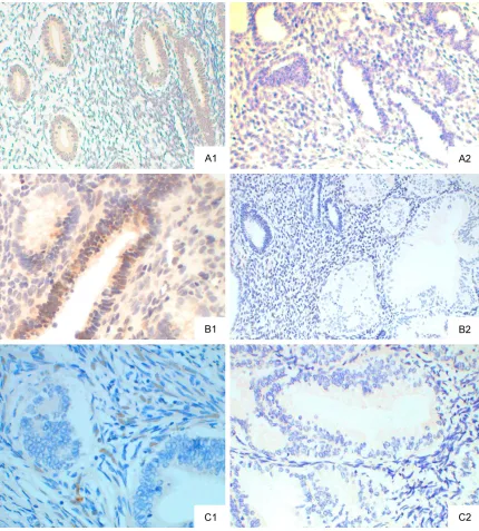
Figure 2. p-ERK1/2 protein expression (SP method; ×20). A: normal endometrium; A1: p-ERK1/2 positive; A2: ERK1/2 negative; B: EAH; B1: ERK1/2 positive; B2: ERK1/2 negative; C: EEC; C1: ERK1/2 positive; C2: p-ERK1/2 negative.
Statistical analysis
Statistical analysis was conducted with SPSS version 17.0. Qualitative data are expressed as rate and compared with chi square test. Correlation analysis was performed with Spearman rank correlation analysis with r=0 as a criterion for correlation. A value of P<0.05 was considered statistically significant.
Results
GPR30 and p-ERK1/2 expression in the endo-metrium
the endoplasmic reticulum or Golgi bodies [10]. In EEC, GPR30 expression was mainly found in the cytoplasm and cell membrane of EEC cells, cells strong positive for GPR30 were brown, and a lot of positive cells were found in EEC. In EAH, GPR 30 expression was mainly found in the cytoplasm and membrane (predominantly in cytoplasm), and moderately positive was found. In normal endometrium, weak GPR30 expression was found, cells were mainly light yellow, and GPR30 expression was predomi-nantly found in the cytoplasm (Figure 1). The positive rate of GPR30 expression was 15.0% (3/20), 40% (8/20) and 72.7% (16/22) in nor-mal endometrium, EAH and EEC, respectively. The positive rate of GPR30 expression in EEC was significantly higher than in other groups (P<0.05). The positive rate of GPR30 expres-sion in EAH group also increased markedly as compared to control group (P<0.05).
In normal endometrium, weak p-ERK1/2 expression was noted, cells were light yellow, and p-ERK1/2 expression was mainly found in the cytoplasm. In EAH, p-ERK1/2 expression increased significantly, cells were yellow-brown or brown, and p-ERK1/2 expression was found in the cytoplasm and nucleus. In EEC, p-ERK1/2 expression was mainly found in the nucleus and cytoplasm (predominantly in nucleus), and positive cells were yellow-brown and showed spotty distribution. In normal endometrium, p-ERK1/2 expression was not observed in the cytoplasm and nucleus (Figure 2). The positive rate of p-ERK1/2 expression was 30% (6/20), 65% (13/20) and 45.5% (10/22) in normal endometrium, EAH and EEC, respectively. In normal endometrium, low p-ERK1/2 expres-sion was found although there was still p-ERK1/2 expression. In EAH group, p-ERK1/2 expression increased significantly as compared to normal endometrium, (P<0.05). In EEC group,
erent in EEC patients with different depths of myometrial invasion: the deeper the myometri-al invasion, the higher the positive rate of GPR30 expression was (Spearman correlation analysis; P=0.003). However, GPR30 expres-sion had no relationship with other clinicopath-ological characteristics. (P>0.05). In addition, p-ERK1/2 had no correlation with any clinico-pathological characteristic (Table 2).
Correlation between GPR30 expression and p-ERK1/2 expression
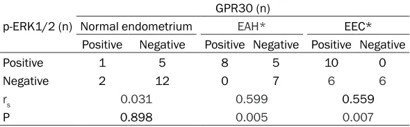
In normal endometrium, the proportion of GPR30 and p-ERK1/2 co-expression was 5.0% (1/20), and there was no correlation between GPR30 expression and p-ERK1/2 expression (rs=0.031, P>0.05). In EAH, the proportion of GPR30 and p-ERK1/2 co-expression was 35.0% (7/20), and positive relationship was found between GPR30 expression and p-ERK1/2 expression (rs=0.599, P<0.05). In EEC group, the proportion of GPR30 and p-ERK1/2 co-expression was 45.6% (10/22), and positive relationship was noted between GPR30 expression and p-ERK1/2 expression (rs=0.559, P<0.05) (Table 3).
Discussion
[image:5.612.90.371.98.174.2]GPR30 was independently identified and cloned in 1990s by different groups. GPR30 gene is mapped to 7p22 and its full length mRNA is 2604 bp. The full length mRNA includes a long open reading frame of 1128 bp which encodes GPR30 protein of 375 amino acids. Studies have revealed that GPR30 has a high homology with G Protein-Coupled Receptors (GPCRs) [11, 12]. GPR30 is a func-tional membrane receptor of estrogen [13, 14]. GPR30 mediates the non-genomic effects of estrogen which are involved in a variety of phys-iological and pathological processes in cells.
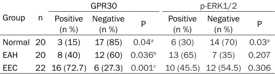
Table 1. GPR30 and p-ERK1/2 expression in the endometrium of different groups (n %)
Group n
GPR30 p-ERK1/2
Positive
(n %) Negative (n %) P Positive (n %) Negative (n %) P
Normal 20 3 (15) 17 (85) 0.04a 6 (30) 14 (70) 0.03a
EAH 20 8 (40) 12 (60) 0.036b 13 (65) 7 (35) 0.207
EEC 22 16 (72.7) 6 (27.3) 0.001c 10 (45.5) 12 (54.5) 0.306 Note: a. P<0.05 vs. EAH group; b. P<0.05 vs. EEC group; c. P<0.05 vs. normal control group.
the p-ERK1/2 expression was moderate and comparable to that in other groups (P>0.05) (Table 1).
Correlation of GPR30 and p-ERK1/2 expression with clinicopathological characteris-tics of EEC patients
There is evidence showing that GPR30 plays important roles in the follicular development, adrenal gland development and protection against liver injury as well as regulation of cell proliferation. Of important, GPR30 is also involved in the occurrence and progression of cancers. E2 and selective agonist of GPR30 (G1) may activate GPR30 to promote the prolif-eration of ovarian cancer cells and thyroid can-cer cells. The binding of E2 to GPR30 may acti-vate GPR30 and the intracellular downstream cascade, which leads to Ca2+ influx, production
found the GPR30 expression and the biological activities of GPR30 in EC cell lines (HEC50, HEC-1A, Ishikawa, KLE, RL95-2 and H38) [17, 18]. Lin et al [19] postulated that GPR30 could mediate the estrogen induced rapid activation of PI3K or MAPK in EC cells and had crosstalk with nuclear hormone receptor SF-1 or LRH-1 signaling pathway. GPR30 mediated the E2 or tamoxifen induced activation of EGFR-MAPK/ Erk1/2 signaling pathway in HEC-1A cells and Ishikawa cells, in which the c-fos expression was up-regulated, leading to the proliferation of
Table 2. Correlation of GPR30/p-ERK1/2 expression with clinicopathological characteristics of EEC patients
GPR30 p-ERK1/2
Clinicopathological characteristics Positive (n %) Negative (n %) P Positive (n %) Negative (n %) P
Age (yr) 0.432 0.122
≤55 10 (71.4) 4 (28.6) 5 (62.5) 3 (37.5)
>55 6 (75) 2 (25) 5 (35.7) 9 (64.3)
Menstruation 0.220 0.122
Premenopausal 5 (62.5) 3 (37.5) 9 (75) 3 (25)
Menopausal 11 (78.6) 3 (21.4) 5 (50) 5 (50)
FIGO stage 0.068 0.149
I 6 (60) 4 (40) 4 (40) 6 (60)
II 5 (71.4) 2 (28.6) 2 (28.6) 5 (71.4)
III 5 (100) 0 (0) 4 (80) 1 (20)
Histological grade 0.500 0.500
G1 4 (80) 1 (20) 2 (40) 3 (60)
G2 8 (66.7) 4 (33.3) 6 (50) 6 (50)
G3 4 (80) 1 (20) 2 (40) 3 (60)
Myometrial invasion 0.003* 0.115
≤1/2 6 (50) 6 (50) 4 (33.3) 8 (66.7)
>1/2 10 (100) 0 (0) 6 (60) 4 (40)
Lymph node metastasis 0.348 0.241
Yes 4 (80) 1 (20) 3 (60) 2 (40)
No 12 (70.6) 5 (29.4) 7 (41.2) 10 (58.8)
[image:6.612.91.386.459.551.2]Note: *P<0.05 (Spearman correlation analysis).
Table 3. Correlation between GPR30 expression and p-ERK1/2 expression in the endometrium of different groups
p-ERK1/2 (n)
GPR30 (n)
Normal endometrium EAH* EEC*
Positive Negative Positive Negative Positive Negative
Positive 1 5 8 5 10 0
Negative 2 12 0 7 6 6
rs 0.031 0.599 0.559
P 0.898 0.005 0.007
Note: *P<0.05 (Spearman correlation analysis).
EC cells. The crosstalk between GPR30 and SF-1 leads to the focal aggregation of estrogen and together with classic ER signaling pathway regulates the TAM induced proliferation of EC cells and even the tumorogenesis. Smith et al [20] detected GPR30 expression in 46 patients with pathologically proven EC, and their results showed GPR30 expression was very low in benign endometrial lesions and EC with good differentiation, but GPR30 expression in- creased markedly in EC with poor differentia-tion or deep invasion (such as uterine papillary serous adenocarcinoma, clear cell carcinoma and carcinosarcoma).
Our results showed GPR30 was mainly expressed in the cytoplasm and membrane of glandular epithelium. In normal endometrium and EAH, the GPR30 expression was moderate to low. However, most EEC tissues had a high GPR30 expression, and the proportion of GPR30 positive patients among EEC patients was significantly higher than in patients with normal endometrium and EAH patients (P<0.05). In addition, GPR30 expression in EAH group also increased markedly as compared to patients with benign endometrial lesions (P<0.05). These findings were consistent with previously reported. Our results showed the GPR30 expression increased gradually from normal endometrium to EAH and then to EEC, suggesting that GPR30 plays an important role in the estrogen induced tumorogenesis of endometrium. In patients with high GPR30 expression, trace estrogen binds to GPR30 to exert its non-genomic effects, leading to the sustained hyperplasia of endometrium or even tumorogenesis. Correlation analysis showed GPR30 expression was positively related to the depth of myometrial invasion (P<0.05): the deeper the myometrial invasion, the higher the GPR30 expression was. This suggests that GPR30 expression is closely related to the inva-sion of EEC. Further analysis revealed that GPR30 expression had no relationship with pathological stage and lymph node metastasis (P>0.05). These findings were different from those reported by Smith et al, but consistent with findings in some other studies. This indi-cates that GPR30 is not directly involved in the biobehaviors of EEC. On the basis of above find-ings, we speculate that GPR30 plays crucial roles in the occurrence and development of EEC, and GPR30 is involved in the estrogen induced activation of several signaling
path-gression of cancers [21]. However, the complex mechanisms are required to be elucidated in future studies.
Few studies have been conducted to investi-gate whether ERK1/2 signaling pathway is involved in the tumorogenesis of endometrium and whether abnormal activation of ERK1/2 signaling pathway is related to the pathogene-sis of EC. Some investigators proposed that estrogen could rapidly activate MAPK/ERK1/2 signaling pathway via the non-genomic mecha-nism, leading to the increase in p-ERK expres-sion, elevated proliferation of cells, inhibition of cell apoptosis, which are involved in the occur-rence and development of EC. Treeck et al pro-posed that estrogen could stimulate the prolif-eration of EC cells via the ERK1/2 signaling pathway [22]. Desouki et al [23] detected ERK1/2, C-Jun and P38 expressions in 33 patients with EC and 38 patients with benign endometrial lesions. Their results showed the progression from normal endometrium into EC had not relationship with MAPK signaling path-way activation. Mizumoto et al [24] detected p-ERK expression in 63 patients with EC, and results showed 63.5% of patients had p-ERK expression of different extents. Vantaggiato et al [25] found that cells with ERK2 knock out showed a poor proliferation, suggesting that ERK mediates the cell proliferation. The acti-vated ERK enters the nucleus and then activate transcription factors such as NF-κB, leading to the increase in Cyclin D1 expression and the subsequent proliferation of cells [26, 27]. Carcamo et al [28] revealed that reduction in ERK2 expression could decrease Cyclin D1 expression, arrest cells in G0/G1 phase and reduce cell proliferation.
positive patients in EEC patients (45.5%) was slightly lower than in EAH patients (65%) (P>0.05). Moreover, the p-ERK1/2 expression was comparable between EAH and normal endometrium (P>0.05). Desouki et al postulat-ed that the increaspostulat-ed activatpostulat-ed ERK in EEC might be related to the anti-estrogen effects of TAM in the endometrium, but had no relation-ship with the progression of EEC. There is evi-dence showing that estrogen may rapidly acti-vate MAPK/ERK signaling pathway via the non-genomic mechanism, to increase p-ER- K1/2 expression, stimulate cell proliferation, and inhibit cell apoptosis, which are involved in the occurrence and development of EEC. We speculate that there were two different regula-tory mechanisms of ERK1/2 in the EAH and EEC: in EAH, aberrant ERK1/2 activation may promote the atypical hyperplasia. When the ERK1/2 expression reaches a threshold, the negative feedback is initiated, causing the irre-versible malignant transformation as demon-strated by reduced p-ERK1/2 expression in EEC in the present study. ERK1/2 signaling pathway is one of pathways involved in the reg-ulation of EEC cells, but there is no non-hor-mone dependent aberrant activation as observed in breast cancer. In the present study, correlation analysis showed p-ERK1/2 expres-sion had no relationship with age, menstrua-tion state, FIGO stage, histological differentia-tion, depth of myometrial invasion and lymph node metastasis, which was similar to findings of Mizumoto et al. Thus, we speculate that ERK1/2 signaling pathway is not directly involved in the progression of EEC.
In vitro experiments showed GPR30 mediated the activation of MAPK/Erk1/2 signaling path-way in ER positive or negative endometrial cell lines [30]. In the present study, the GPR30 expression was positively related to p-ERK1/2 expression in EAH and EEC (r=0.599 and 0.559, respectively; P<0.05). This correlation might play an important role in the progression of EAH and might belong to an early molecular event in tumorogenesis. We speculate that ERK1/2 may exert synergistic effect with GPR30 in the occurrence and development of EEC, and p-ERK1/2 may be a key point in the GPR30 related estrogen induced activation of signaling pathway. However, the specific me- chanism underlying the interaction between GPR30 and ERK1/2 are required to be further investigated. To study the correlation between GPR30 and p-ERK1/2 may provide new targets for the therapy of EEC.
Disclosure of conflict of interest
None.
Address correspondence to: Dr. Weijuan Xin, De- partment of Obstetrics and Gynecology, Obstetrics and Gynecology Hospital of Fudan University, Shang- hai, China. E-mail: [email protected]; Dr. Qianyu Wu, Department of Obstetrics and Gy- necology, East Hospital, Tongji University, School of Medicine, Shanghai, China. E-mail: 5185874@qq. com
References
[1] Siegel R, Naishadham D and Jemal A. Cancer statistics, 2013. CA Cancer J Clin 2013; 63: 11-30.
[2] De Souza Nunes LS, De Oliveira RV, Holgado LA, Nary Filho H, Ribeiro DA and Matsumoto MA. Use of bovine hydroxyapatite with or with-out biomembrane in sinus lift in rabbits: histo-pathologic analysis and immune expression of core binding factor 1 and vascular endotheli-um growth factor. J Oral Maxillofac Surg 2011; 69: 1064-1069.
[3] Thomas P, Pang Y, Filardo EJ and Dong J. Iden-tity of an estrogen membrane receptor cou-pled to a G protein in human breast cancer cells. Endocrinology 2005; 146: 624-632. [4] Boonyaratanakornkit V and Edwards DP.
Re-ceptor mechanisms of rapid extranuclear sig-nalling initiated by steroid hormones. Essays Biochem 2004; 40: 105-120.
[5] Ajenjo N, Aaronson DS, Ceballos E, Richard C, Leon J and Crespo P. Myeloid leukemia cell growth and differentiation are independent of mitogen-activated protein kinase ERK1/2 acti-vation. J Biol Chem 2000; 275: 7189-7197. [6] Rice PL, Goldberg RJ, Ray EC, Driggers LJ and
Ahnen DJ. Inhibition of extracellular signal-reg-ulated kinase 1/2 phosphorylation and induc-tion of apoptosis by sulindac metabolites. Can-cer Res 2001; 61: 1541-1547.
[7] Acconcia F, Barnes CJ and Kumar R. Estrogen and tamoxifen induce cytoskeletal remodeling and migration in endometrial cancer cells. En-docrinology 2006; 147: 1203-1212.
[8] Filardo EJ, Graeber CT, Quinn JA, Resnick MB, Giri D, DeLellis RA, Steinhoff MM and Sabo E. Distribution of GPR30, a seven membrane-spanning estrogen receptor, in primary breast cancer and its association with clinicopatho-logic determinants of tumor progression. Clin Cancer Res 2006; 12: 6359-6366.
protocol. UK Receptor Group, UK NEQAS, The Scottish Breast Cancer Pathology Group, and The Receptor and Biomarker Study Group of the EORTC. J Clin Pathol 2000; 53: 634-635. [10] Lapensee EW, Tuttle TR, Fox SR and
Ben-Jona-than N. Bisphenol A at low nanomolar doses confers chemoresistance in estrogen receptor-alpha-positive and -negative breast cancer cells. Environ Health Perspect 2009; 117: 175-180.
[11] Feng Y and Gregor P. Cloning of a novel mem-ber of the G protein-coupled receptor family related to peptide receptors. Biochem Biophys Res Commun 1997; 231: 651-654.
[12] O’Dowd BF, Nguyen T, Marchese A, Cheng R, Lynch KR, Heng HH, Kolakowski LF Jr and George SR. Discovery of three novel G-protein-coupled receptor genes. Genomics 1998; 47: 310-313.
[13] Filardo E, Quinn J, Pang Y, Graeber C, Shaw S, Dong J and Thomas P. Activation of the novel estrogen receptor G protein-coupled receptor 30 (GPR30) at the plasma membrane. Endo-crinology 2007; 148: 3236-3245.
[14] Revankar CM, Cimino DF, Sklar LA, Arterburn JB and Prossnitz ER. A transmembrane intra-cellular estrogen receptor mediates rapid cell signaling. Science 2005; 307: 1625-1630. [15] Prossnitz ER, Arterburn JB, Smith HO, Oprea TI,
Sklar LA and Hathaway HJ. Estrogen signaling through the transmembrane G protein-coupled receptor GPR30. Annu Rev Physiol 2008; 70: 165-190.
[16] Prossnitz ER, Arterburn JB and Sklar LA. GPR30: A G protein-coupled receptor for estro-gen. Mol Cell Endocrinol 2007; 265-266: 138-142.
[17] Filardo EJ, Quinn JA and Sabo E. Association of the membrane estrogen receptor, GPR30, with breast tumor metastasis and transactivation of the epidermal growth factor receptor. Ste-roids 2008; 73: 870-873.
[18] Otto C, Rohde-Schulz B, Schwarz G, Fuchs I, Klewer M, Brittain D, Langer G, Bader B, Prelle K, Nubbemeyer R and Fritzemeier KH. G pro-tein-coupled receptor 30 localizes to the endo-plasmic reticulum and is not activated by es-tradiol. Endocrinology 2008; 149: 4846-4856. [19] Lin BC, Suzawa M, Blind RD, Tobias SC, Bulun
SE, Scanlan TS and Ingraham HA. Stimulating the GPR30 estrogen receptor with a novel tamoxifen analogue activates SF-1 and pro-motes endometrial cell proliferation. Cancer Res 2009; 69: 5415-5423.
[20] Smith HO, Leslie KK, Singh M, Qualls CR, Re-vankar CM, Joste NE and Prossnitz ER. GPR30: a novel indicator of poor survival for endome-trial carcinoma. Am J Obstet Gynecol 2007; 196: 386.e381-389; discussion 386.e389-311.
[21] Maggiolini M and Picard D. The unfolding sto-ries of GPR30, a new membrane-bound estro-gen receptor. J Endocrinol 2010; 204: 105-114.
[22] Treeck O, Diedrich K and Ortmann O. The acti-vation of an extracellular signal-regulated ki-nase by oestradiol interferes with the effects of trastuzumab on HER2 signalling in endometri-al adenocarcinoma cell lines. Eur J Cancer 2003; 39: 1302-1309.
[23] Desouki MM and Rowan BG. SRC kinase and mitogen-activated protein kinases in the pro-gression from normal to malignant endometri-um. Clin Cancer Res 2004; 10: 546-555. [24] Mizumoto Y, Kyo S, Mori N, Sakaguchi J, Ohno
S, Maida Y, Hashimoto M, Takakura M and In-oue M. Activation of ERK1/2 occurs indepen-dently of KRAS or BRAF status in endometrial cancer and is associated with favorable prog-nosis. Cancer Sci 2007; 98: 652-658. [25] Vantaggiato C, Formentini I, Bondanza A,
Bo-nini C, Naldini L and Brambilla R. ERK1 and ERK2 mitogen-activated protein kinases affect Ras-dependent cell signaling differentially. J Biol 2006; 5: 14.
[26] Yang C, Klein EA, Assoian RK and Kazanietz MG. Heregulin beta1 promotes breast cancer cell proliferation through Rac/ERK-dependent induction of cyclin D1 and p21Cip1. Biochem J 2008; 410: 167-175.
[27] Marampon F, Casimiro MC, Fu M, Powell MJ, Popov VM, Lindsay J, Zani BM, Ciccarelli C, Watanabe G, Lee RJ and Pestell RG. Nerve Growth factor regulation of cyclin D1 in PC12 cells through a p21RAS extracellular signal-regulated kinase pathway requires cooperative interactions between Sp1 and nuclear factor-kappaB. Mol Biol Cell 2008; 19: 2566-2578. [28] Carcamo-Orive I, Tejados N, Delgado J,
Gaz-telumendi A, Otaegui D, Lang V and Trigueros C. ERK2 protein regulates the proliferation of human mesenchymal stem cells without af-fecting their mobilization and differentiation potential. Exp Cell Res 2008; 314: 1777-1788. [29] Sebolt-Leopold JS and Herrera R. Targeting the
mitogen-activated protein kinase cascade to treat cancer. Nat Rev Cancer 2004; 4: 937-947.