Original Article
Correlation of expression of ADAR1 in oral squamous
cell carcinoma with clinicopathologic parameters
Jingping Zhou1, Daofang Tang1, Detao Tao2, Zhenlin Gao3, Qing Xu1
1School of Stomatology, Wannan Medical College, Wuhu, Anhui 241002, China; 2Department of Oral and
Maxillo-facial Surgery, The First Hospital of Wannan Medical College, Wuhu, Anhui 241001, China; 3Department of
Oncol-ogyIV, First Hospital of Shijiazhuang, Shijiazhuang, 050011, China
Received December 13, 2015; Accepted February 18, 2016; Epub March 1, 2016; Published March 15, 2016
Abstract: The aim of the study is to determine the expression Double-stranded RNA-specific adenosine deami -nase-1 (ADAR1) in subjects with and without oral squamous cell carcinoma, and to explore the correlation between expression of ADAR1 and clinicopathologic parameters, so as to confirm the role and significance of ADAR1 expres -sion in development of oral squamous cell carcinoma. Immunohistochemical staining was performed to detect the expression of ADAR1 in oral mucosa of 23 case normal tissues and 124 cases with oral squamous cell carcinoma. We found that ADAR1 positive expressed in all subjects with normal oral mucosa, high expressed in 13 out of 23 subjects. In subjects with oral squamous cell carcinoma, ADAR1 is positive expression in 118 out of 124 subjects, high expression in 79 out 129 subjects. No significance differences was found between positive of ADAR1 and age, gender, tumor location and lymph node metastasis, except for tumor diameter, lclinical stage , histopathologic grade and relapse (P<0.05). Our results suggested that RNA editing of ADAR1 may be not linked to oral cancer occur but to progress of oral cancer, which indicated that the regulation of ADAR1 protein existed in the development of oral cancer. Thus, the detection of ADAR1 expression may have certain reference to evaluate prognosis of patients with oral cancer.
Keywords: Oral squamous cell carcinoma, immunohistochemistry, RNA-editing enzyme, ADAR1
Introduction
Head and neck squamous cell carcinoma (HNSCC) ranks sixth among the most common cancers worldwide with an incidence of over 500,000 new cases each year [1]. Oral squa-mous cell carcinoma (OSCC) is the most preva-lent malignancy in oral cavity [2], which is a disease found particularly in low income com-munities and mainly a problem of older men, 90% being in >45 year age group who are exposed to the known risk factors of tobacco and/or alcohol [3].
DNA mutations play an important role in the occurrence and development of tumor. In recent years, many scholars have found that the imbalance of RNA editing is an important factor to accelerate tumor progression. RNA editing biology diversity and complexity have important role to guarantee, the most common RNA editing is adenine into hypoxanthine I,
which carry genetic information and its coding protein structure and function change. In recent years, the study found that many diseases occurrence and RNA editing is closely related to the abnormal, in the process of occurrence and development of tumor, RNA editing disorders may lead to anti oncogene inactivation of living or oncogene activation and promote tumor pro-gression. ADAR1 is widespread in human tis-sues and its role in the substrate are glutamate receptors and serotonin receptor mRNA, when
in the presence of specific substrates, ADAR1
will from the nucleus into plasma cells play a catalytic role [4], thus, the function of ADAR1 is closely related with intracellular localization. It has been found that there are a variety of ADAR1 genes regulating the intracellular local-ization of the related sequences, which affect the intracellular localization of ADAR1, resulting in a different role in the outcome of different substrate. Study found that RNA editing
response plays an important role [5]. Mean- while, abnormal of RNA editing in many
malig-nant tumors also have influence on tumor
occurrence and progress [6].
RNA editing is a common phenomenon that exists in organism, which can make genetic information change, and RNA editing enzyme plays an important role in the process. ADAR1 is a double chain RNA editing enzyme, one of the members of the ADAR family; so far the family has found three members: ADAR1, ADAR2 and ADAR3. ADAR1 is located on human chromosome 1q21.1-q21.2, with a total length of about 30 KB, with 15 exons [7]. After selec-tive cutting, multiple transcripts can be gener-ated, which contain P150 and P110, P150 are distributed in the cytoplasm and the nucleus, P110 only concentrated in the nucleus [8]. Increasing number of study show that the disor-der of RNA editing may be closely related to the occurrence and progression of tumor. Oral can-cer is a squamous cell carcinoma (cell carcino-ma squamous) which occurs in the oral muco-sa, accounting for 90% of the oral malignant
tumors. At present, the five year survival rate is
only maintained at 50%-60%, and most of the patients eventually died due to tumor metasta-sis and recurrence. Currently about ADAR1 and cancer research focus in brain tumor, liver can-cer, laryngeal cancan-cer, blood disease and gastric cancer, colon cancer and other tumors of the digestive tract. Little is known about the expres-sion and function of ADAR1 in oral cancer worldwide. Thus, the purpose of present study is to detect the expression of ADAR1 in normal oral mucosa and oral cancer by immunohisto-chemical staining method, to analysis the rela-tionship between the expression degree of ADAR1 and clinical pathological character. Moreover, exploring the role of ADAR1 in the development of oral cancer, which will offer cer-tain reference for the mechanism of metasta-sis and recurrence and prognometasta-sis of oral cancer in future research.
Methods
Sources of material
A total of 124 OSCC specimens were taken
from department of pathology affiliated stoma -tology hospital of nanjing medical university (Jiangsu province dental hospital) from August, 2008 to April, 2012, which consist of 72 male
and 52 female. The age of the study patients was from 34 to 88 years old at the time of diag-nosis. Of these 142 patients, 99 case over 50 years old and 25 case under 50 years old, 52 cases of tongue cancer, 31 patients with buc-cal carcinoma, 23 cases with gingival carcino-ma, 18 cases with other type of oral cancer
(mouth floor carcinoma, palate carcinoma, lip
and chin carcinoma).
According to the tumor diameter, 21 cases in T1 phase, in 68 cases in T2 stage , 25 cases and 10 cases in T3 and T4,respectively. 52 cases with lymph node metastases, and72 cases without lymph node metastasis. Only 1 out of 124 patients with distant metastasis. According to the degree of clinical stage, 12 cases, 42 cases, 34 cases and 36 cases in stage I, II, III and IV. Depending on the degree of tumor differentiation, 75, 41 and 8 cases in high, medium and low differentiated of squa-mous cell carcinoma (SCC), respectively. Followed-up of all patients in the March, 2015 found, 2 out of 124 patients lost to follow-up. Among the remaining 122 cases, 41 cases with tumor recurrence (including 32 cases of death because of recurrence, 9 cases of recurrence but is still alive).
Another 23 cases of normal oral mucosa sues as control, all the normal oral mucosa tis-sues were derived from oral mucosa which was more than 2 cm from the tumor edge, and
con-firmed by pathological examination of oral
mucosal negative. Two investigators assessed the slides without knowledge of the clinicopath-ological features and were blinded to each oth-er’s evaluation. They were in agreement on all the slides examined. The diagnosis, differential
diagnosis and pathological classification of oral
squamous cell carcinoma are based on the sev-enth edition “oral histopathology and patholo-gy” [9].
Antibodies
reagent were purchased from Nanjing Jian- cheng Bioengineering Institute.
Experimental methods
Super sensitive two step (Polink-2 Plus) immu-nohistochemical staining method was used in our research. Sliced the specimen under 65°C oven, bake for 30 min. Then, taking slice into solution of dimethyl benzene and alcohol for regular dewaxing and gradient dehydration. In citrate buffer salt (PH 6.0), after the microwave antigen repair kit in 3% H2O2 deionized water incubation for 10 min, to block endogenous peroxidase; Add a resistance to 1:30 0 (con-centration) in 4°C refrigerator overnight. The next day, in turn, add kit of reagent 1 (polymer auxiliary agent), reagent 2 (horseradish enzyme mark goat IgG resistant polymer), the incuba-tion 10-20 min; After the DAB chromogenic redyeing, dehydration, transparent sealing piece. Every step between 2 min is washed with PBS buffer was used washing for three times ,every time last 2 min. repeat 3 times. Using known hippocampus slices as positive control, with PBS instead of one as a negative control.
Result determination
ADAR1 staining was mainly localized in the cell nuclear and cell cytoplasm, the positive expression of yellow or brown/brown coloring. Immunohistochemical staining results were determined by semi quantitative scoring meth-od, based on the expression of intensity inte-gration: no coloring is 0; the yellow is 1; brown
is 2; Black-brown is 3. At low magnification,
selected specimens positive cells and uniform distribution area, the mean percentage of ran-dom counting 5 unique, non overlapping high
power field of positive cells, according to the
percentage of positive cell integral: 0-5% to 0; 6-25% to 1; 26-50% to 2; 51-75% to 3, more than 75% to 4. Finally the two integral multipli-cations, the integral is greater than or equal to 1 divided into positive. 0 score means “-”; 1-4 score means “+”; 5-8 score means “++”; 6-12 score means “+++”; “-“ or “+” means low expression, “++” or “+++” means - ~; low high expression.
Statistical analysis
Statistical analysis was performed by R pro-gram language, percent of gender, age, tumor
location, lymph node metastasis, differential degree, tissue location and positive expression of ADAR1 between two groups was compared by chi-square test. A P-value of less than 0.05
was considered to be statistically significant.
Results
Expression of ADAR1 in the normal oral mu-cosa
ADAR1 positive expression in epithelial tissue of normal oral mucosa of 23 cases, the posi- tive expression rate was 100% (23/23), the high expression rate was 56.5% (13/23). Positive staining located in the nuclei and cytoplasm, basal epithelial cell layer negative expression; nuclear staining positive cells scattered in distribution in the epithelium, which coloring number of stratum spinosum cell nuclei above the basal layer was, brownish yellow or brown; near surface epithelial prickle cells in the cytoplasm of mainly was yellow brown or yellow.
Expression of ADAR1 in oral cancer
The positive expression rate of ADAR1 in 124 cases of oral cancer was 95.2% (118/124), the high expression rate was 63.7% (79/124). The expression of positive expression rate and expression intensity of ADAR1 in normal
oral mucosa was not statistically significant.
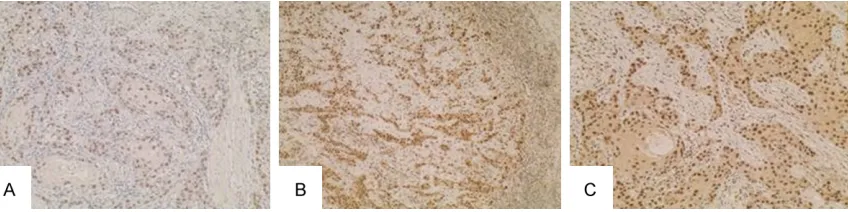
Positive staining also positioning in the nuclei and cytoplasm, scattered uniformly in the can-cer tissues, yellow, brown or yellow brown, some cancer nests of peripheral cells was neg-ative (Figures 1-3).
ADAR1 expression in oral squamous cell carci-noma and its relation to the clinic pathological characteristics
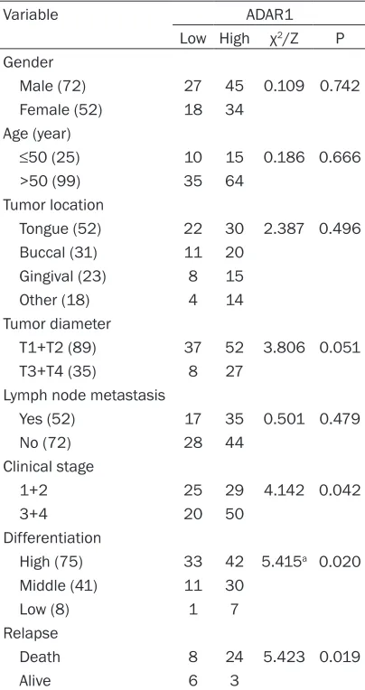
Analysis results revealed that no correlation existed between ADAR1 expression level and patients age, sex, site of lesion and lymph node metastasis (P>0.05), but tumor size, clinical staging, pathological grading and recurrence. The high expression rate of ADAR1 in T1 and T2 stage and in T3 and T4 stage of oral cancer was 58.4% (52/89) and 77.1% (27/35), respectively.
53.7% (29/54) and 71.4% (50/70). Chi-square test results showed that ADAR1 expression level increased with increasing of clinical stages (chi-square=4.142, P<0.05). ADAR1 high expression rate in high, middle and low differentiation of oral cancer was 56.0% (42/75), 73.2% a (30/41) and 87.5% (7/8). Chi square test results show that ADAR1 expres-sion rate increases with decreasing the degree of differentiation of oral cancer (chi-square =5.415, P<0.05). Because of recurrent cancer of the oral cavity lead to death rate of the high
expression of ADAR1 in died cases with re-
current was 75% (24/32), which is
signifi-cantly higher than in survival case with recur-rent 33.3% (3/9) (chi-square=5.423, P<0.05) and that poor clinical prognosis of patients with ADAR1 expression was enhanced (Table 1).
Discussion
[image:4.612.90.523.73.179.2]Our research results show that no statistically difference was found in ADAR1 expression
[image:4.612.96.520.252.358.2]Figure 1. The expression of ADAR1 in highly differentiated oral cancer; positive staining was localized in the nucleus and cytoplasm. A: ADAR1 negative expression (A×200); B: ADAR1 weak positive expression (B×200), cancer nest surrounding cells was negative expression; C: ADAR1 strong positive expression, positive staining of nuclear stain-ing (C×200).
Figure 2. Expression of ADAR1 in moderately differentiated oral cancer, positive staining was localized in the nu-cleus and cytoplasm. A: The weak positive expression of ADAR1 (A×200); B: Strong positive expression of ADAR1 (B×100); C: Strong positive expression of ADAR1 (C×200).
[image:4.612.95.521.420.527.2]between oral squamous cell carcinoma and normal oral mucosa and oral cancer occur-rence may be not associated with catalysis of aberrant RNA editing. Some scholars have shown that the study of bladder cancer, cancer tissue and normal bladder tissue encoding and non encoding District RNA editing are not
sig-nificantly different, but also shows that the
occurrence of bladder cancer and RNA is not related to the abnormal editing [10]. Although no correlation was found between ADAR1 expression level and tumor size in present research (p=0.051), properly link maybe exist when we enlarge the sample size in future studies.
This study also found that ADAR1 expression is associated with tumor size, clinical stage,
degree of differentiation and prognosis of oral squamous cell carcinoma. High expression of ADAR1 is positive associated with the tumor diameter, tumor TNM stage and tumor low dif-ferentiation degree. ADAR1 expression
intensi-ty is significantly higher in recurrence patients
(already died) than in recurrence patients (surviving).
All of the above results suggest that the high expression of ADAR1 is closely related to the progression and prognosis of oral cancer. Thus, we hypothesis that abnormal phenomenon of RNA editing maybe occur in oral cancer. Tumor occurrence, development and recurrence and metastasis is a very complex process, in the gene expression process, there are a large number of transcription, reverse transcription and translation.RNA editing disorders may affect tumor progression. for example, pleo-morphic malignant glioma in glur-b Q/R site of low RNA editing, will change the nature of the ion channels between cells, resulting in the imbalance of intracellular and extracellular ion concentration, leading to tumor growth and produced a series of clinical symptoms of rea-son [11]; Levanon et al. found in neuroblasto-ma in the existence of a large number of new RNA editing phenomenon [12]; nearly a quarter
of multiple neurofibromatosis patients with
abnormal RNA editing [13]. Meanwhile, the bal-ance of RNA editing could increase the cell apoptosis and inhibit its proliferation, and reverse the growth and migration of malignant cells [14].
[image:5.612.91.293.96.478.2]The present study showed that the expression of ADAR1 in different tumors showed a double side effect. ADAR1 showed low expression in tumors, such as glioma of the ADAR family of three molecular mRNA levels decreased [15]; Gu Yong et al. Research results showed that the expression of ADAR1 in digestive tract malignant tumor of gastric cancer, liver cancer and colon cancer than in normal tissues and cancer adjacent tissues [13], suggesting that ADAR1 may play an inhibitory role in cancer gene. On the other hand, ADAR1 expression enhancements will lead to a large number of apoptotic genes such as bcl2 and bcl10 reduced, enhanced the anti apoptotic effect of the body, leading to cell proliferation caused by cancer [16], ADAR1 seemed plays the role of oncogene.
Table 1. The expression of ADAR1 in oral squamous cell carcinoma
Variable ADAR1
Low High χ2/Z P
Gender
Male (72) 27 45 0.109 0.742 Female (52) 18 34
Age (year)
≤50 (25) 10 15 0.186 0.666
>50 (99) 35 64 Tumor location
Tongue (52) 22 30 2.387 0.496 Buccal (31) 11 20
Gingival (23) 8 15
Other (18) 4 14
Tumor diameter
T1+T2 (89) 37 52 3.806 0.051
T3+T4 (35) 8 27
Lymph node metastasis
Yes (52) 17 35 0.501 0.479
No (72) 28 44
Clinical stage
1+2 25 29 4.142 0.042
3+4 20 50
Differentiation
High (75) 33 42 5.415a 0.020
Middle (41) 11 30
Low (8) 1 7
Relapse
Death 8 24 5.423 0.019
Alive 6 3
Conclusion
From the point of our results, ADAR1 expres-sion is not associated with occur of oral cancer but prognosis of oral cancer, which suggested that high expression of ADAR1 plays a role in
prognosis of oral cancer. We should confirm
that whether RNA editing disorder of ADAR1 catalyze activate of ontogenesis and tumor growth or inactivate of tumor suppressor genes and the progression of cancer in further research. In addition, ADAR1 high expression is not associated with lymph node metastasis in oral cancer but with tumor grade staging. In particular, the high expression of ADAR1 in patients with recurrent death is worthy of fur-ther study. It is expected to provide a new way to evaluate the prognosis of patients with oral cancer.
Acknowledgements
This work was supported by Provincial Natural Science Research Project of Anhui Colleges (No. KJ2014A269).
Disclosure of conflict of interest
None.
Address correspondence to: Dr. Jingping Zhou, Department of Oral Medicine, School of Stomatology, Wannan Medical College, 22 Wenchangxi Road, Yijiang District, Wuhu, Anhui 241002, China. E-mail: [email protected]
References
[1] Parkin DM, Bray F, Ferlay J and Pisani P. Global cancer statistics, 2002. CA Cancer J Clin 2005; 55: 74-108.
[2] Cao ZG and Li CZ. A single nucleotide polymor-phism in the matrix metalloproteinase-1 pro-moter enhances oral squamous cell carcinoma susceptibility in a Chinese population. Oral On-col 2006; 42: 32-38.
[3] Scully C and Bagan J. Oral squamous cell carci-noma: overview of current understanding of aetiopathogenesis and clinical implications. Oral Dis 2009; 15: 388-399.
[4] Wang Q. RNA editing catalyzed by ADAR1 and its function in mammalian cells. Biochemistry (Mosc) 2011; 76: 900-911.
[5] Casey JL. Control of ADAR1 editing of hepatitis delta virus RNAs. Curr Top Microbiol Immunol 2012; 353: 123-143.
[6] Galeano F, Tomaselli S, Locatelli F and Gallo A. A-to-I RNA editing: the “ADAR” side of human cancer. Semin Cell Dev Biol 2012; 23: 244-250.
[7] Weier HU, George CX, Greulich KM and Samuel CE. The interferon-inducible, double-stranded RNA-specific adenosine deaminase gene (DSRAD) maps to human chromosome 1q21.1-21.2. Genomics 1995; 30: 372-375.
[8] Patterson JB and Samuel CE. Expression and regulation by interferon of a double-stranded-RNA-specific adenosine deaminase from hu -man cells: evidence for two forms of the de-aminase. Mol Cell Biol 1995; 15: 5376-5388. [9] Shifeng Y. Oral histopathology and pathology.
Beijing: People’s medical publishing house, 2012.
[10] Zilberman DE, Safran M, Paz N, Amariglio N, Simon A, Fridman E, Kleinmann N, Ramon J and Rechavi G. Does RNA editing play a role in the development of urinary bladder cancer? Urol Oncol 2011; 29: 21-26.
[11] Cenci C, Barzotti R, Galeano F, Corbelli S, Rota R, Massimi L, Di Rocco C, O’Connell MA and Gallo A. Down-regulation of RNA editing in pe-diatric astrocytomas: ADAR2 editing activity inhibits cell migration and proliferation. J Biol Chem 2008; 283: 7251-7260.
[12] Levanon EY, Eisenberg E, Yelin R, Nemzer S, Hallegger M, Shemesh R, Fligelman ZY, Shoshan A, Pollock SR, Sztybel D, Olshansky M, Rechavi G and Jantsch MF. Systematic iden-tification of abundant A-to-I editing sites in the human transcriptome. Nat Biotechnol 2004; 22: 1001-1005.
[13] Mukhopadhyay D, Anant S, Lee RM, Kennedy S, Viskochil D and Davidson NO. C-->U editing of neurofibromatosis 1 mRNA occurs in tumors that express both the type II transcript and apobec-1, the catalytic subunit of the apolipo-protein B mRNA-editing enzyme. Am J Hum Genet 2002; 70: 38-50.
[14] Ishiuchi S, Yoshida Y, Sugawara K, Aihara M, Ohtani T, Watanabe T, Saito N, Tsuzuki K, Oka-do H, Miwa A, Nakazato Y and Ozawa S. Ca2+-permeable AMPA receptors regulate growth of human glioblastoma via Akt activation. J Neu-rosci 2007; 27: 7987-8001.
[15] Paz N, Levanon EY, Amariglio N, Heimberger AB, Ram Z, Constantini S, Barbash ZS, Adam-sky K, Safran M, Hirschberg A, KrupAdam-sky M, Ben-Dov I, Cazacu S, Mikkelsen T, Brodie C, Eisenberg E and Rechavi G. Altered adenosine-to-inosine RNA editing in human cancer. Ge-nome Res 2007; 17: 1586-1595.