Original Article
Expression of IL-33 in colorectal cancer and
its association with clinical features
Quanqin Xie, Dachuan Zhang, Wenwei Hu, Xu Deng, Jingting Jiang, Changping Wu
Department of Tumor Biological Treatment, The Third Affiliated Hospital of Soochow University, Changzhou, Jiangsu, China
Received January 7, 2016; Accepted March 20, 2016; Epub July 1, 2016; Published July 15, 2016
Abstract: Tumor immunotherapy has strong potential worldwide. IL-33 mostly acts as a danger signal from damaged
cells, and has recently been discovered as a influencing factor in the process of tumor immunity. The aim of the
study is to discover the expression of IL-33 in colorectal cancer (CRC) and its association with clinical features and
prognosis. We found that the positive expression rate of IL-33 in tumor tissues was significantly lower than that in peritumoral tissues (38.78% vs. 63.75%, Pearson χ2 = 10.989 P = 0.001). In addition, serum IL-33 levels in moder-ate or poor differentiation CRC cases were significantly higher than that in well differentiation cases. Serum IL-33 level in colon cancer cases was also significantly higher than that in rectal cancer cases (P < 0.01). Moreover, that
combination of serum IL-33, CEA and CA19-9 may improve diagnostic efficiency for colorectal cancer. In conclusion,
IL-33 may associate with tumor progress in CRC, and showed improved value in CRC diagnosis.
Keywords: Interleukin-33, colorectal cancer, clinical features
Introduction
Colorectal cancer (CRC) is the third most com-mon type of cancer in the world. The present mainstream methods of treatment include sur-gery, chemotherapy and radiation [1]. The con-ventional therapies have greatly induce tumor inhibition, but with side effects and drug resis-tance in the long term. So the improvements of overall survival and quality of life of CRC patients have certain limitations [2]. In recent years, tumor immunotherapy has put up the characteristics with strong potential and wide-spread acceptance worldwide [3]. A deeper understanding of the mechanisms of tumori-genesis and tumor prognosis may facilitate the achievement of tumor immunotherapy.
Interleukin-33 (IL-33) is a member of the inter-leukin-1 (IL-1) cytokine family. IL-33 is mainly expressed in epithelial cells, endothelial cells, and smooth muscle cells [4]. IL-33 mostly acts as a danger signal from damaged cells, and is identified as an inflammatory cytokine. Inhi- bition of IL-33 involved pathway may have anti-inflammatory effects [5]. IL-33 has recently been discovered as an influencing factor in the
process of tumor immunity, and is implicated in cancer pathogenesis. The aim of the study is to discover the expression of IL-33 in colorectal cancer and its association with clinical features and prognosis.
Materials and methods
Tissue microarray
in citrate solution (10 mmol/L, pH 6.0). Then the tissue microarray was incubated for 15 min in 0.3% hydrogen peroxide solution to block endogenous peroxidase activity, followed by overnight incubat ion with the polyclonal rabbit antibody against human IL-33 in a humidified chamber at 4 degrees centigrade. After incuba-tion for 30 min with the sec ondary antibody at room temperature 25°C, Tissue microarray was stained with DAB, counterstained with hema-toxylin. Finally, tissue microarray was dehydrat-ed and mountdehydrat-ed. Negative controls were treat-ed similarly except for phosphate buffer solu-tion instead of primary antibody.
Evaluation of IL-33 expression
IL-33 immunostaining intensities were asse- ssed by professional pathologists in the Third Affiliated Hospital of Soochow University. Tissues with medium brown or blue staining were considered as positive. Every sample in the tissue microarray with more than 50 per-cent positive staining area was considered as positive ultimately.
Serum samples
Serum samples of colorectal cancer patients from the third affiliated hospital of Soochow University were collected. 121 Samples were collected during Feb 2014 and May 2015, including 72 male patients and 49 female patients, with the age of (62.55 ± 11.70) years old. 53 samples were from colon cancer, and 68 were from rectal cancer. Inclusion criteria: The patients in this study didn’t accept prior hormonal therapy, chemotherapy or radiothera-py, with normal cardiac and renal function. Exclusion criteria: The patients were complicat-ed with primary tumors locatcomplicat-ed in other part of body, or internal diseases that out of control. Meanwhile, 81 serum samples from healthy
ation manual. The microplate reader (Multiskan GO, Thermo Fisher Scientific, Massachusetts, USA) was used for the measurement of absor-bance of all samples. The standard curve was drawn by the standard concentration as the abscissa, and absorbance as the ordinate. So the serum IL-33 concentrations were worked out from the standard curve. The CEA (carcino-embryonic antigen) and CA19-9 (carbohydrate antigen 19-9) of the samples were detected by Cobas e601 automatic electrochemical lumi-nescence immunity analyzer (Roche Company, Basel, Switzerland). All the operation sequenc-es were according to the operating instructions for the equipment.
Statistical analyses
SPSS Statistics 19.0 (IBM Company, Chicago, USA) was used in data analysis. Pearson’s chi-square test was utilized to compare the con-stituent ratio in order to analyze the relation-ship of IL-33 or serum IL-33 with clinical fea-tures. Overall survival (OS) of various clinical features was compared using Kaplan-Meier and log-rank tests. The Cox proportional haz-ards model was performed to estimate hazard risk (HR) with 95% confidence intervals (CI) of the linking strength between different clinical characteristics, IL-33 expression death risks. The P-value of < 0.05 was considered statisti-cally significant. All statistical tests were two-sided. GraphPad Prism 6.02 (CA 92037, USA) was utilized for creating statistical graphs. Results
IL-33 expression in tumor and peritumoral co-lon tissues
no or weak staining was observed in the tumor tissues (Figure 1A and 1B). As is shown in Table 1, Among 98 cases of tumor tissues, 38 cases had positive IL-33 staining. Among 80 cases of peritumoral tissues, 51 cases had positive IL-33 staining. The positive expression rate of IL-33 in tumor tissues was significantly lower than that in peritumoral tissues (38.78% vs. 63.75%, Pearson χ2 = 10.989 P = 0.001).
Relationship of IL-33 expression and clinico-pathological parameters
To study the relationships between IL-33 expre- ssion and patients’ clinicopathological
param-eters, we divided cases into subgroups by vari-ous clinicopathological parameters, respective-ly. The various clinicopathological parameters consist of patients’ gender, age, tumor diame-ter, pathological grading, tumor location, depth of invasion, lymphatic metastasis, distant metastasis and AJCC TNM Staging (7th edition).
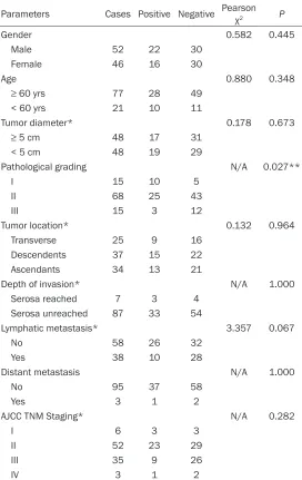
[image:3.612.92.523.71.395.2]The positive and negative expression counts were worked out in each subgroup respectively. Pearson’s chi-square test or Fisher’s exact test were used to compare the positive rates of IL-33 in each subgroup. As is shown in Table 2, cases with pathological grading I, II and III were 10/15, 25/68 and 3/15, respectively, with corresponding positive expression rate of 66.67%, 36.76% and 20%, respectively, with a statistically significant difference (P = 0.027). Conclusion could be drawn from the data that the higher level of pathological grading, the lower expression of IL-33 in tumor tissues. Nevertheless, there were not any relationships between IL-33 expression and patients’ any other clinicopathological parameter (P > 0.05). Figure 1. Immunohistochemical staining of IL-33 in tumor tissue (A and B) and peritumoral tissue (C and D)
Magni-fication: (A, C) 100 ×, (B, D): 200 ×.
Table 1. Comparison of IL-33 expression in colon tumor tissues and peritumoral tissues
Cases PositiveIL-33 NegativeIL-33 Tumor tissues 98 38 (38.78%) 60 Peritumoral tissues 80 51 (63.75%) 29
[image:3.612.91.294.471.523.2]Prognostic values of IL-33 and other clinico-pathological parameters in colon caner
As is shown in Figure 2, patients with TNM stages of III & IV showed significantly worse sur-vival compared with those of I & II (P = 0.0003, log-rank test) (Figure 2A). Patients with TNM stages of I and II had a 60% reduction in risk of death (Hazard ratio 0.3999, 95% confiden- ce interval: 0.1922~0.6014), compared with those with TNM stages of III and IV.
significantly higher than that in intestinal polyp and healthy control group (Z = 4.938, P < 0.01 and Z = 8.921, P < 0.01, respectively). The serum IL-33 level in intestinal polyp group had no significantly difference in comparing with healthy control group (Z = 1.015, P = 0.31). Serum IL-33 level in CRC patients and its cor-relation with clinical features
The CRC group was divided into various sub-groups by different clinical features, such as
< 5 cm 48 19 29
Pathological grading N/A 0.027**
I 15 10 5
II 68 25 43
III 15 3 12
Tumor location* 0.132 0.964
Transverse 25 9 16
Descendents 37 15 22
Ascendants 34 13 21
Depth of invasion* N/A 1.000
Serosa reached 7 3 4 Serosa unreached 87 33 54
Lymphatic metastasis* 3.357 0.067
No 58 26 32
Yes 38 10 28
Distant metastasis N/A 1.000
No 95 37 58
Yes 3 1 2
AJCC TNM Staging* N/A 0.282
I 6 3 3
II 52 23 29
III 35 9 26
IV 3 1 2
Notes: *Very few cases censored because parameters cannot retrieved,
**sta-tistical significance. Abbreviations: AJCC, American Joint Committee on Cancer.
N/A, not applicable, because Fisher’s exact test was applicable only.
tant metastasis was a negative prognostic factor for OS (overall survival), [P < 0.01, HR (hazard ratio) = 13.246, 95% CI (confi-dence interval): 3.721~47.152]. AJCC TNM staging III/IV versus I/II was also a negative prognos-tic factor for OS (P < 0.01, HR = 2.396, 95% CI: 1.430~4.016). Multivariate analysis showed that distant metastasis was a negative factor for OS (P = 0.002, HR = 7.841, 95% CI: 2.095~29.352), AJCC TNM stag-ing III/IV versus I/II was also a negative factor for OS (P = 0.003, HR = 2.594, 95% CI: 1.394~4.827).
Serum IL-33 level in colorectal cancer and control group
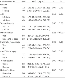
[image:4.612.92.364.97.528.2]TNM staging, tumor location and histological type. As is shown in Table 4, serum IL-33 levels in moderate or poor differentiation CRC cases were significantly higher than that in well differ-entiation cases (Z = 6.25, P < 0.01). Serum IL-33 level in colon cancer cases were also sig-nificantly higher than that in rectal cancer cases (Z = 2.90, P < 0.01). Other clinical fea-tures showed no significant differences when compared with serum IL-33 levels.
Logistic regression model with IL-33 involved in for CRC diagnosis
[image:5.612.92.515.73.227.2]The CRC group was set as disease group. Similarly, the intestinal polyp control group and healthy control group was set as control group. Each case in disease and control group had serum IL-33, CEA (carcinoembryonic anti-gen) and CA 19-9 (carbohydrate antigen 19-9) Figure 2. Kaplan-Meier survival curves of colon cancer patients based on TNM stages and IL-33 expressions.
Pa-tients with TNM stages of III/IV showed significantly worse survival compared with those of I/II (P = 0.0003, log-rank test) (A). Patients with positive IL-33 expressions have no significant different overall survival compared with those
[image:5.612.92.524.310.443.2]with negative IL-33 expressions (P = 0.5400, log-rank test) (B).
Table 3. Univariate and multivariate analysis of clinicopathological parameters for overall survival
Clinicopathological parameters Univariate Multivariate
HR (95% CI) P HR (95% CI) P
Gender (male vs. female) 1.033 (0.619~1.725) 0.900 0.807 (0.449~1.449) 0.473
Age (≥ 60 yrs vs. < 60 yrs) 1.541 (0.780~3.044) 0.213 1.323 (0.652~2.682) 0.438
Tumor diameter (≥ 5 cm vs. < 5 cm) 1.051 (0.631~1.752) 0.847 1.227 (0.709~2.123) 0.465 Pathological grading (Grade III vs. Grade I/II) 1.552 (0.785~3.067) 0.206 1.556 (0.741~3.267) 0.243 Serosa invasion (reached vs. unreached) 0.656 (0.262~1.642) 0.368 0.591 (0.228~1.529) 0.278 Distant metastasis (Yes vs. No) 13.246 (3.721~47.152) < 0.01* 7.841 (2.095~29.352) 0.002* AJCC TNM staging (III/IV vs. I/II) 2.396 (1.430~4.016) < 0.01* 2.594 (1.394~4.827) 0.003* IL-33 expression (positive vs. negative) 1.178 (0.695~1.997) 0.543 0.904 (0.515~1.586) 0.724
Notes: *Statistical significance. **Parameter “Lymphatic metastasis” was censored because of its strongly linearly association with parameter “AJCC TNM staging”. Abbreviations: HR, hazard ratio. CI, confidence interval.
Figure 3. Serum IL-33 level in colorectal cancer and control group. Abbreviations: CRC, colorectal cancer. IP, intestinal polyp. HC, healthy control.
[image:5.612.91.286.490.634.2]detected. Binary regression model method was used to generate a formula for diagnosis. The logistic regression analyze output was shown in Table 5. In accordance with the output, the for-mula was log [p (1-p)] =0.015*a+0.498*b+ 0.046*c-4.01. Each case had morbidity proba-bility (p) worked out derived from the formula. The new parameter, morbidity probability, was identified as a combined biomarker integrated
off values of CEA and CA19-9 were set accord-ing to standard criterion, that is, 5 ng/mL and 37 ng/mL, respectively. As a consequence, the sensitivity, specificity, PPV (positive predictive value), NPV (negative predictive value) and accuracy were calculated out. As is shown in Table 6, CEA and CA19-9 had higher specificity and lower sensitivity for CRC diagnosis. The sensitivity of serum IL-33 was higher than CEA and CA19-9, while it had lower sensitivity than CEA and CA19-9. The combined diagnosis had maximum sensitivity and accuracy, and the specificity was at a high level.
Discussion
Cancer development is strongly influenced by innate and adaptive immunity. The immune sys-tem consists of complex structures, some of which may promote tumorigenesis, and some has inhibiting effect on tumorigenesis [6]. Interleukin-33 (IL-33), a cytokine of the IL-1
Differentiation 6.25 < 0.01*
Well 68 114.64 (86.84, 196.04) Moderate or poor 53 278.51 (182.28, 425.88)
Lymphatic metastasis 0.06 0.95
Yes 44 168.94 (112.61, 283.81) No 77 175.45 (104.46, 356.34)
AJCC TNM staging 0.06 0.95
I/II 77 175.45 (104.46, 356.34) III/IV 44 168.94 (112.61, 283.81)
Tumor location 2.90 < 0.01*
Colon 53 211.40 (135.49, 417.55) Rectum 68 142.05 (88.67, 249.15)
Histological type 1.52 0.13
Ulcerative 98 183.82 (112.69, 352.03) Protrude 23 139.82 (91.33, 215.44)
[image:6.612.91.290.487.555.2]Notes: *statistical significance.
Table 5. Binary logistic regression model output
Variable Alias CR SE Wald P
Serum IL-33 a 0.015 0.003 32.29 < 0.01 CEA b 0.498 0.114 19.122 < 0.01 CA19-9 c 0.046 0.019 6.245 0.012 Constant n/a -4.01 0.53 57.357 < 0.01
Abbreviations: CR, coefficient of regression. SE, standard
error. Wald, Wald chi-square value. P, P value. n/a, not applicable.
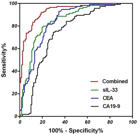
in Figure 4, The AUC (Area Under roc Curve) of combined diagnosis was 0.9334 (95% CI: 0.9026~0.9643), higher than that of serum IL-33 (0.8446, 95% CI: 0.7938~0.8954), CEA (0.8394, 95% CI: 0.7882~ 0.8905), and CA19-9(0.7390, 95% CI: 0.6736~0.8043), re- spectively.
cut-family, was identified as a legend for the ST2. The IL-33/ST2L axis stimulates the generation of cytokines and type 2 immune response [7]. Recently, several studies have found the asso-ciation between expression of IL-33 and differ-ent types of solid tumors. Yang et al. demon-strated that serum concentration of IL-33 and ST2 were significantly higher in patients with breast cancer than in healthy volunteers, respectively. IL-33 and sST2 may serve as non-invasive diagnosis markers for breast cancer [8]. Kim et al. found that serum IL-33 levels are associated with progression of lung cancer. Serum IL-33 levels were significantly lower in cancer patients than normal controls, and were higher at stage I and markedly lower at stages III and IV than those of controls [9]. Ye et al. did a study suggesting that the level of serum IL-33 should be detected during the treatment of gastric cancer, particularly when using
[image:7.612.91.332.72.304.2]plati-serum IL-33 in lung cancer [9], but in the pres-ent study of serum, no similar result was found. Patients with positive IL-33 expressions have no significant different overall survival com-pared with those with negative IL-33 expres-sions in the present study, the same as the result in the study of hepatocellular carcinoma [12]. We also found that serum IL-33 levels in moderate or poor differentiation CRC cases were significantly higher than that in well differ-entiation cases. Serum IL-33 level in colon can-cer cases was also significantly higher than that in rectal cancer cases. These findings sug-gested that serum IL-33 level may suggest malignancy degree and location of colorectal cancer. Finally, we established a logistic regres-sion model involving serum IL-33 and conven-tional tumor markers for colorectal diagnosis. Diagnosis result analysis suggested that com-bination of serum IL-33, CEA and CA19-9 may improve diagnostic efficiency for colorectal Table 6. Comparison of different markers for CRC
diagnosis
[image:7.612.91.332.383.452.2]Markers Sensitivity Specificity PPV NPV Accuracy CEA 46.28 97.37 94.92 63.07 71.06 CA19-9 18.18 98.25 91.67 53.08 57.02 Serum IL-33 79.34 79.82 80.67 78.45 79.57 Combined 80.99 92.11 91.59 82.03 86.38
Figure 4. ROC of serum IL-33, CEA, CA19-9 and combination for CRC diagnosis.
Disclosure of conflict of interest
None.
Address correspondence to: Drs. Changping Wu and Jingting Jang, Department of Tumor Biological
Treatment, The Third Affiliated Hospital of Soochow
University, 185 Juqian Street, Changzhou 213003, Jiangsu Province, China. Tel: +86-519-68871129; Fax: +86-519-68871129; E-mail: [email protected] (CPW); [email protected] (JTJ)
References
[1] Chen H, Werner S, Tao S, Zornig I, Brenner H. Blood autoantibodies against tumor-associat-ed antigens as biomarkers in early detection of colorectal cancer. Cancer Lett 2014; 346: 178-187.
[2] Brenner H, Stock C, Hoffmeister M. Colorectal cancer screening. Ther Umsch 2013; 70: 231-235.
[3] Karimi S, Chattopadhyay S, Chakraborty NG. Manipulation of regulatory T cells and
antigen-specific cytotoxic T lymphocyte-based tumour
immunotherapy. Immunology 2015; 144: 186-196.
[4] Ohno T, Morita H, Arae K, Matsumoto K, Nakae S. Interleukin-33 in allergy. Allergy 2012; 67: 1203-1214.
[5] Nakae S, Morita H, Ohno T, Arae K, Matsumoto K, Saito H. Role of interleukin-33 in innate-type immune cells in allergy. Allergol Int 2013; 62: 13-20.
[6] Shalapour S, Karin M. Immunity, inflammation, and cancer: an eternal fight between good and
evil. J Clin Invest 2015; 125: 3347-3355.
[10] Ye XL, Zhao YR, Weng GB, Chen YC, Wei XN, Shao JP, Ji H. IL-33-induced JNK pathway acti-vation confers gastric cancer chemotherapy resistance. Oncol Rep 2015; 33: 2746-2752. [11] Schmieder A, Multhoff G, Radons J. Interleu-
kin-33 acts as a pro-inflammatory cytokine and
modulates its receptor gene expression in highly metastatic human pancreatic carcino-ma cells. Cytokine 2012; 60: 514-521. [12] Bergis D, Kassis V, Ranglack A, Koeberle V,
Piiper A, Kronenberger B, Zeuzem S, Waidmann O, Radeke HH. High Serum Levels of the Interleukin-33 Receptor Soluble ST2 as a Negative Prognostic Factor in Hepatocellular Carcinoma. Transl Oncol 2013; 6: 311-318. [13] Chen SF, Nieh S, Jao SW, Wu MZ, Liu CL, Chang
YC, Lin YS. The paracrine effect of
cancer-asso-ciated fibroblast-induced interleukin-33 regu -lates the invasiveness of head and neck squa-mous cell carcinoma. J Pathol 2013; 231: 180-189.
[14] Zhao J, Zhang H, Liu SB, Han P, Hu S, Li Q, Wang ZF, Mao-Ying QL, Chen HM, Jiang JW, Wu GC, Mi WL, Wang YQ. Spinal interleukin-33 and its receptor ST2 contribute to bone cancer-in-duced pain in mice. Neuroscience 2013; 253: 172-182.