Original Article
Overexpression of EIF5A2 correlates with
tumor progression and predicts poor
prognosis in cervical cancer
Hu Li1, Yan Xu1, Li Yang1, Lei He2
1Department of Gynecology, Panyu Central Hospital, Guangzhou, China; 2Department of Neurology, Sun Yat-sen
Memorial Hospital of Sun Yat-sen University, Guangzhou, China
Received January 4, 2016; Accepted March 18, 2016; Epub May 1, 2016; Published May 15, 2016
Abstract: Background: Eukaryotic translation initiation factor 5A2 (EIF5A2) has been demonstrated to play impor-tant roles in tumor progression. However, the role of EIF5A2 in cervical cancer remains unknown. This study aimed to investigate the expression and clinicopathological significance of EIF5A2 in cervical cancer. Methods: The mRNA expression level of EIF5A2 was analyzed in 10 pairs of cervical cancer and adjacent non-cancerous tissues by semi-quantitative real time-PCR assay. Immunohistochemistry was performed to assess EIF5A2 protein expression in 124 paraffin-embedded, archived cervical cancer samples. Survival analysis and Cox regression analysis were performed to investigate the clinicopathological significance of EIF5A2 expression. Results: Our data revealed that EIF5A2 was frequently overexpressed in cervical cancer tissues compared to normal adjacent noncancerous cervi-cal tissues. Moreover, high expression of EIF5A2 in is strongly associated with advanced FIGO stage (P=0.005) and parametrial invasion (P=0.041). Cervical cancer patients with higher Parametrial invasion expression had substan-tially shorter overall (P=0.038) and recurrence-free (P<0.001) survival times than patients with lower parametrial invasion expression. Multivariate analysis revealed that parametrial invasion might be an independent prognostic factor for breast cancer patients (P=0.017). Conclusions: Our study showed that EIF5A2 expression was positively associated with progression and aggressiveness of cervical cancer, and that EIF5A2 could serve as an independent prognostic marker in cervical cancer patients.
Keywords: EIF5A2, cervical cancer, progression, prognosis
Introduction
Human cervical cancer is one of the most com-mon malignances in female reproductive sys-tem, and the third leading cause of cancer related mortality in women, with approximately 500,000 new cancer cases among women and 274,000 cancer related mortality, representing a serious health threat to women worldwide [1]. Although various treatments for cervical can-cer, such as chemotherapy, radiation and radi-cal surgery, have been used and have been improved in the past decades, the clinical out-come of patients with advanced-stage diseas-es remains unsatisfactory [2]. This is largely because of a lack of effective and specific bio-markers that predict the progression of cervical cancer [3]. Thus, it is important to identify new genes and molecules that can effectively
gene has two isoforms, EIF5A1 and EIF5A2. The EIF5A2 is located on chromosome 3q26, a region where amplification is observed in differ-ent human malignancies [7]. It is suggested that EIF5A2 expression s elevated in multiple cancers, and that up-regulation of EIF5A2 expression contributes to proliferation of can-cer cells [8]. Additionally, EIF5A2 overexpres-sion promotes tumorigenesis both in vitro and in vivo and is also positively correlated to an advanced stage of the disease [9]. Recently, EIF5A2 was shown to contribute to tumor metastasis and angiogenesis via hypoxia or gene amplification [10]. However, the expres-sion and clinical relevance of EIF5A2 in cervical cancer have not been determined.
In the present study, we aimed to investigate the expression of EIF5A2 in cervical cancer and its relationship with clinical parameters and prognosis in cervical cancer patients. The results showed that EIF5A2 is significantly upregulated in cervical cancer, and overexpres-sion of EIF5A2 is closely associated with the FIGO stage and parametrial invasion in cervical cancer. Multivariate Regression analysis revealed that EIF5A2 might be considered as an independent biomarker for cervical cancer prognosis. Collectively, our findings strongly suggested that EIF5A2 plays an important role in the development and progression of human cervical cancer, and might be a useful predic-tive marker of prognosis in cervical cancer patients.
Materials and methods
Tissue specimens and patient information
A total of 124 cervical cancer paraffin-embed-ded specimens from female patients, which had been histopathologically and clinically diagnosed as cervical cancer at the Panyu Central Hospital from 2005-2009, were used in the present study. Tumor stage was defined according to the pathological classification system of the International Federation of Gynecology and Obstetrics (FIGO). For the use of these clinical materials for research purpos-es, prior patients’ consents and approval from Panyu Central Hospital Institutional Review Board were obtained. Clinical information on the samples is summarized in Table 1. The median age at the time of surgery was 49 years (range 21-77 years). The percentage of tumor purity in sections adjacent to the regions used for RNA extraction was estimated during rou-tine histopathological analysis.
RNA extraction, reverse transcription and real-time PCR
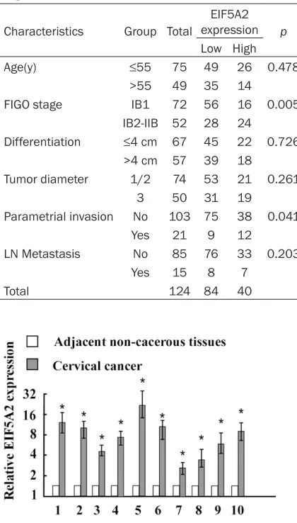
[image:2.612.90.300.94.463.2]Total RNA from fresh surgically obtained tumor tissues and their adjacent noncancerous tis-sues was extracted using the Trizol reagent (Invitrogen, Carlsbad, CA) following the manu-facturer’s recommendations. The RNA was tre- ated with DNase, and 2 ug of total RNA was used for cDNA synthesis using random hexam-ers. Real-time reverse transcription polymerase chain reaction (qRT-PCR) was carried out using SYBER green kit in a Light Cycler system (Roche Table 1. Correlation between the
clinicopatho-logic features and expression of EIF5A2
Characteristics Group Total
EIF5A2 expression p Low High
Age(y) ≤55 75 49 26 0.478
>55 49 35 14
FIGO stage IB1 72 56 16 0.005
IB2-IIB 52 28 24
Differentiation ≤4 cm 67 45 22 0.726 >4 cm 57 39 18
Tumor diameter 1/2 74 53 21 0.261
3 50 31 19
Parametrial invasion No 103 75 38 0.041
Yes 21 9 12
LN Metastasis No 85 76 33 0.203
Yes 15 8 7
Total 124 84 40
Applied Science). For the evaluation of the rela-tionship between EIF5A2 and b-actin (internal control), the primer selected were as follows: EIF5A2, forward, 5’-CCCTGCTGAC AGAAACT- GGT-3’ and reverse, 5’-TTGCACACATGACAGA CACC-3; b-actin, forward, 5’-GCTCTTTTCCAGC- CTTCCTT-3’, and reverse, 5’-CGGATGTCAAC- TTCACACTT-3’.
Immunohistochemistry
Immunohistochemical analysis was done to measure EIF5A2 protein expression in 124 human cervical cancer tissues. Briefly, paraffin embedded specimens were cut into 4 μm sec-tions and baked at 60°C for 2 hours, followed by deparaffinized with xylenes and rehydrated. Antigenic retrieval was done by submerging
[image:3.612.89.525.73.234.2]the Sections into EDTA antigenic retrieval buf-fer and microwaving. The sections were then treated with 3% hydrogen peroxide in methanol to quench the endogenous peroxidase activity, followed by incubation with 1% bovine serum albumin to block the nonspecific binding. Sections were then incubated with anti-EIF5A2 rabbit polyclonal antibody (1:100, Abcam) over-night at 4°C. For negative controls, the primary antibody was replaced by normal goat serum. After washing, the tissue sections were treated with biotinylated rabbit secondary anti-body (Abcam), followed by a further incubation with streptavidin-horseradish peroxidase com-plex (Abcam). The tissue sections were imm- ersed in 3-amino-9-ethylcarbazole and coun-terstained with 10% Mayer’s hematoxylin, dehydrated and mounted in Crystal Mount.
Figure 2. Expression of EIF5A2 protein in tissue by immunohistochemistry. A. Positive expression of EIF5A2 in cervi-cal cancer tissues. B. Weak staining of EIF5A2 in cervicervi-cal cancer tissues.
[image:3.612.92.521.287.468.2]The degree of immunostaining of formalin-fixed, paraffin-embedded sections was review- ed and scored by two independent observers. The proportion of the stained cells and the extent of the staining were used as criteria of evaluation. One score was given according to the percent of positive cells as: ≤5% of the cells, 1 point; 6-35% of the cells, 2 point; 36-70% of the cells, 3 point; ≥71% of the cells, 4 point. Another score was given according to the intensity of staining as: 0 point, negative, 1 point, weak staining (light yellow); 2 point, positive staining (yellowish brown); and 3 point, strong staining (brown). A final score was then calculated by multiple the above two scores. If the final score was equal or bigger than four, the tumor was considered high expression; otherwise, the tumor was considered low expression.
Statistical analysis
All statistical analyses were carried out using the SPSS 13.0 statistical software package. The chi-square test and Fisher’s exact test were used to analyze the correlation between EIF5A2 expression and the clinicopathologic characteristics. Survival curves were plotted by the Kaplan Meier method and compared by the log-rank test. The significance of various vari-ables for survival was analyzed by the Cox pro-portional hazards model in the multivariate analysis. P<0.05 in all cases was considered statistically significant.
Results
EIF5A2 is upregulated in cervical cancer tis-sues
To explore the potential role of EIF5A2 in the tumorigenesis of cervical cancer, the
expres-sion of the EIF5A2 mRNA in 10 pairs of cervical cancer and adjacent non-cancerous tissues was determined by real time-PCR. These results indicated that EIF5A2 is upregulated in cervical cancer tissues (Figure 1).
EIF5A2 expression is associated with cervical cancer progression
To further explore whether EIF5A2 upregulation is associated with clinicopathological charac-teristics of cervical cancer, we examined the EIF5A2 expression status in 124 paraffin-embedded, archived cervical cancer tissues by immunohistochemistry (IHC) (Figure 2). IHC revealed that increasing EIF5A2 staining was positively correlated with advancing FIGO stage (P=0.005) and parametrial invasion (P=0.041). Taken together, these results suggested that upregulation of EIF5A2 is associated with cervi-cal cancer progression.
EIF5A2 expression level is associated with the patient survival
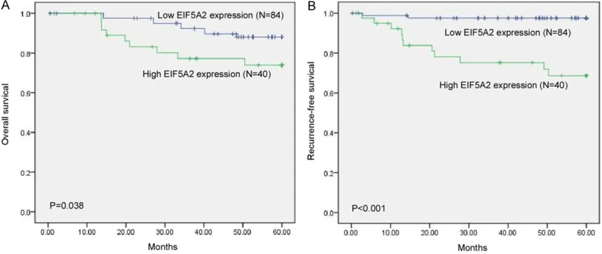
Assessment of patient survival by the Kaplan-Meier analysis indicated an adverse correlation between EIF5A2 expression and overall surviv-al (P=0.038) and recurrent-free survivsurviv-al time (P<0.001) of patients with cervical cancer (Figure 3). Multivariate Cox proportional haz-ards model analysis revealed that EIF5A2 expression was an independent prognostic fac-tor for overall survival (P=0.017) (Table 2). Discussion
In the present study, we provided the first evi-dence that overexpression of EIF5A2 protein is associated with poor prognosis in cervical can-cer patients. Our data showed that EIF5A2 is upregulated in cervical cancer specimens in Table 2. Multivariate analysis of different prognostic parameters in patients with cervical cancer
Overall survival Recurrence-free survival Hazard Ratio (95% CI) P Hazard Ratio (95% CI) P Age (>55 y vs ≤55 y)
FIGO Stage (IB2-IIB vs IB1) 4.429 (0.214-12.577) 0.004 4.507 (1.115-10.097) 0.015 Tumor diameter (>4 cm vs ≤4 cm)
Tumor grade (Grade 3 vs 1/2) Parametrial invasion (yes vs no)
compared with that in adjacent non-cancerous tissues. Moreover, analysis of 124 archived cervical cancer samples revealed that EIF5A2 expression is significantly correlated with FIGO stage, parametrial invasion, and clinical prog-nosis in these patients. Furthermore, Cox regression analysis showed that higher EIF5A2 expression was an independent prognostic indicator of shorter survival in cervical cancer patients. These findings strongly suggested that lower expression of EIF5A2 would provide a selective advantage in prognosis for cervical cancer patients.
EIF5A is an abundant, small, highly conserved, RNA-binding protein that associates with ribo-somes and with the cytoskeleton, and shuttles between the nucleus and cytoplasm. It con-tains the amino acid hypusine, which appears to be essential to most of EIF5A’s functions [11]. EIF5A2 is an isoform of EIF5A, and the expression of EIF5A2 is rare in most normal tis-sues but conspicuous in many malignancies. The association and functions of EIF5A2 have been studied in several tumors. EIF5A2 was first identified as an elevated expressed gene by analysis of an amplified chromosomal region from ovarian cancer [7]. Aberrant EIF5A2 expression is associated with tumor progres-sion, increased likelihood of recurrence after surgery or radiotherapy [12]. EIF5A2 was reported to be upregulated by Hh signaling pathway via cis-acting elements in prostate cancer cells [13]. EIF5A2 promotes tumorigen-esis, tumor cell proliferation and apoptosis of various cancers, such as esophageal squa-mous cell carcinoma, hepatocellular carcino-ma, and bladder cancer [4], indicating its onco-genic role in tumor development. Thus, we assumed that EIF5A2 might promote cervical cancer progression through a similar mecha-nism. The present study showed that EIF5A2 is overexpressed in cervical cancer in compared with that in adjacent non-cancerous tissues, and that high EIF5A2 expression is correlated with advanced tumor stage, indicating that EIF5A2 is involved in the progression of cervi-cal cancer.
Lymph node invasion and remote metastasis are EIF5A2-related pathological features that have attracted attention from a mechanistic point of view. EIF5A2 overexpression is detect-ed in hepatocellular carcinoma margins [14]. Interestingly, EIF5A2 is one of only three genes
found to be predictive of lymph node metasta-sis in gastric cancer, while downregulation of EIF5A2 by miR-30b targeting suppresses tumor migration and invasion in gastric cancer cells [15]. Association of EIF5A2 with metastasis is also seen in hepatocellular carcinoma. Tang et al. found that ectopic expression of EIF5A2 could enhance cancer cell migration and inva-sion of hepatocellular carcinoma cells, while inhibition of EIF5A2 by small interfering RNA or deoxyhypusine synthase inhibitor GC7 decrease cell motility in vitro [14]. Therefore, we specu-lated that EIF5A2 might be correspecu-lated with tumor invasion in cervical cancer. Although we revealed that EIF5A2 expression was not cor-related with lymph nodes metastasis, which may be due to the small sample size, high expression of EIF5A2 was associated with parametrial invasion. Thus, EIF5A2 might be a positive regulator of cervical cancer develop-ment and progression; however, the exact mechanism needs further investigation.
Remarkably, our study indicated that EIF5A2 expression was significantly correlated with clinical prognosis in cervical cancer patients. Our results were supported by previous stud-ies. Wei et al. demonstrated that EIF5A2 over-expression predicts tumor metastatic potential in patients with localized invasive bladder can-cer [16]. In addition, Bao et al. suggested that EIF5A2 promoted the chemoresistance to doxorubicin via regulation of EMT in colon can-cer cells [17]. Li et al. showed that overexpres-sion of EIF5A2 is associated with up-regulation of HIF1a, metastasis, and shorter survival times of patients with esophageal squamous cell carcinoma [10]. These studies together suggested that EIF5A2 overexpression predicts poor clinical outcomes in cancer patients. In conclusion, we found that EIF5A2 overex-pression is correlated with cervical cancer pro-gression and progressive phenotype. Moreover, our data indicated that EIF5A2 might be an independent prognostic marker in cervical can-cer patients. Thus, EIF5A2 protein expression might be a useful marker for stratifying cervical cancer patients’ prognosis as well as an effec-tive novel criterion for selection of therapeutic options.
Acknowledgements
Disclosure of conflict of interest
None.
Address correspondence to: Dr. Lei He, Department of Neurology, Sun Yat-sen Memorial Hospital of Sun Yat-sen University, Guangzhou 510000, China. Tel: +86 1382-4453-603; Fax: +86 21-8133-2199; E-mail: [email protected]
References
[1] Pfaendler KS and Tewari KS. Changing para-digms in the systemic treatment of advanced cervical cancer. Am J Obstet Gynecol 2016; 214: 22-30.
[2] Harkenrider MM, Alite F, Silva SR and Small W Jr. Image-Based Brachytherapy for the Treatment of Cervical Cancer. Int J Radiat Oncol Biol Phys 2015; 92: 921-934.
[3] Robova H, Rob L, Halaska MJ, Pluta M and Skapa P. Review of neoadjuvant chemotherapy and trachelectomy: which cervical cancer pa-tients would be suitable for neoadjuvant che-motherapy followed by fertility-sparing sur-gery? Curr Oncol Rep 2015; 17: 446.
[4] Caraglia M, Marra M, Giuberti G, D’Alessandro AM, Budillon A, del Prete S, Lentini A, Beninati S and Abbruzzese A. The role of eukaryotic ini-tiation factor 5A in the control of cell prolifera-tion and apoptosis. Amino Acids 2001; 20: 91-104.
[5] Park MH, Nishimura K, Zanelli CF and Valentini SR. Functional significance of eIF5A and its hypusine modification in eukaryotes. Amino Acids 2010; 38: 491-500.
[6] Reubi JC, Lamberts SW and Palacios JM. Hormone dependency of meningiomas. Lancet 1989; 2: 1099-1100.
[7] Guan XY, Sham JS, Tang TC, Fang Y, Huo KK and Yang JM. Isolation of a novel candidate on-cogene within a frequently amplified region at 3q26 in ovarian cancer. Cancer Res 2001; 61: 3806-3809.
[8] Griffin CA, Morsberger L, Hawkins AL, Haddadin M, Patel A, Ried T, Schrock E, Perlman EJ and Jaffee E. Molecular cytogenetic characteriza-tion of pancreas cancer cell lines reveals high complexity chromosomal alterations. Cytogenet Genome Res 2007; 118: 148-156. [9] Guan XY, Fung JM, Ma NF, Lau SH, Tai LS, Xie
D, Zhang Y, Hu L, Wu QL, Fang Y and Sham JS. Oncogenic role of eIF-5A2 in the development of ovarian cancer. Cancer Res 2004; 64: 4197-4200.
[10] Li Y, Fu L, Li JB, Qin Y, Zeng TT, Zhou J, Zeng ZL, Chen J, Cao TT, Ban X, Qian C, Cai Z, Xie D, Huang P and Guan XY. Increased expression of EIF5A2, via hypoxia or gene amplification, contributes to metastasis and angiogenesis of esophageal squamous cell carcinoma. Gastroenterology 2014; 146: 1701-1713, e1709.
[11] Rossi D, Kuroshu R, Zanelli CF and Valentini SR. eIF5A and EF-P: two unique translation fac-tors are now traveling the same road. Wiley Interdiscip Rev RNA 2014; 5: 209-222. [12] Zhu W, Cai MY, Tong ZT, Dong SS, Mai SJ, Liao
YJ, Bian XW, Lin MC, Kung HF, Zeng YX, Guan XY and Xie D. Overexpression of EIF5A2 pro-motes colorectal carcinoma cell aggressive-ness by upregulating MTA1 through C-myc to induce epithelial-mesenchymaltransition. Gut 2012; 61: 562-575.
[13] Xu X, Liu H, Zhang H, Dai W, Guo C, Xie C, Wei S, He S and Xu X. Sonic Hedgehog-GLI Family Zinc Finger 1 Signaling Pathway Promotes the Growth and Migration of Pancreatic Cancer Cells by Regulating the Transcription of Eu- karyotic Translation Initiation Factor 5A2. Pancreas 2015; 44: 1252-1258.
[14] Tang DJ, Dong SS, Ma NF, Xie D, Chen L, Fu L, Lau SH, Li Y, Li Y and Guan XY. Overexpression of eukaryotic initiation factor 5A2 enhances cell motility and promotes tumor metastasis in hepatocellular carcinoma. Hepatology 2010; 51: 1255-1263.
[15] Tian SB, Yu JC, Liu YQ, Kang WM, Ma ZQ, Ye X and Yan C. MiR-30b suppresses tumor migra-tion and invasion by targeting EIF5A2 in gastric cancer. World J Gastroenterol 2015; 21: 9337-9347.
[16] Wei JH, Cao JZ, Zhang D, Liao B, Zhong WM, Lu J, Zhao HW, Zhang JX, Tong ZT, Fan S, Liang CZ, Liao YB, Pang J, Wu RH, Fang Y, Chen ZH, Li B, Xie D, Chen W and Luo JH. EIF5A2 predicts out-come in localised invasive bladder cancer and promotes bladder cancer cell aggressiveness in vitro and in vivo. Br J Cancer 2014; 110: 1767-1777.