Original Article
CDK4
amplification is associated with distant
metastasis and poor clinical outcome in breast cancer
Peng Guo1, Tianjie Pu1,2, Shinan Chen1, Yan Qiu1,2, Xiaorong Zhong3,4, Hong Zheng3,4, Lina Chen5, Feng Ye1,
Hong Bu1,2
1Laboratory of Pathology, West China Hospital, Sichuan University, Chengdu, Sichuan Province, China;
2Department of Pathology, West China Hospital, Sichuan University, Chengdu, Sichuan Province, China; 3Cancer
Center, West China Hospital, Sichuan University, Chengdu, Sichuan Province, China; 4Laboratory of Molecular
Diagnosis of Cancer, State Key Laboratory of Biotherapy, National Collaborative Innovation Center for Biotherapy, West China Hospital, Sichuan University, Chengdu, Sichuan Province, China; 5Department of Pediatrics, West
China Second University Hospital, Sichuan University, Chengdu, Sichuan Province, China
Received January 24, 2016; Accepted April 22, 2016; Epub June 1, 2016; Published June 15, 2016
Abstract: Amplification of the cyclin-dependent kinase 4 (CDK4), located at 12q13-q14, has been reported in vari-ous human tumors including breast cancer. The aim of this study was to assess CDK4 gene amplification in invasive ductal carcinoma (IDC) with clinical implications. Quantitative PCR (qPCR) was performed on formalin-fixed and paraffin-embedded (FFPE) tumor samples for gene amplification detection. The clinical histopathological charac-teristics and prognostic significance were analyzed. Of the 157 IDC patients, CDK4 gene amplification was found in 18 (11.5%). CDK4 amplification was associated with distant metastasis (after initial surgery) (P=0.009). In survival analysis, it was also associated with disease-free survival (DFS, P=0.026) and overall survival (OS, P=0.020). With multivariate analysis showed that CDK4 amplification was found to be associated with DFS (amplification vs non-amplification, hazard ratio, 4.456; 95% confidence interval, 1.383-14.353; P=0.012). With respect to treatment regimen, this is also true for DFS (P=0.014 for chemotherapy and P=0.010 for radiotherapy). Patients with CDK4 amplification is associated with distant metastasis after initial surgery and favors poor clinical outcome.
Keywords: Breast cancer, CDK4, gene amplification, metastasis, prognosis
Introduction
Based on the 2014 World Health Organization (WHO) report, breast cancer is the second most life-threatening tumor after lung cancer for women in China [1]. Many of the tumor-sup-pressor genes and oncogenes altered in can- cer are known to affect cell cycle regulation. Disruption of the cell cycle machinery might enhance genomic instability, contribute to un- controlled cell growth, and lead to the develop-ment of cancer [2, 3]. The CDK4 gene encodes a 33-kD protein that plays an important role in the regulation of the G1-S transition of the cell cycle. CDK4, in complex with cyclin D1, can phosphorylate Rb, inactivating the protein and releasing negative control, thus allowing the G1/S transition to precede [2, 4]. CDK4 activity is deregulated in many human tumors [3, 5].
Numerous genes were found to be abnormal in breast cancer with different biological signifi-cance [6]. Gene amplification is an important and common mechanism for oncogene overex-pression in many tumors. Amplification and consequent overexpression of the CDK4 gene, located in the 12q13-q14 region, have been found in various cancers including different types of sarcomas and glioblastomas [7-9]. Previous studies show that CDK4 amplification and overexpression were associated with high-er breast tumor cell prolifhigh-eration rate, but no clinical characteristicwere reported [10]. The
inter-action, potentially indicating a role for targeted therapies. Certain CDK4/6 inhibitors such as PD0332991 (palbociclib), LEE011 (ribociclib), and LY2835219 (abemaciclib) were already in clinical trial [11-13]. In this study we aimed to assess whether there are patients with amplifi-cation of CDK4 in IDC. Further we also analyzed the clinical significance for CDK4 amplification in breast cancer patients, e.g., tumor size, inva-sion, and metastasis, as well as prognostic val-ues and therapy responses.
In our study, we find that CDK4 amplification is associated with distant metastasis after initial surgery and, our data indicate that the CDK4
amplification favors poor clinical outcome and might be a biomarker for breast cancer target therapy.
Materials and methods
Patients and sample preparation
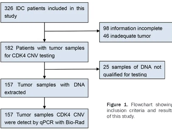
The samples were human breast neoplasm tissue removed during surgery. Patients’ ano-nymity was preserved in all of the cases. Approval for the study was granted by the Ethics Committee of West China Hospital (No. 2013-191). We analyzed formalin-fixed and paraffin-embedded (FFPE) sample from 157 patients with breast cancer who underwent breast mastectomy between 2010 and 2012 at West China Hospital (Figure 1). Surgical specimens were obtained before systemic treatment, and paraffin embedding was performed within the
using a QIAamp DNA FFPE tissue kit (Qiagen, Hilden, Germany) according to the manufactur-er’s instructions. DNA quantitation was per-formed using a nanodrop 2000 (Thermo, Waltham, MA, USA). Finally, DNA purity was confirmed by measuring the A260/280 absor-bance ratios. Good-quality DNA was indicated with the ratio A260 nm/A280 nm=1.70-1.95. Reactions were carried out using a Bio-Rad CFX96 system (Applied Biosystems, Hercules, CA, USA). Each gene was measured in triplicate and normalized relative to a set of two refer-ence genes (GAPDH, TFRC) (Table 1). The rela-tive quantitation of CDK4 gene amplification in IDC was calculated by the 2-ΔΔCT method using
[image:2.612.90.384.69.290.2]the average copy number in 50 normal breast tissues besides tumor as control samples and reference genes (GAPDH, TFRC). The sample was considered positive for CDK4 gene amplifi-cation if the ratio was greater than 2.0, where-as a ratio less than 2.0 indicated that sample was negative [14, 15] (Table 1). We confirmed the cut-off value of qPCR through HER2 in IDC (data unpublished). For detailed quantification method please refers to the previous studies [16, 17].
Statistical analysis
Statistical analyses were conducted using SPSS version 16.0 software (SPSS Inc., Chi- cago, USA), and a 5% two-tailed significance level was considered to be statistically signifi-cant. Associations between the prevalence of Figure 1. Flowchart showing
inclusion criteria and results of this study.
framework of diagnostic procedures. Disease-free survival (DFS) and overall survival (OS) were defined as the time between the initial surgery and local or distant metastatic relapse, and the time between sur-gery and death, respect- ively.
DNA isolation and quan-titative polymerase chain reaction
Tumor areas (at least 1 cm2) from 4.0 μm-thick
CDK4 amplification and clinical parameters were evaluated with a chi-squared test. Uni- variate survival analysis was conducted using the Kaplan-Meier method, and multivariate sur-vival analysis was carried out using the Cox pro-portional hazard model.
[image:3.612.92.403.86.190.2]tively, regarding age, histology grading, tumor size, nodal status, clinical stage, ER, PR, HER2/ neu status, local recurrence and distant metas-tasis. In our study, CDK4 amplification was sig-nificantly associated with distant metastasis (after initial surgery) (P=0.009); additionally, Table 1. qPCR primers of the CDK4 and reference genes
Gene GenBank No. name Oligo sequenceOligo Target size TFRC NC_000003.12 TFRCF 5’-ACTTCCTCTCTCCCTACGTATC-3’ 105 bp
TFRCR 5’-GCAGTTTCAAGTTCTCCAGTAAAG-3’
GAPDF NG_007073.2 GAPDFF 5’-CCTCAAGATCATCAGCAATGCCTC-3’ 100 bp GAPDHR 5’-GTGGTCATGAGTCCTTCCACGATA-3’ CDK4 NG_007726.3 CDK4F 5’-GGGTGGGACTCAAGCAATATAC-3’ 144 bp
CDK4R 5’-CCTCACCTCCTTCACACATTAC-3’
Table 2. Baseline clinical characteristics of the study subjects Total No.
(n=157) Disease-free Survival Overall Survival No. (%) Log-rank P-Value Log-rank P-Value AGE 27-75 (49.1) 0.157 0.692 0.086 0.770 ≤50 years 94 (59.9)
>50 years 63 (40.1)
GRADING† 3.245 0.072 0.257 0.612
G1-G2 46 (30.5) G3 105 (69.5)
TUMOR SIZE† 1.595 0.207 5.112 0.024*
T0-2 146 (94.2) T3-4 9 (5.8)
NODAL STATUS† 3.800 0.051 4.484 0.034*
N0 69 (44.8)
N1-N3 85 (55.2)
CLINICAL STAGE† 3.773 0.052 3.418 0.064
I-II 115 (74.7) III-IV 39 (25.3)
ER STATUS 2.612 0.106 0.484 0.487 ER+ 52 (33.3)
ER- 104 (66.7)
PR STATUS† 3.729 0.053 0.827 0.363
PR+ 61 (39.6) PR- 93 (60.4)
HER2† 6.027 0.049* 1.148 0.563
0-1+ 84 (54.2)
2+ 36 (23.2)
3+ 35 (22.6)
ER=estrogen receptor; PR=progesterone receptor; HER2=human epidermal growth factor receptor 2; †Number differences reflect missing data. *Statistically significant.
OS was 28.3 months. The DFS and OS of the 157 patients are listed in Table 2 with respect to histopathologic characteristics and prognostic factors, includ-ing age, histology gradinclud-ing, tumor size, nodal status, metastasis, and clinical stage, ER, PR and HER2/neu. As expected, HER2/ neu (P=0.049) was found to be significantly correlated with DFS. Her2/neu overexpression was found associated with shorter DFS. However, only tumor size (P=0.024) and nodal metastasis status (0.034) were significant- ly associated with OS. Larger tumor size and a positive node status were found associated with shorter OS (Table 2).
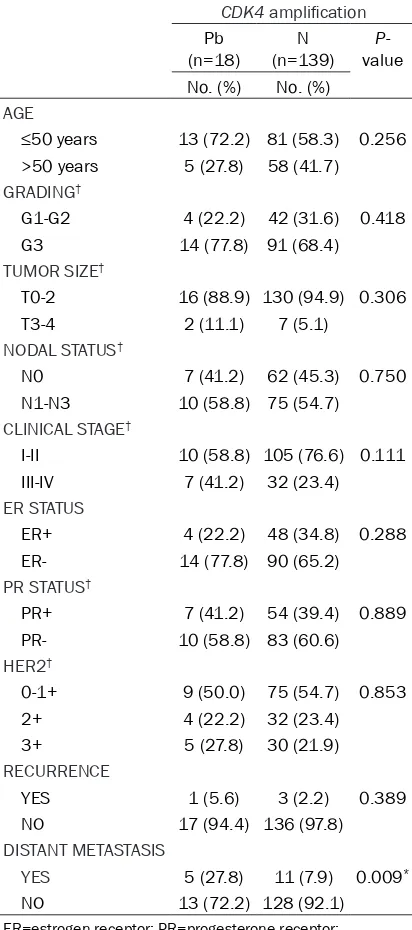
Clinical histopathological fea-tures of CDK4 amplification in breast cancer
In our study, 18 of 157 patients (11.5%) were detected CDK4 amplification with qPCR. To iden-tify any correlation between the gene amplification status of the
CDK4 and clinical characteris-tics (Table 3), we analyzed the correlation between CDK4 amp- lification and clinical features. The patients with CDK4 amplifi-cation were analyzed,
respec-Results
Baseline clinical char-acteristics
[image:3.612.92.367.226.594.2]CDK4 amplification primarily occurred in tu- mors with a high histological grade (Table 3).
CDK4 amplification for IDC prognosis
To further reveal the prognostic value of gene amplification for CDK4 in IDC patients, we eval-uated the CDK4 amplification status with DFS and OS by Kaplan-Meier analysis. In this study,
we analyzed the CDK4 amplification group ver-sus nonamplification group for DFS and OS. We found that the patients with CDK4 amplification had a significantly shorter DFS (P=0.026) and OS (P=0.020) than nonamplification (Figure 2). Furthermore, multivariate analysis showed that CDK4 amplification was found to be associat- ed with DFS (amplification vs nonamplification, hazard ratio, 4.456; 95% confidence interval, 1.383-14.353; P=0.012). But we found that
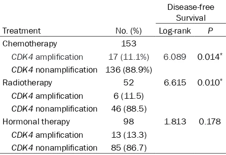
[image:4.612.323.521.72.428.2]CDK4 amplification did not associated with OS (Table 4). Concerning the treatment regimen, we found that the CDK4 amplification patients were also significantly correlated with poor DFS, regarding both chemotherapy (P=0.014) and radiotherapy (P=0.010) (Table 5).
Table 3. Prevalence of CDK4 amplification in breast tumors stratified according to clinical characteristics
CDK4 amplification
Pb
(n=18) (n=139)N value P-No. (%) No. (%)
AGE
≤50 years 13 (72.2) 81 (58.3) 0.256 >50 years 5 (27.8) 58 (41.7)
GRADING†
G1-G2 4 (22.2) 42 (31.6) 0.418 G3 14 (77.8) 91 (68.4)
TUMOR SIZE†
T0-2 16 (88.9) 130 (94.9) 0.306 T3-4 2 (11.1) 7 (5.1)
NODAL STATUS†
N0 7 (41.2) 62 (45.3) 0.750 N1-N3 10 (58.8) 75 (54.7)
CLINICAL STAGE†
I-II 10 (58.8) 105 (76.6) 0.111 III-IV 7 (41.2) 32 (23.4)
ER STATUS
ER+ 4 (22.2) 48 (34.8) 0.288 ER- 14 (77.8) 90 (65.2)
PR STATUS†
PR+ 7 (41.2) 54 (39.4) 0.889 PR- 10 (58.8) 83 (60.6)
HER2†
0-1+ 9 (50.0) 75 (54.7) 0.853 2+ 4 (22.2) 32 (23.4) 3+ 5 (27.8) 30 (21.9)
RECURRENCE
YES 1 (5.6) 3 (2.2) 0.389
NO 17 (94.4) 136 (97.8)
DISTANT METASTASIS
YES 5 (27.8) 11 (7.9) 0.009* NO 13 (72.2) 128 (92.1)
ER=estrogen receptor; PR=progesterone receptor; HER2=human epidermal growth factor receptor 2; †Number differences reflect missing data. *Statistically significant.
[image:4.612.91.297.110.575.2]Discussion
The copy number of CDK4 gene has been deter-mined in a group of 157 invasive ductal carci-noma patients with an average follow-up of 28.3 months and compared with clinical patho-logical features. The 11.5% (18/157) patients were detected with CDK4 amplification of IDC in the present study. This subgroup was signifi-cantly correlated with a higher possibility dis-tant metastasis (after initial surgery, P=0.009), The CDK4 amplification was found to be signifi-cantly associated with DFS (P=0.026) and OS (P=0.020). Concerning the treatment regimen, we found that the CDK4 amplification patients were significantly correlated with poor DFS, regarding chemotherapy (P=0.014) and
radio-stem cell theory, EMT theory, special somatic tumor cell mutation [21-23]. Because CDK4
amplified tumor cells were abnormal in corre-sponding cell cycle, the patients may response quite different for the treatment regimens. In our study, patients with CDK4 amplification show poor clinical outcome for both DFS and OS. Interestingly, this is also true with DFS with respect to treatment regimen for chemo-therapy (P=0.014) and radiochemo-therapy (P=0.010). Further we wonder how this subgroup of patients’ response for the target therapy, e.g. Herceptin treatment for HER2 amplified pati- ents. However, only 9 patients in this study received Herceptin treatment. Only two patients were CDK4 amplified. One patient was found with distant metastasis (brain) after initial sur-Table 4. Multivariate Cox Analysis of the
histopatho-logic characteristics and CDK4 amplification Rela-tionship with the Likelihood of DFS and OS
DFS OS
Variable P-value P-value
Age ≤50 years vs >50 years 0.445 0.545 Histologic grade I, II, r 0.283 0.894 PT ≤5 cm vs >5 cm 0.615 0.091 Stage I, II vs III, IV 0.936 0.292
PN 0 VS 1, 2, 3 0.059 0.070
ER protein 0.727 0.974
PR protein 0.277 0.432
HER2 protein 0.316 0.675
co-amplification vs no co-amplification 0.012* 0.100
[image:5.612.92.320.108.254.2]ER=estrogen receptor; PR=progesterone receptor; HER2=human epidermal growth factor receptor 2. *Statistically significant.
Table 5. Prevalence of CDK4 amplification and treat-ment response
Disease-free Survival
Treatment No. (%) Log-rank P
Chemotherapy 153
CDK4 amplification 17 (11.1%) 6.089 0.014*
CDK4 nonamplification 136 (88.9%)
Radiotherapy 52 6.615 0.010*
CDK4 amplification 6 (11.5)
CDK4 nonamplification 46 (88.5)
Hormonal therapy 98 1.813 0.178
CDK4 amplification 13 (13.3)
CDK4 nonamplification 85 (86.7)
*Statistically significant.
therapy (P=0.010) Thus, CDK4 amplifica-tion can be an independent prognostic indicator.
In our study, the CDK4 amplification rate was 11.5%, a value similar to that in a previ-ous study (15%) [6, 10]. It was previprevi-ously shown that CDK4 amplification and overex-pression were associated with high breast tumor cell proliferation, but without any other clinical characteristic [10]. We further investigated the relationship between CDK4
[image:5.612.90.319.325.482.2]gery and then dead in two years, but there is no statistically significant data shows the resis-tance of Herceptin treatment in CDK4 amplified patients. We also further invested CDK4 ampli-fication subgroup response to the hormonal therapy, 99 patients received hormonal thera-py. 13 (13.3%) patients were CDK4 amplified. However there are no statistically significant data shows the resistance of hormonal treat-ment in CDK4 amplified patients due to limited patients we included in this study. Maybe CDK4
is a key regulator of cell cycle, so the CDK4
amplification may be sensitive to the endocrine therapy. Further researches on the treatment response of different treatment on this special subgroup of patients should be carried out. Certain CDK4/6 inhibitors such as PD0332991 (palbociclib), LEE011 (ribociclib), and LY2835- 219 (abemaciclib) were already in clinical trial [11, 12]. The CDK4/6 inhibitor (palbociclib, ribociclib and abemaciclib) may be the CDK4
amplification target medicine [24].
In summary, we detected CDK4 gene amplifica-tion using qPCR, and the results suggest that
CDK4 amplification has considerable prognos-tic relevance regarding the clinical outcome in breast cancer. CDK4 amplification can be a novel special subgroup in invasive ductal breast cancer that can be considered predictive of poor clinical outcomes. Regarding treatment regimen analysis, the result of this study indi-cates that patients with CDK4 amplification show resistance to chemotherapy, radiothera-py and may be sensitive to hormonal theraradiothera-py. Special treatment regimens may be required for this special subgroup of patients.
Acknowledgements
This work was funded by grants from the National Natural Science Foundation of China (31000601, 81200461) and Young Investigator Scholar in Sichuan University (2012SCU04A14). Disclosure of conflict of interest
None.
Address correspondence to: Bu Hong, Laboratory of Pathology, West China Hospital, Sichuan University, 37 Guo Xue Xiang, Chengdu 610041, Sichuan Pro- vince, China. E-mail: molecularpathology@hotmail. com
References
[1] WHO. Cancer country profiles 2014. http:// www.who.int/cancer/country profiles/chn_ en.pdf?ua=1.
[2] Hartwell LH and Kastan MB. Cell cycle control and cancer. Science 1994; 266: 1821-1828. [3] Ortega S, Malumbres M and Barbacid M.
Cyclin D-dependent kinases, INK4 inhibitors and cancer. Biochim Biophys Acta 2002; 1602: 73-87.
[4] Matsushime H, Ewen ME, Strom DK, Kato JY, Hanks SK, Roussel MF and Sherr CJ. Identi- fication and properties of an atypical catalytic subunit (p34PSK-J3/cdk4) for mammalian D type G1 cyclins. Cell 1992; 71: 323-334. [5] Sherr CJ and McCormick F. The RB and p53
pathways in cancer. Cancer Cell 2002; 2: 103-112.
[6] Cancer Genome Atlas N. Comprehensive mo-lecular portraits of human breast tumours. Nature 2012; 490: 61-70.
[7] Khatib ZA, Matsushime H, Valentine M, Shapiro DN, Sherr CJ and Look AT. Coampli- fication of the CDK4 gene with MDM2 and GLI in human sarcomas. Cancer Res 1993; 53: 5535-5541.
[8] Collins VP. Gene amplification in human glio-mas. Glia 1995; 15: 289-296.
[9] Malumbres M and Barbacid M. Cell cycle, CDKs and cancer: a changing paradigm. Nat Rev Cancer 2009; 9: 153-166.
[10] An HX, Beckmann MW, Reifenberger G, Bender HG and Niederacher D. Gene amplification and overexpression of CDK4 in sporadic breast car-cinomas is associated with high tumor cell pro-liferation. Am J Pathol 1999; 154: 113-118. [11] Dukelow T, Kishan D, Khasraw M and Murphy
CG. CDK4/6 inhibitors in breast cancer. Anti- cancer Drugs 2015; 26: 797-806.
[12] Beaver JA, Amiri-Kordestani L, Charlab R, Chen W, Palmby T, Tilley A, Zirkelbach JF, Yu J, Liu Q, Zhao L, Crich J, Chen XH, Hughes M, Bloomquist E, Tang S, Sridhara R, Kluetz PG, Kim G, Ibrahim A, Pazdur R and Cortazar P. FDA Approval: Palbociclib for the Treatment of Postmenopausal Patients with Estrogen Receptor-Positive, HER2-Negative Metastatic Breast Cancer. Clin Cancer Res 2015; 21: 4760-4766.
[13] Murphy CG and Dickler MN. The Role of CDK4/6 Inhibition in Breast Cancer. Oncologist 2015; 20: 483-490.
[15] Bernardi CC, Ribeiro Ede S, Cavalli IJ, Chautard-Freire-Maia EA and Souza RL. Amplification and deletion of the ACHE and BCHE cholines-terase genes in sporadic breast cancer. Cancer Genet Cytogenet 2010; 197: 158-165. [16] Wang TL, Maierhofer C, Speicher MR, Lengauer
C, Vogelstein B, Kinzler KW and Velculescu VE. Digital karyotyping. Proc Natl Acad Sci U S A 2002; 99: 16156-16161.
[17] Wang TL, Diaz LA Jr, Romans K, Bardelli A, Saha S, Galizia G, Choti M, Donehower R, Parmigiani G, Shih Ie M, Iacobuzio-Donahue C, Kinzler KW, Vogelstein B, Lengauer C and Velculescu VE. Digital karyotyping identifies thymidylate synthase amplification as a mech-anism of resistance to 5-fluorouracil in meta-static colorectal cancer patients. Proc Natl Acad Sci U S A 2004; 101: 3089-3094. [18] Diver EJ, Foster R, Rueda BR and Growdon WB.
The Therapeutic Challenge of Targeting HER2 in Endometrial Cancer. Oncologist 2015; 20: 1058-68.
[19] Feldinger K and Kong A. Profile of neratinib and its potential in the treatment of breast cancer. Breast Cancer (Dove Med Press) 2015; 7: 147-162.
[20] Black JC, Atabakhsh E, Kim J, Biette KM, Van Rechem C, Ladd B, Burrowes PD, Donado C, Mattoo H, Kleinstiver BP, Song B, Andriani G, Joung JK, Iliopoulos O, Montagna C, Pillai S, Getz G and Whetstine JR. Hypoxia drives tran-sient site-specific copy gain and drug-resistant gene expression. Genes Dev 2015; 29: 1018-1031.
[21] Jaspers JE, Sol W, Kersbergen A, Schlicker A, Guyader C, Xu G, Wessels L, Borst P, Jonkers J and Rottenberg S. BRCA2-deficient sarcoma-toid mammary tumors exhibit multidrug resis-tance. Cancer Res 2015; 75: 732-741. [22] Zhang P, Sun Y and Ma L. ZEB1: at the
cross-roads of epithelial-mesenchymal transition, metastasis and therapy resistance. Cell Cycle 2015; 14: 481-487.
[23] Wang S, Mou Z, Ma Y, Li J, Li J, Ji X, Wu K, Li L, Lu W and Zhou T. Dopamine enhances the response of sunitinib in the treatment of drug-resistant breast cancer: Involvement of eradicating cancer stem-like cells. Biochem Pharmacol 2015; 95: 98-109.