Understanding the biophysical mechanism(s) underlying geomagnetic field sensitivity in animals is one of the most exciting challenges in sensory biology. The use of geomagnetic cues for spatial orientation appears to be a fairly ubiquitous trait in animals (Wiltschko and Wiltschko, 1995a). Yet, the physiological mechanisms that underlie magnetic orientation have not been conclusively identified. Research has focused on two classes of biophysical models for magnetoreception: light-dependent, photoreceptor-based models and magnetite-based mechanisms. Specific wavelengths of light have been shown to influence magnetic orientation in a variety of animals including flies, amphibians and birds (Phillips and Borland, 1992a; Phillips and Sayeed, 1993; Munro et al., 1997b; Wiltschko et al., 1993, 1997; Wiltschko and Wiltschko, 1995b, 1998b). In addition, some neurophysiological studies suggest that the avian visual system and photosensitive pineal gland may be sensitive to the geomagnetic field (Semm et al., 1984; Demaine and Semm, 1985; Semm and Demaine, 1986b). These data have been interpreted as support for
photoreceptor-based mechanisms of magnetoreception (Leask, 1977; Schulten, 1982; Schulten and Windemuth, 1986; Edmonds, 1996). However, the effects of light on magnetic orientation responses (e.g. shifts in the direction of orientation or disorientation) and the wavelength-dependence of these effects vary considerably among newts, flies and birds. In addition, several animals are able to orient using magnetic cues in the absence of light (Arendse, 1978; Lohmann, 1991; Lohmann and Lohmann, 1993; Marhold and Wiltschko, 1997), and numerous experiments have provided evidence for a non-light-dependent mechanism of magnetoreception involving permanently magnetic material, possibly biogenic magnetite (e.g. Kirschvink et al., 1993; Wiltschko et al., 1994; Beason et al., 1995; Beason and Semm, 1996; Marhold and Wiltschko, 1997; Walker et al., 1997). Therefore, many questions still need to be addressed concerning the nature of the influence of light on magnetic orientation. For example, does the influence of light on magnetic orientation represent a primary interaction with a light-sensitive biophysical mechanism mediating JEB1907
Light-dependent models of magnetoreception have been proposed which involve an interaction between the magnetic field and either magnetite particles located within a photoreceptor or excited states of photopigment molecules. Consistent with a photoreceptor-based magnetic compass mechanism, magnetic orientation responses in salamanders, flies and birds have been shown to be affected by the wavelength of light. In birds and flies, it is unclear whether the effects of light on magnetic orientation are due to a direct effect on a magnetoreception system or to a nonspecific (e.g. motivational) effect of light on orientation behavior. Evidence from shoreward-orienting salamanders, however, demonstrates that salamanders perceive a 90 ° counterclockwise shift in the direction of the magnetic field under long-wavelength (⭓500 nm) light. A simple physiological model based on the antagonistic interaction between two magnetically sensitive spectral
mechanisms suggests one possible way in which the wavelength-dependent effects of light on the salamander’s magnetic compass response might arise. Assuming that the wavelength-dependent characteristics of the avian magnetic response can be attributed to an underlying magnetoreception system, we discuss several hypotheses attempting to resolve the differences observed in the wavelength-dependent effects of light on magnetic orientation in birds and salamanders. By considering the evidence in the context of photoreceptor- and non-photoreceptor-based mechanisms for magnetoreception, we hope to encourage future studies designed to distinguish between alternative hypotheses concerning the influence of light on magnetoreception.
Key words: magnetic orientation, geomagnetism, salamander, bird, navigation, photoreceptor, orientation.
Summary
REVIEW
THE CASE FOR LIGHT-DEPENDENT MAGNETIC ORIENTATION IN ANIMALS
MARK E. DEUTSCHLANDER*, JOHN B. PHILLIPS ANDS. CHRIS BORLAND
Indiana University, Department of Biology (and the Center for the Integrative Study of Animal Behavior), Bloomington, IN 47405, USA
*Present address: Department of Biology, PO Box 3020, University of Victoria, Victoria, British Columbia, Canada V8W 3N5 (e-mail: [email protected])
Accepted 27 January; published on WWW 22 March 1999
magnetoreception, or is the influence of light due to an interaction between other visually dependent behaviors and a non-light-dependent magnetoreception system? Furthermore, if a light-dependent magnetoreception mechanism underlies wavelength-dependent magnetic responses, do qualitatively different, taxon-specific effects of light on magnetic orientation suggest that there is more than one type of light-dependent mechanism? In this article, we will review the evidence for light-dependent magnetic compass orientation. We will then compare the effects of light on magnetic orientation and consider several hypotheses concerning the different patterns of wavelength-dependence observed in the two organisms for which we have the most data, newts and birds.
Light-dependent models of magnetoreception: functional characteristics and contraints
Three different biophysical models for magnetoreception have been proposed which involve photoreceptor-based mechanisms. All the models invoke biophysical interactions that could result in an effect of the magnetic field on the efficiency of photon absorption or phototransduction. Leask (1977, 1978) proposed a double-resonance process in which the population of triplet substates in a photo-excited molecule, such as rhodopsin, could be altered by changes in the alignment and/or intensity of an external magnetic field. The relative population of the triplet substate and, hence, the magnetic field’s effect could be determined by the polarization properties of light resulting from the radiative decay of the different triplet substates back to the ground state. However, Leask’s ‘optical pumping’ mechanism requires an internal source of energy in the radio-frequency (RF) range. Such an RF energy source may not occur in biological systems (Edmonds, 1994; but see Prato et al., 1996b). In addition, the linewidth of the optical emissions from triplet substates of suitable biological molecules are often much larger than the splittings that could be produced by an earth-strength magnetic field (Edmonds, 1994). Overlapping linewidths would eliminate the magnetic-dependence of transitions between triplet substates.
Schulten (1982) proposed that a magnetic field could alter the recombination rate, or subsequent biochemistry, of photoinduced radical pairs within a specialized photoreceptor. The alignment of an external magnetic field can influence the spin of the unpaired electrons in a radical pair, thereby changing the probability of transitions between the triplet excited state and the singlet excited state. In the triplet excited state, the two members of the radical pair are less likely to recombine than if they are in a singlet excited state (Edmonds, 1994). In addition, if the two triplet state radicals do recombine, the recombination product will be in a triplet excited state rather than in the singlet state (Schulten and Windemuth, 1986). A molecule in the triplet excited state typically exhibits very different chemical properties from the same molecule in a singlet excited state. For example, the singlet excited state of rhodopsin may be more efficient than the triplet excited state at initiating the phototransduction
cascade that triggers a change in the membrane potential of a photoreceptor. For an external magnetic field to have an appreciable effect on the singlet/triplet character of a radical pair, the internal hyperfine magnetic fields created by nuclear spins of the molecule must be relatively weak (i.e. of the same order as, or less than, the external magnetic field), which is generally not the case for most biological molecules (Grissom, 1995). Also, the separation of the two members of the radical pair must be maintained within a narrow spatial range for long enough for state conversions to occur. The highly structured environment of membrane-bound photopigment molecules within a photoreceptor seems ideally suited to meet the geometric and temporal requirements necessary for the geomagnetic field to affect the biochemistry of a radical pair (Edmonds, 1994; Phillips and Deutschlander, 1997).
Finally, Edmonds (1996) developed a model for photoreceptor-based magnetodetection in which freely rotating, single-domain (SD) particles of magnetite (Fe3O4)
could modulate the intensity of light reaching the photopigment-containing outer segment of a vertebrate photoreceptor. The absorption efficiency of elongated light-absorbing molecules, such as β-carotene, depends on the angle of the E-vector of incident light relative to the axis of the molecule (Fein and Szuts, 1982). In a liquid crystal containing needle-shaped particles of magnetite, the alignment of light-absorbing molecules can be influenced by the strong local magnetic field produced by the magnetite (Edmonds, 1996). Freely rotating SD magnetite will ‘track’ the alignment of an external earth-strength field, producing a corresponding change in the alignment of light-absorbing molecules in the liquid crystal. Consequently, the spectral transmission of a liquid crystal containing SD magnetite can be altered by the alignment of an external magnetic field. Many animals, including birds, possess carotenoid-pigment-containing oil droplets in the inner segment of their photoreceptors (e.g. Goldsmith et al., 1984; Bowmaker et al., 1997). If some of these oil droplets were to contain SD particles of magnetite that produced the type of spectral properties demonstrated by Edmonds (1996), the amount and/or spectral composition of light reaching the photopigments in the outer segment would depend on the axial alignment of an earth-strength magnetic field.
magnetoreception since some magnetite-based mechanisms are expected to exhibit similar properties (Kirschvink and Gould, 1981; Kirschvink and Walker, 1985). Furthermore, the magnetic compass of a few organisms does appear to be sensitive to the polarity of the magnetic field (Quinn et al., 1981; Marhold and Wiltschko, 1997), a property exhibited by only certain types of magnetite-based models of magnetoreception (Kirschvink and Gould, 1981; Kirschvink and Walker, 1985; Kirschvink et al., 1993; see also Phillips and Deutschlander, 1997).
Several lines of evidence indicate that birds, salamanders and possibly other migratory animals may use the geomagnetic field to obtain both compass and ‘map’, or geographic position, information (for reviews, see Rodda and Phillips, 1992; Wiltschko and Wiltschko, 1995a; Phillips, 1996). The use of the geomagnetic field for ‘map’ information would require that an animal be sensitive to very small changes in the inclination, or intensity, of the magnetic field in order to sense the spatial variation over their home range or migratory route. Since a photoreceptor-based mechanism is likely to be insensitive to small changes in magnetic field parameters, a photoreceptor-based magnetoreceptor could not be used to derive to ‘map’ information from the magnetic field. In contrast, some magnetite-based mechanisms could theoretically exhibit the high level of sensitivity that would be required for a magnetic ‘map’ sense (Kirschvink and Gould, 1981; Kirschvink and Walker, 1985; Yorke, 1979, 1981). Biogenic magnetite has been implicated in a magnetite-based magnetic ‘map’ system in birds (Semm and Beason, 1990; Beason and Semm, 1996; Beason et al., 1997; Munro et al., 1997a) and, possibly, other vertebrates (Phillips, 1986a; Phillips and Borland, 1994; Walker et al., 1997).
In this paper, we first analyze the evidence for light-dependent effects on magnetic orientation behaviors in amphibians, flies and birds. Experiments on a shoreward-orienting amphibian, the Eastern red-spotted newt Notophthalmus viridescens, provide the strongest case for a photoreceptor-based magnetic compass (Phillips and Borland, 1992a, M. E. Deutschlander, J. B. Phillips and S. C. Borland, in preparation). We review the evidence for a simple physiological model which can explain the effect of light on magnetic compass orientation in the newt. We then consider whether the wavelength-dependent effects in birds (1) are indicative of a light-dependent magnetoreception system and (2) could be mediated by a light-dependent process similar to that proposed for newts. By presenting the data in the context of specific photoreceptor-based and non-photoreceptor-based physiological mechanisms of magnetoreception, we hope to encourage future experiments designed to distinguish between alternative hypotheses concerning the influence of light on magnetoreception.
The case for photoreceptor-based magnetoreception The first attempt to establish the role of photoreceptors in magnetic orientation involved the homing response of young,
inexperienced pigeons Columba livia. Inexperienced homing pigeons have been shown to rely on route-based compass cues obtained during transport to a release site to determine the homeward direction (Wiltschko, 1983; Wiltschko and Wiltschko, 1995a). Young pigeons deprived of celestial cues appear to use magnetic compass information. Disruption of the magnetic field during displacement has been shown to result in random orientation at the release site when birds were subsequently released under normal skylight (Wiltschko and Wiltschko, 1978; Wiltschko et al., 1978). Transporting young pigeons to the release site in the ambient geomagnetic field, but in the absence of light, resulted in similar disorientation (Wiltschko and Wiltschko, 1981, 1985, 1998b). Magnetic compass orientation in newts has also been shown to be abolished in the absence of visible light (Phillips and Borland, 1992b). These findings from pigeons and newts are consistent with a light-dependent magnetic compass mechanism. However, experimental manipulations which produce disorientation could also result from nonspecific, possibly motivational, effects on the animals’ behavior (e.g. DelSeppia et al., 1996; Luschi et al., 1996; but see Wiltschko and Wiltschko, 1998a). In young pigeons, motivational effects due to transportation in the dark seem unlikely since experienced pigeons, which do not appear to rely on path integration, were unaffected by the same treatment (Wiltschko and Wiltschko, 1985). However, considering that several animals appear to exhibit magnetic orientation in the dark (Arendse, 1978; Lohmann, 1991; Lohmann and Lohmann, 1993; Marhold and Wiltschko, 1997), neither the pigeon nor the newt ‘dark’ tests provide a compelling case for photoreceptor-based magnetoreception.
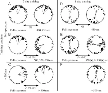
counterclockwise shift observed in the newts’ orientation was due to a direct effect of light on the newts’ perception of the magnetic field, exposing newts to long-wavelength light in the outdoor tanks should cause their perception of the magnetic field, and hence of the magnetic direction of shore, to be rotated by 90 ° counterclockwise (Fig. 2A). When tested under long-wavelength light, the newts’ perception of the direction of the magnetic field in the arena would be the same as it was in the outdoor tank, and they should orient in the correct shoreward direction (Fig. 2C). In contrast, when tested under full-spectrum light, the newts’ perception of the field would be rotated 90 ° clockwise from their perception of the field in the outdoor tank and they should orient 90 ° clockwise of the direction of shore (Fig. 2B). These predictions were indeed what was observed after long-wavelength training (Fig. 1C).
Using a new ‘1 day’ training protocol, we recently replicated the effect of the wavelength of light on magnetic compass orientation in the newt. When newts are allowed to walk down a steep incline into the training tank, they exhibit a shoreward magnetic compass response when tested the very next day (Deutschlander et al., 1999a). In contrast to the unimodal shoreward orientation displayed by newts held in the training tanks for 5 days (Phillips and Borland, 1992a,b), newts
‘trained’ using the shorter period exhibited bimodal orientation along the shoreward axis. Despite the bimodality of the response, wavelength experiments using the ‘1 day’ training protocol resulted in the same pattern of wavelength-dependence as in previous experiments. Newts trained under natural skylight and tested under either full-spectrum or short-wavelength (450 nm) light exhibited bimodal orientation parallel to the shoreward axis learned in the outdoor training tanks (Fig. 1D; Table 1). In contrast, newts trained under natural skylight and tested under long-wavelength (550 or
⭓500 nm) light oriented along an axis perpendicular to the shoreward axis (Fig. 1E). Once again, training newts under long-wavelength light abolished the 90 ° shift in newts tested under long-wavelength (Fig. 1F). Together with previous results (Phillips and Borland, 1992a), these data indicate that the newts’ perception of the direction of the magnetic field under long-wavelength light is rotated 90 ° from their perception of the magnetic field under full-spectrum or short-wavelength light.
Phillips and Borland (1992a) proposed a simple physiological model to explain how the magnetic field might alter the response of a photoreceptor-based magnetoreception system to produce the wavelength-dependent effects observed
5 day training
A
B
NS
Full-spectrum 400, 450 nm
P < 0.001
Full-spectrum 500, 550, 600 nm
P < 0.001
Full-spectrum ⭓500 nm
C
⭓
500
nm
Full-spectrum
Training condition
1 day training
D
E
NS
Full-spectrum 450 nm
P < 0.001
Full-spectrum 550 (■),⭓500 (●) nm
[image:4.609.210.558.74.372.2]⭓500 nm
F
Fig. 1. The effect of the wavelength of light on shoreward-orienting newts. (A–C) Results from Phillips and Borland (1992a) using a 5 day training protocol. (D–F) Previously unpublished results from experiments using a ‘1 day’ training protocol. Each pair of circular distributions shows the results from one set of experiments in which individual newts were tested under either full-spectrum light or the designated portion of the spectrum. Each data point represents the magnetic bearing of one newt. All data are plotted with respect to the magnetic direction of shore in the training tank (i.e. the shore direction is rotated to the top of each circle plot). Single-headed and double-headed arrows at the center of each plot indicate the mean vector, or axis, for unimodal and bimodal distributions, respectively. The mean vector length is proportional to the strength of orientation (r) with the radius in A–C, and the diameter in D–F, corresponding to r=1. Dashed lines indicate the 95 % confidence intervals for the mean vector. Each distribution is significant at P<0.05 or less. P values under the double-headed arrows between circle plots indicate
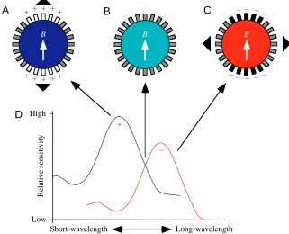
in the shoreward magnetic compass response of newts (Fig. 3). In their model, Phillips and Borland (1992a) suggested that each receptor cell contains two spectral mechanisms, a short-wavelength absorption mechanism (peak absorption <450 nm) and a long-wavelength absorption mechanism (peak absorption >500 nm) that act antagonistically on the neural output of the cell (as shown in Fig. 3D). Alternatively, the two spectral mechanisms could occur in separate classes of photoreceptors that provide antagonistic inputs to a second-order neuron. [Whether the two absorption mechanisms are actually two different photopigments or two different active states of the same photopigment is unclear from the experiments. We use the term absorption mechanism, or spectral mechanism, instead of photopigment, since the two different absorption mechanisms could theoretically be the same photopigment in two different functional states (e.g. see Dodt and Heerd, 1962; Eldred and Nolte, 1978).] In Fig. 3, we have arbitrarily labeled the short-wavelength absorption mechanism as excitatory (+) and the long-wavelength absorption mechanism as inhibitory (−), but these signs could just as well be reversed. The model does not explicitly implicate a particular biophysical mechanism for magnetotransduction. However, the basic assumption of the model is that the receptor cells’ response to
light is enhanced (or reduced) when the receptors are in a particular alignment relative to the magnetic field. Because all three of the proposed light-dependent mechanisms of magnetoreception are insensitive to the polarity of the magnetic field (Leask, 1977; Schulten, 1982; Edmonds, 1996), the pattern of response exhibited by this type of receptor will exhibit axial sensitivity. For example, under short-wavelength light, receptors aligned parallel to the axis of the magnetic field (B) might show an increase in excitation relative to receptors not aligned parallel to the magnetic field (Fig. 3A). In contrast, the same receptors would show a decreased response under long-wavelength light (Fig. 3C). [Alternatively if the two spectral mechanisms occur in separate classes of photoreceptor, then long-wavelength receptors in the same alignment as the magnetic field would show an enhanced response and, subsequently, have an antagonistic (in this case, inhibitory) effect on second-order cells.] As a consequence of these antagonistic responses, the axis exhibiting the highest level of response under short-wavelength light is perpendicular to the axis exhibiting the highest level of response under long-wavelength light. If newts followed the simple rule that the direction of B corresponds to the axis exhibiting the highest response, their perception of the alignment of B under long-Table 1. Statistical analysis of 1 day training data (Fig. 1D–F)
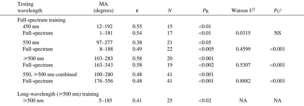
Testing MA
wavelength (degrees) r N PR Watson U2 PU2
Full-spectrum training
450 nm 12–192 0.55 15 <0.01
Full-spectrum 1–181 0.54 17 <0.01 0.0315 NS
550 nm 97–277 0.38 21 <0.05
Full-spectrum 8–188 0.49 22 <0.005 0.4599 <0.001
⭓500 nm 103–283 0.58 20 <0.001
Full-spectrum 163–343 0.58 19 <0.002 0.5307 <0.001
550, ⭓500 nm combined 100–280 0.48 41 <0.001
Full-spectrum 176–356 0.48 41 <0.001 0.8882 <0.001
Long-wavelength (⭓500 nm) training
⭓500 nm 5–185 0.41 25 <0.02 NA NA
The magnetic bearings of the newts have been pooled relative to the direction of shore, so that shore=0 °. Individual bearings were doubled
prior to calculating the bimodal mean axis for each distribution and comparing the distributions using the Watson U2-test (Batschelet, 1981).
MA, mean axis for the bimodal distribution; r, mean vector length; N, sample size; PR, probability by the Rayleigh test; Watson U2, test for
differences between two distributions; PU2 , probability by the Watson U2-test; NS, not significant at P<0.05; NA, not applicable.
A detailed description of the methods for 1 day training and testing has been presented in Deutschlander et al. (1999a). Individual newts were tested only once under either full-spectrum light or the specified region of the spectrum. In individual tests, an approximately equal number of newts was tested under either full-spectrum light (as controls) or the wavelength in question (except in long-wavelength training tests, in which newts were only tested under long-wavelength light). Each newt was tested in one of four symmetrical alignments of the magnetic field (magnetic north at geographic north, east, west or south). Data were pooled relative to the magnetic direction of shore to test for significant magnetic shoreward orientation. Full-spectrum light in the testing arena was produced using a 150 W xenon lamp. Long-wavelength
stimuli (550 nm and 艌500 nm) were the same as described by Phillips and Borland (1992a). The short-wavelength stimulus (450 nm) was
created by placing a short-wavelength-transmitting gel filter (Lee no. HT120, 450 nm peak, approximately 40 nm HBW) in the light path to the arena. Quantal flux at 450 and 550 nm was within 0.2 log units of the values used by Phillips and Borland (1992a). For long-wavelength training tests, newts were exposed to long-wavelength light in circular buckets for at least 3 h prior to being introduced into the training tank. Both the tank and the training incline (on which the newts walked down into the training tank; see Deutschlander et al., 1999a) were covered
[image:5.609.55.565.87.266.2]wavelength light would be perpendicular to their perception of the alignment of B under short-wavelength light (Phillips and Borland, 1992a). This model does not address how newts distinguish between the two ends of the magnetic axis, which is necessary to exhibit a unimodal response towards shore (as in newts trained for 5 days). Shoreward-orienting newts, however, use a magnetic inclination compass (Phillips, 1986b), i.e. newts determine the inclination of the magnetic field with respect to an external reference, such as the gravity vector or the horizon, to determine ‘polarity’.
To test their model, Phillips and Borland (1992a) tested newts under 475 nm light, intermediate to the regions of the spectrum that produced shifted (⭓500 nm) and unshifted (⭐450 nm) orientation. In a light-dependent magnetodetector with the properties proposed in Fig. 3, intermediate wavelengths should activate both spectral mechanisms equally, causing the opposing effects of the magnetic field on the two mechanisms to cancel out (Fig. 3B). As predicted, the distribution of magnetic bearings of newts tested under 475 nm light was indistinguishable from random (data not shown).
D
Short-wavelength Long-wavelength
High
Low
C
B B
B
B
A
−− − −−− − − − −
+ + + ++
+ + + ++
Relative sensitivity
+
− Fig. 3. A model for the wavelength-dependent
effects of light on a magnetically sensitive photoreceptor organ in the newt. A hypothetical magnetoreception system is shown in A, B and C to consist of a circular array of receptors (small rectangles). (D) The absorption spectra of hypothetical short-wavelength and long-wavelength absorption mechanisms proposed to mediate the newt’s light-dependent magnetic compass response. The two spectral mechanisms could be contained in the same receptor cell or could occur in two separate classes of magnetically sensitive photoreceptors that provide input to the same second-order cell (see text). In either case, the two spectral mechanisms have antagonistic effects on the output of the detector as indicated by the plus and minus signs under the absorption curves in D. (A) Under short-wavelength light, receptors within certain alignments relative to the magnetic axis, B, exhibit an increase in response (open rectangles) relative to receptors in alignments that are not affected by the magnetic field (grey-filled rectangles). Arrowheads at the edge of the circular array indicate the axis that will appear to have the
highest level of response. (B) Under intermediate wavelengths of light that activate the two spectral mechanisms more or less equally, the effect of the magnetic field on the two spectral mechanisms cancels out so that the net response of the receptors is unaffected by the magnetic field alignment (grey-filled rectangles). (C) Under long-wavelength light, receptors in alignments that are affected by the magnetic field exhibit a decrease in response (black-filled rectangles) relative to receptors in alignments that are unaffected by the magnetic field (grey-filled rectangles). The axes with the highest level of response (indicated by the arrowheads) differ by 90 ° in A and C (modified from Phillips and Deutschlander, 1997).
mN
mN′
B
C
mN
mN′
[image:6.609.235.556.409.669.2]A
Fig. 2. Predicted orientation of newts after training under long-wavelength light (yellow shading) if the wavelength-dependent 90 ° shift is due to a direct effect of light on the underlying magnetoreception mechanism. (A) Schematic diagram of a training tank with a shore towards magnetic north. If
long-wavelength light (⭓500 nm) causes the newts’
perception of magnetic north (mN′) to be rotated 90 ° counterclockwise from true magnetic north (mN), then the direction of shore will appear to the newts to
Phillips and Borland (1992a) also proposed that the two spectral mechanisms differ in relative sensitivity (Fig. 3D). If the two mechanisms had similar sensitivities, natural broadband light would activate both mechanisms similarly, causing the magnetic field’s effect to be canceled out. Although, in principle, a reduction in the sensitivity of either mechanism would avoid the problem of equal activation, the short-wavelength mechanism appears be more sensitive in newts. Newts tested under short-wavelength light show the same orientation as newts tested under full-spectrum light. Hence, the short-wavelength mechanism must be preferentially activated under full-spectrum light. Spectral mechanisms located in separate cells that were equally sensitive, but that had different weighting of their inputs onto a second-order neuron, would also prevent the effects of the magnetic field from canceling out under broad-spectrum light.
A similar wavelength-dependent 90 ° shift in magnetic compass orientation has been observed in experiments on Drosophila melanogaster (Phillips and Sayeed, 1993). D. melanogaster can be trained to orient in the direction of a light source. When tested under diffuse, non-directional light, D. melanogaster use magnetic cues in an attempt to orient towards the previously experienced light source. Male D. melanogaster trained and tested under ultraviolet light (365 nm) oriented towards the magnetic direction from which the light emanated during training. In contrast, males trained to an ultraviolet light source and tested under 500 nm light oriented 90 ° clockwise from the direction of the light during training. Despite the difference in the direction of the 90 ° shift (clockwise in flies versus counterclockwise in newts), the similarity of the effects of light on magnetic compass orientation in newts and D. melanogaster is striking. However, further experiments are needed to determine whether the effect of light on D. melanogaster orientation is due to a direct effect on the flies’ perception of the magnetic field.
Four species of birds, three migratory passerines and homing pigeons, have been tested for wavelength-dependent effects of light on their magnetic orientation responses. The pattern of wavelength-dependence observed in birds, although consistent among avian species, appears to be qualitatively different from the wavelength-dependence of shoreward-orienting newts. Migratory responses were tested in orientation funnels under various wavelengths of equal intensity. The peak wavelengths of the spectral stimuli reported in the original papers have recently been remeasured (see Wiltschko and Wiltscko, 1998b). We report the more recent measurements here. Adult silvereyes Zosterops l. lateralis, tested under diffuse ‘white’ light, blue light (443 nm) and green light (565 nm), oriented in a seasonally appropriate direction when given access to magnetic cues only (Wiltschko et al., 1993). However, both adult and juvenile silvereyes failed to maintain a consistent direction when tested under red light (630 nm: Wiltschko et al., 1993; Munro et al., 1997b). Young adult European robins Erithacus rubecula and juvenile garden warblers Sylvia borin also oriented in a seasonally appropriate direction under blue and green light, but were disoriented under both red and yellow
light (630 and 590 nm, respectively; Wiltschko and Wiltschko, 1995b; Wiltschko et al., 1997). Preliminary studies on the orientation of young homing pigeons suggest a wavelength-dependence similar to that found in passerines. Wiltschko and Wiltschko (1998b) transported inexperienced, young pigeons to a release site under various wavelengths of light and released all the birds under natural skylight. After transport to the release site under full-spectrum light and green light (565 nm), the pigeons were well-oriented relative to the home direction. In contrast, the vanishing bearings of pigeons transported under red light (630 nm and 660 nm) were indistinguishable from random.
As discussed above, experimental treatments that result in randomly distributed directional bearings are difficult to interpret. Therefore, motivational or other nonspecific behavioral effects of long-wavelength light on birds are hard to rule out. One example of a behavioral effect of light on orientation behavior that is not mediated by a magnetic compass occurs in the escape behavior of frogs. Some ranid frogs appear to orient towards visual fields that are relatively rich in short-wavelength light (such as light from open sky or reflected by water) and away from visual fields that are relatively rich in long-wavelength light (such as light reflected by foliage) (Muntz, 1962, 1966; for species-dependent variation, see Hailman and Jaeger, 1974). By analogy, migratory birds may perceive an overhead visual field that is rich in long-wavelength light as an obstruction. Hence, their activity could represent an unoriented attempt to escape from beneath the obstruction, rather than an attempt to initiate oriented flight. However, young pigeons were exposed to experimental lighting conditions in holding cages only during displacement to the release site (Wiltschko and Wiltschko, 1998b). Pigeons transported in cages are typically rather docile during displacement (J. B. Phillips, personal observation); it is therefore unlikely that an attempt by the pigeons to escape from beneath an obstruction while being exposed to red light would produce disorientation when they were subsequently released or when their vanishing bearings were recorded at a distance of 1 km or more from the release site. Furthermore, pigeons exposed to full-spectrum and green light were also held in cages during transport to the release site and so were subject to a similar ‘caged’ treatment prior to release. The differences in the behavioral context in which magnetic compass information was utilized by migratory passerines and homing pigeons suggest that nonspecific effects of light on avian orientation are unlikely explanations for their disorientation under long-wavelength light.
full-spectrum and 400 nm light oriented towards home. Newts tested under 450 nm light also exhibited a tendency to orient in the home direction. However, newts tested under long-wavelength (550, 600 nm) light failed to exhibit consistent orientation with respect to the home direction (Phillips and Borland, 1994). Homing newts appear to derive both map and compass information from the magnetic field to determine the homeward direction (Phillips, 1986b; Phillips et al., 1995, J. Fischer, M. J. Freake, S. C. Borland and J. B. Phillips, in preparation). In contrast, newts orienting towards shore need only compass cues to determine the direction of shore. Phillips and Borland (1994) suggest that the qualitative difference between the wavelength-dependence of shoreward-orienting and homing newts is due to the additional ‘map’ step required for true navigation. We will discuss this hypothesis in more detail below.
Long-wavelength training experiments with shoreward-orienting newts provide the strongest evidence for a direct effect of light on a magnetic compass mechanism. Despite the congruent patterns of wavelength-dependence observed in the orientation responses of homing pigeons and migratory birds, there are no data for birds that conclusively demonstrate that light has a direct effect on their perception of the magnetic field. Moreover, if birds do have a light-dependent magnetic compass, the evidence presented thus far suggests that birds may have a different type of photoreceptor-based magnetoreceptor from that proposed to occur in newts (see below). In the following discussion, however, we propose several speculative hypotheses which suggest how a single type of light-dependent magnetic compass mechanism (like that proposed to occur in newts) could produce the different behavioral responses observed in newts and birds. We hope that tests of these hypotheses will help to determine whether the wavelength-dependence in birds is due to a photoreceptor-based magnetoreceptor and whether the different patterns of wavelength-dependence in newts and birds represent biophysical and/or physiological differences in the underlying magnetoreception mechanism.
Resolving the discrepancies in the wavelength-dependent effects of light on magnetic orientation in birds and newts As discussed above, the wavelength-dependence of the newt’s shoreward magnetic compass response suggests that two antagonistic spectral mechanisms are involved in the magnetic compass. As a result of the antagonistic interaction that is inferred to occur between these two mechanisms, reduction in the sensitivity (or relative weighting) of one of the spectral mechanisms would appear to be necessary for the newt’s magnetic compass to function under normal lighting conditions. Why do newts retain a ‘two-pigment’ system? Having a single spectral mechanism would seem to be a more parsimonious solution than involving two antagonistic spectral mechanisms. One possible explanation is that the two spectral mechanisms are mediated by two molecules that are both essential to the magnetoreception process (Phillips and
Deutschlander, 1997). For example, the magnetoreception mechanism may involve an interaction between two members of a photoinduced radical pair (Schulten, 1982; Schulten and Windemuth, 1986), both of which are excited by light in the visible or near-ultraviolet range of the spectrum. Although excitation of only one of the two molecules that interact to form the radical pair may be necessary for magnetoreception to occur (Schulten, 1982), preferential photoexcitation of the second molecule under long-wavelength (⭓500 nm) light might initiate a radical pair process with somewhat different (i.e. antagonistic) properties. Regardless of the specific mechanism, if the two spectral mechanisms are part of the biophysical process required for magnetoreception in the newt, both the short-wavelength and long-wavelength mechanism may be present in the same cell (Phillips and Deutschlander, 1997).
There is little evidence in either vertebrates or invertebrates for photoreceptors that contain two antagonistic photopigments or spectral mechanisms in the same cell. However, antagonistic spectral mechanisms have been described in single photoreceptors in the pineal complex of lizards and amphibians (Eldred and Nolte, 1978; Solessio and Engbretson, 1993). Interestingly, we have recently conducted experiments which implicate pineal photoreceptors as the source of the wavelength-dependence in the newt’s magnetic compass. Small gel filters were attached to the dorsal surface of the newts’ heads to alter the wavelengths of light reaching the area of the brain containing the pineal gland, without altering the spectral composition of light reaching the eyes. Covering the head of a newt with long-wavelength (⭓500 nm)-transmitting filters during training under ambient light was sufficient to mimic the effect of long-wavelength training on the newt’s shoreward compass response (Deutschlander et al., 1999b).
photopigment typically do not show a sharp cutoff over such a narrow wavelength range (Fein and Szuts, 1982). Therefore, if the avian magnetic compass is based on a single photopigment, the strength of orientation would be expected to decrease gradually at wavelengths above the peak absorption of the photopigment. Interestingly, newts also exhibit an abrupt transition from well-oriented magnetic compass orientation at 450 nm to disorientation at 475 nm (Phillips and Borland, 1992a). The sharp spectral cutoff in the newt’s magnetic compass response, however, appears to be the result of an antagonistic interaction between a short-wavelength and a long-wavelength spectral mechanism (Fig. 3B).
The sharp cutoff in the magnetic orientation of birds at long wavelengths suggests another possible explanation: that red light may actively disrupt magnetic orientation in birds, causing disorientation. In a magnetoreceptor based on optical pumping, Leask (1977) suggested that long-wavelength light might modify energy transfer to the lowest triplet state, thereby interfering with magnetoreception. However, since the requirements of optical pumping do not seem to be satisfied in biological systems, this is an unlikely explanation. Alternatively, monochromatic long-wavelength light might disrupt orientation in birds by preferentially activating a second, less-sensitive spectral mechanism that interacts antagonistically with the shorter-wavelength mechanism. Consistent with the involvement of two different spectral mechanisms in birds, Semm and Demaine (1986b) reported evidence for two spectral classes of magnetically sensitive units in the nucleus of the basal optic root (nBOR) of pigeons. One class of units exhibited a peak response to magnetic field changes when illuminated with 500 nm light and the other when illuminated with 580 nm light. The units with peak sensitivity near 580 nm also exhibited magnetic sensitivity under 674 nm light. Moreover, even units from the nBOR with peak sensitivities around 500 nm exhibited appreciable magnetic sensitivity at wavelengths near 580 nm. These neurophysiological experiments from birds have proved difficult to replicate. Until successful replications are carried out, comparisons between behavioral and neurophysiological findings must remain tentative. However, if neurons in the nBOR carry magnetic compass information in birds, one would predict that birds would be able to obtain directional information from the magnetic field under long-wavelength light (i.e. 590 nm and possibly also 630 nm). The contrast between the physiological and behavioral findings in birds is puzzling, but it does give credibility to the hypothesis that red light may indeed ‘disrupt’ orientation in birds by activating a second (i.e. long-wavelength-absorbing) antagonistic mechanism. Taken together, the behavioral and neurophysiological data from birds are consistent with a ‘two-pigment’ model consisting of a short- to mid-wavelength mechanism and a less-sensitive, antagonistic long-wavelength mechanism (e.g. Fig. 4A). However, it is possible that, even if Semm and Demaine’s (1986b) neurophysiological findings are correct, the neurophysiological data from the nBOR may not be directly related to magnetic orientation behavior. Although
magnetic responses from the nBOR exhibit a number of properties found in magnetic orientation in birds (e.g. sensitivity to magnetic inclination, failure to respond to the magnetic field in total darkness; Semm et al., 1984), these physiological findings have never been conclusively linked to magnetic orientation behavior. Below we explore three alternative explanations for differences in the wavelength-dependence of magnetic orientation in newts and birds and for the conflicting information from behavioral and neurophysiological studies of magnetoreception in birds.
The neural processing hypothesis: ‘different rules for the same game’
Birds and newts could respond differently to preferential activation of a second, less-sensitive, long-wavelength mechanism because of differences in how the two groups of organisms process magnetic compass information. That is, birds and newts may exhibit different behavioral responses under long-wavelength light even if their underlying magnetoreceptors are affected similarly by light. The impetus for this hypothesis came, in part, from experiments examining the effect of changes in magnetic field intensity on orientation in European robins (Wiltschko, 1978). Therefore, before considering whether perceptual differences can account for the different effect(s) of light on magnetic orientation in newts and birds, we first discuss (1) how a photoreceptor-based magnetic compass organ might respond to changes in field intensity and (2) the spatial scales over which an animal would have to cope with changes in magnetic intensity.
Leask (1977, 1978), Schulten (1982) and Schulten and Windemuth (1986) argued that the response of an array of magnetically sensitive photoreceptors should vary depending on both the alignment and the intensity of the magnetic field. For a particular alignment and intensity, specific subsets of photoreceptors will be in alignments that are affected by the magnetic field. Therefore, a given intensity of the magnetic field should generate a specific axially symmetrical three-dimensional pattern of affected photoreceptors in a light-dependent magnetoreceptive organ (e.g. across the retina). Different field intensities would affect photoreceptors in different alignments relative to the magnetic field and, hence, produce a new three-dimensional pattern of response.
caught at 46 000 nT and held in a 150 000 nT field were oriented when tested in a 46 000 nT or 150 000 nT field, but not when tested in a 81 000 nT field). If a photoreceptor-based mechanism similar to Leask’s or Schulten’s models underlies the magnetic compass response of birds, Wiltschko’s results suggest that European robins may at first be disoriented by the unfamiliar pattern of response produced in the magnetodetector organ by the new field intensities. After several days of exposure to a new field intensity, the birds may have ‘adapted’ to the new response pattern and become able to orient. According to this interpretation, the birds would still have been able to orient at the original field intensity since it produced a pattern of response with which they were already familiar. However, the response pattern produced at intermediate intensities may have been sufficiently different from that produced at either higher or lower intensities so that it would be novel to the birds.
Because of spatial variation in magnetic intensity, any animal that moves over a large distance would experience a wide range of magnetic field intensities. For example, some birds migrate from field strengths as high as 60 000–65 000 nT near the poles to field strengths as low as 25 000–30 000 nT near the equator (Skiles, 1985; Wiltschko and Wiltschko, 1995a). Therefore, long-distance migrants may have to derive directional information from a magnetic compass in which the pattern of response changes over the bird’s migratory range. The effect of changes in magnetic intensity on the pattern of response in a photoreceptor-based magnetodetector could be one reason that birds rely on multiple directional cues for orientation (for a review, see Wiltschko and Wiltschko, 1995a). Under natural migratory conditions, birds may calibrate the response of the magnetic compass with respect to celestial cues.
In shoreward-orienting newts, exposure to long-wavelength (⭓500 nm) light appears to cause a change in the pattern of response exhibited by a photoreceptor-based magnetic compass which is perceived by newts as a 90 ° rotation in the direction of the magnetic field. Phillips and Borland (1992a) suggested that newts may follow a simple rule to determine compass information from the response pattern in their magnetodetector; e.g. the axis that yields the highest (or lowest) level of response in the detector organ corresponds to the axial direction of the magnetic field (Fig. 3). Such a simple rule is likely to be sufficient since newts, in contrast to birds, do not move over large distances and under normal circumstances would never experience large enough changes in magnetic intensity to affect the response of a photoreceptor-based magnetodetector. A similar rule is also likely to be sufficient for D. melanogaster, which exhibit a similar wavelength-dependent 90 ° shift in magnetic compass orientation (Phillips and Sayeed, 1993) and are likely to have
A
D
C
B
Relative absorbance
Wavelength (nm)
[image:10.609.368.511.305.727.2]350400 450500 550 600 650
a small home range. However, since many species of birds may have to cope with changes in their perception of the magnetic field due to variation in intensity encountered during long-distance migration, they may have more complex rules for determining magnetic compass direction. The effects of magnetic field intensity on orientation in European robins suggest that birds are initially confused, or disoriented, by manipulations that alter the response of their magnetic compass (at least when denied access to other directional cues). It would not be surprising, therefore, if the failure of birds to exhibit consistent magnetic compass orientation under long-wavelength light results from a similar qualitative change in their perception of the magnetic field. In the wavelength experiments conducted with migratory birds, the birds experienced the experimental light conditions only in the orientation funnels during the 1–2 h of testing each night. In some experiments, the birds were exposed to different wavelength conditions each night, so that one night a bird might be exposed to blue light and the next night to red or green light (e.g. Wiltschko et al., 1993). Between orientation tests, the birds were kept in holding cages under full-spectrum light. If long-wavelength light does alter the birds’ perception of the magnetic field, they would have had little time to ‘adapt’ to the novel pattern of response. Furthermore, if red light produced a pattern of response in the magnetic compass that differed from that produced by full-spectrum, blue or green light, alternating the birds between lighting conditions may have increased the birds’ confusion. Similarly, pigeons transported under red light to the release site may have been confused by an altered response pattern of the magnetic compass and have been unable to obtain useful directional information from their magnetic compass on the outward journey to the release site.
Experiments in which migratory birds are exposed to long-wavelength (630 nm) light during both holding and testing may help resolve these issues. By analogy with the behavior of European robins exposed to changes in magnetic field intensity (Wiltschko, 1978), exposing birds to the new lighting conditions for a period prior to testing may allow them to adapt to the new pattern of response produced by the magnetic compass. If birds are able to orient under long-wavelength light after a period of adaptation, not only would it suggest that birds and newts have more similar underlying detection mechanisms than previously realized, but it could also help to explain the apparent discrepancies between the wavelength-dependence of neurophysiological and behavioral data in birds.
The navigation hypothesis: interactions between multiple magnetoreceptors
A growing body of evidence suggests that some animals use spatial variation in one or more components of the geomagnetic field, such as intensity or inclination, to determine their geographical position during migration and/or homing (for a review, see Wiltschko and Wiltschko, 1995a). Spatial variation in the geomagnetic field could be used in at least two ways. Through experience, an animal could develop a
cognitive representation, or ‘map’, of the spatial gradients of one or more magnetic parameters around its home range or along its migratory route. The animal could then use information from the magnetic gradient(s) to determine its geographical position relative to a goal, such as its home or its wintering grounds (Gould, 1980; Wiltschko and Wiltschko, 1982, 1995a; Walcott, 1991; Walraff, 1991; Rodda and Phillips, 1992; Phillips, 1996). This type of orientation has been called magnetic ‘map’ orientation and is an example of true, or site-based, navigation. Specific values of the magnetic field could also be used to trigger specific adaptive orientation responses at particular geographic locations (‘sign-post’ navigation). For example, juvenile pied flycatchers Fidecula hypoleuca on their first migration appear to respond to specific values of the magnetic field as an indicator, or releaser stimulus, to change compass headings during migration (Beck and Wiltschko, 1988). Young loggerhead sea turtles Caretta caretta may be able to recognize specific magnetic values associated with the boundaries of their range in the North Atlantic gyre (Lohmann and Lohmann, 1994, 1996; however, see Courtillot et al., 1997). Since these responses in flycatchers and sea turtles are expressed in juveniles with no migratory experience, they appear to be innate and, therefore, are unlikely to be due to a true magnetic ‘map’.
The use of the geomagnetic field for ‘map’ or ‘sign-post’ information would require that an animal be sensitive to the small changes in the inclination, or intensity, of the magnetic field that result from changes in geographical position. Because the photoreceptor-based mechanisms discussed above are unlikely to be sensitive enough to derive magnetic ‘map’ information (Leask, 1977, 1978; Schulten, 1982; Edmonds, 1996), magnetite-based magnetoreception mechanisms which, theoretically, could exhibit sensitivity to magnetic intensity changes of less than 100 nT are the most likely candidates for the basis of a ‘map’ sense (Kirschvink and Gould, 1981; Kirschvink and Walker, 1985, Kirschvink et al., 1993; Yorke, 1979, 1981). In birds and fish, concentrations of single-domain biogenic magnetite have been found in the ethmoid region of the skull (Beason and Nichols, 1984; Walker et al., 1984; Mann et al., 1988) and appear to be associated with the ophthalmic branch of the trigeminal nerve (Beason and Semm, 1996; Semm and Beason, 1990; Walker et al., 1997). Semm and Beason (1990) reported that the ophthalmic nerve in birds is sensitive to magnetic intensity changes of as little as 200 nT (<1/100 of the total field strength of the earth).
(Wiltschko et al., 1994; Beason et al., 1995; Wiltschko and Wiltschko, 1995b) and also of adult homing pigeons (Beason et al., 1997). In the bobolink Dolichonyx oryzivorus, the magnetic information altered by pulse remagnetization appears to be conveyed by the ophthalmic nerve (Beason and Semm, 1996). When anesthetic was applied to the ophthalmic nerve, the effect of the magnetic pulse was abolished. Bobolinks in which the nerve was anesthetized, however, oriented in a seasonally appropriate magnetic direction, indicating that their magnetic compass was still intact despite being denied information from the trigeminal system. These results are consistent with the interpretation that a magnetite-based magnetoreception system associated with the ophthalmic nerve is used for geographical positioning information, whereas compass information is provided by another channel (e.g. an input associated with the visual system).
Phillips and Borland (1994) suggested that the effect of long-wavelength light on homing newts differs from the effect on shoreward-orienting newts (i.e. random versus 90 °-shifted orientation) because of the interaction between a magnetite-based ‘map’ detector and the light-dependent magnetic compass. Newts are capable of site-based homing, which requires the use of both a ‘map’ and a compass sense (Phillips et al., 1995). The responses of homing newts to small changes in magnetic inclination (±2 °) are consistent with the possibility that newts use magnetic inclination to derive information about geographical position while homing (J. Fischer, M. J. Freake, S. C. Borlamd and J. B. Phillips, in preparation). Furthermore, Phillips (1986b) found that homing newts respond to the polarity of the magnetic field, while shoreward-orienting newts exhibit only axial sensitivity. Although photoreceptor-based mechanisms are not expected to be sensitive to the polarity of the magnetic field, some magnetite-based mechanisms are expected to exhibit polar sensitivity (Kirschvink and Gould, 1981; Kirschvink and Walker, 1985; Kirschvink et al., 1993). The polar sensitivity of homing newts and their sensitivity to small changes in magnetic inclination therefore implicate a second, possibly magnetite-based, magnetoreception mechanism in the newt’s homing response. Phillips and Borland (1994) proposed that newts use their light-dependent magnetic compass to position a magnetite-based magnetic inclination (or ‘map’) detector in a consistent alignment relative to the magnetic field in order to determine accurately the magnetic field value(s) necessary for geographic positioning. They argue that the 90 ° shift in the directional response of the magnetic compass under long-wavelength light prevents newts from properly aligning their map detector, resulting in disorientation. Whether the specific argument of Phillips and Borland (1994) is correct, at some point (whether in the detection process or at a higher level of processing), compass information and map information must be combined for site-based navigation (Wiltschko and Wiltschko, 1988). As a consequence of combining map and compass information, an effect on the compass could influence the ‘map-step’ of navigation, or vice versa.
If an interaction between the neural inputs from a magnetic
compass and a ‘map’ detector produces disorientation in homing newts under long-wavelength light, it may be possible that birds show random orientation under long-wavelength light for a similar reason. Since a magnetic ‘map’ presumably needs to be constructed by an individual through experience, only adults should be capable of using geomagnetic spatial variation for ‘map-based’ navigation (Wiltschko and Wiltschko, 1982, 1995a; Rodda and Phillips, 1992; Munro et al., 1997a). Therefore, if pulse remagnetization affects a magnetite-based ‘map’ detector, only adult birds would be expected to show a change in orientation, e.g. if they interpret a change in the response of a ‘map’ detector as some type of change in geographical position. This prediction is supported by findings in silvereyes showing that pulse remagnetization changed the orientation of adult birds but not that of juveniles (Munro et al., 1997a). In contrast, both juveniles and adults silvereyes showed the same responses (i.e. disorientation) when tested under long-wavelength (630 nm) light, suggesting that light affects only the magnetic compass that is used by both juveniles and adults (Munro et al., 1997b; Wiltschko et al., 1997). The similarity between the effect of light on orientation by young and adult birds and the age-dependent responses to pulse remagnetization appears to rule out the possibility that an interaction between a magnetic ‘map’ detector and the magnetic compass accounts for random orientation in birds under long-wavelength light. However, if on their first migratory journey, young birds are learning the geographical distribution of magnetic field values in order to construct a magnetic map of their migratory route, they must already have the receptor capabilities necessary to sense geomagnetic variation. In addition, any disruption of the magnetic field, or of the detector underlying the acquisition of magnetic ‘map’ information, may simply be recorded by juveniles as geographical variation without causing a change in their directional heading. The lack of an effect of pulse remagnetization on juveniles, therefore, does not rule out the possibility that young birds have a functional ‘map’ detector system. Indeed, juvenile pied flycatchers on their first migration appear to respond to geographically significant values of the magnetic field for ‘sign-post’ information, suggesting that they already have the sensory apparatus required to detect spatial variation in the magnetic field (Beck and Wiltschko, 1988). Consequently, exposure to long-wavelength light may result in random orientation in both adult and juvenile migratory birds as a result of the interaction between a light-dependent magnetic compass and a magnetite-based geographical positioning sense.
migration, it is assumed that, during early training flights and releases, young pigeons construct a ‘map’ at least partially based on spatial variation in the magnetic field (Wiltschko and Wiltschko, 1982; Wiltschko, 1983). Hence, young inexperienced pigeons might also be combining information from two types of magnetoreception systems, despite the evidence that they primarily rely on route-based directional information to determine the homeward direction.
Admittedly, the navigation hypothesis is based on many inferences for which we still need more evidence and a vague idea of how map and compass information might be combined during the development of young birds. In addition, one disparity is that homing newts are sensitive to the polarity of the magnetic field, while migratory birds and pigeons exhibit axial sensitivity. If the polar sensitivity exhibited by homing newts is really indicative of an interaction between two magnetic systems required for site-based homing, then birds may not be using the same mechanisms as newts for true navigation. Nevertheless, the similarities in the wavelength-dependence of homing newts, homing pigeons and migratory passerines warrant consideration of this hypothesis.
The navigation hypothesis may also be difficult to test. In newts, we are fortunate to be able to compare the use of magnetic information for true navigation with its use for shoreward compass orientation. Developing an orientation assay for birds that involves the magnetic compass only (i.e. an orientation response that does not involve a magnetic system used for geographical positioning) would help elucidate the effect of long-wavelength light on the magnetic compass in birds. Magnetic compass information may be used by some birds to remember the location of food caches (Wiltschko and Wiltschko, 1995a). Perhaps an experimental design similar to that used to reveal the involvement of the the sun compass in food caching (Wiltschko and Balda, 1989) could be used to develop a test of magnetic compass orientation in birds. Comparisons between the wavelength-dependence of a caching magnetic compass response and the orientation of young migratory birds could help determine whether the orientation of juvenile migrants is affected only by manipulations that influence the magnetic compass. Alternatively, if the ophthalmic nerve does convey ‘map’ information to birds, a nerve block experiment (similar to that conducted by Beason and Semm, 1996), in which birds were tested under long-wavelength light, might uncouple the ‘map’ and compass to reveal a shift in compass orientation (R. C. Beason, personal communication).
The ultraviolet light hypothesis
Another possible explanation for the lack of a 90 ° shift in birds is that the wavelengths that would produce a shift in the magnetic compass orientation of birds have not yet been tested. Tests of wavelength-dependence in both newts and birds have been limited to the portion of the spectrum visible to humans (400–700 nm). Birds, salamanders, flies and a variety of other animals, however, possess ultraviolet-sensitive photopigments and can sense near-ultraviolet light between 340 and 400 nm
(Jacobs, 1992; Goldsmith, 1994; Dodt and Heerd, 1962; Deutschlander and Phillips, 1995; Hardie and Kirschfeld, 1983). The data collected thus far from newts do not distinguish whether the short-wavelength mechanism of the magnetic compass exhibits a peak sensitivity in the ultraviolet or the blue portion of the spectrum. Since, in D. melanogaster, a 90 ° shift in magnetic compass orientation was evident in tests in which flies were trained using a 360 nm light and tested under 500 nm light (Phillips and Sayeed, 1993), an ultraviolet-absorbing photopigment may be involved in the magnetic compass response of at least one species.
We have argued that, in a light-dependent magnetoreceptor based on an interaction between two antagonistic spectral mechanisms, the two mechanisms should differ appreciably in sensitivity so that the magnetic effect is not degraded or eliminated as a consequence of equal activation of the two mechanisms (see discussion above). There is no a priori reason, however, to predict whether the less-sensitive pigment will occur at longer wavelengths (Fig. 4A) or at shorter wavelengths (Fig. 4B–D). In other words, birds could have a light-dependent magnetoreceptor based on two spectral mechanisms neither of which is activated by long-wavelength (⭓590 nm) light. The type of interaction envisioned in Fig. 3 would work just as well if an animal had a short-wavelength mechanism in the ultraviolet that was less sensitive than the longer-wavelength mechanism (Fig. 4B). If the avian magnetic compass involves such a mechanism, the only experimental treatment that could produce a 90 ° shift in orientation would be exposure to ultraviolet light.
the longer-wavelength photopigment (Fig. 4D), then the birds should show normal orientation under ultraviolet light.
Although shifted orientation in birds under ultraviolet light would be consistent with the involvement of two spectral mechanisms, any other result would be ambiguous. If birds do not show a change in orientation under ultraviolet light, it would not be possible to determine whether an ultraviolet-absorbing mechanism was present (but less sensitive than the beta peak of the longer-wavelength mechanism) or absent. In other words, unshifted orientation would not rule out the possibility of a two-pigment system such as that shown in Fig. 4D. Photoreceptors with a ‘hidden’ short-wavelength mechanism that is antagonistic to a more sensitive long-wavelength mechanism have been described in at least one vertebrate (Solessio and Engbretson, 1993). As stated above, a magnetoreception mechanism such as the photoinduced radical pair mechanism may require an interaction between two molecules. Even though activation of only one of the two molecules may be required for magnetoreception to occur (Schulten, 1982), wavelength-dependence might arise if both molecules can be excited by light in the visible or near-ultraviolet range of the spectrum. Preferential activation of the second molecule may only be possible using monochromatic light in the laboratory or may not be possible at all (if the second molecule is less sensitive under all wavelengths of light; as in Fig. 4D). Therefore, birds would only be expected to show a change in orientation if certain wavelengths of monochromatic light can preferentially excite the less-sensitive spectral mechanism.
Random orientation under ultraviolet light would also yield ambiguous conclusions. Random orientation could result from equal activation of two spectral mechanisms (as in Fig. 4C). However, random orientation could also result from lack of activation of a mechanism involving a spectral mechanism absorbing at longer wavelengths. Although the lens of the avian eye transmits ultraviolet light, ultraviolet-absorbing oil droplets are found in the inner segments of some photoreceptors that contain mid- to long-wavelength photopigments (Goldsmith et al., 1984; Bowmaker et al., 1997). If such an ultraviolet-absorbing oil droplet were associated with the photoreceptor underlying the magnetic compass of birds, then ultraviolet light would be prevented from reaching the photopigment-containing outer segment. Thus, ultraviolet light could be prevented from activating either the beta peak of the mid-wavelength-absorbing photopigment or a second spectral mechanism which absorbs ultraviolet light.
We have suggested three hypotheses to help elucidate the role of light in avian magnetic orientation and to help resolve the apparent difference in the wavelength-dependence of newts and birds. In contrast to the other two hypotheses, the ultraviolet hypothesis, although plausible, does not resolve the conflict between the wavelength-dependence of the neurophysiological and behavioral findings in birds. Moreover, the characteristics of photopigments and avian photoreceptors described above would make it difficult to interpret any result
other than shifted orientation under ultraviolet light. The most readily testable hypothesis is the neural processing hypothesis. A simple experiment on migratory birds, similar to those already conducted, could test the effect of adaptation to long-wavelength light on the birds’ ability to orient. To test the navigation hypothesis, either a new magnetic compass assay is needed (e.g. a caching assay) or perhaps experiments involving manipulation of the ophthalmic nerve. Since manipulations of the ophthalmic nerve have already been successfully performed (Beason and Semm, 1996), an experiment examining the effects of light on migratory orientation in birds with their ophthalmic nerve blocked may be the quickest way to test the navigation hypothesis (R. C. Beason, personal communication). Developing a new behavioral assay could be time-consuming, but there would be several considerable advantages to studying magnetic compass behavior in caching birds. Like the shoreward response of newts, a magnetic compass response based on foraging would be a learned response (i.e. birds are trained to learn the direction of food). Birds could be trained to the magnetic direction of caches under certain wavelength conditions and then be tested under others wavelength conditions to test for a direct effect of light on the magnetic compass (as in long-wavelength training experiments in newts; Phillips and Borland, 1992a).
Additional evidence for a light-dependent mechanism of magnetic field sensitivity from non-orientation responses
to magnetic fields