Earlier work showed that the hexactinellid sponge Rhabdocalyptus dawsoni can arrest its feeding current, presumably by stopping the beating of the flagella in the flagellated chambers. Arrests were found to follow local tactile or electrical stimulation and to propagate through the entire sponge on an all-or-none basis (Lawn et al., 1981). The conduction velocity was measured at 0.26 cm s−1, two orders of magnitude faster than Ca2+waves in astrocytes (Nedergaard, 1994) and three orders of magnitude faster than fast axoplasmic transport in squid giant axons (Allen et al., 1982). It therefore seemed likely that the propagated signal, though slow by the standards of nervous conduction, was an electrical impulse, but attempts to record such an event were unsuccessful until very recently. Hexactinellids ‘might have been designed expressly to defeat the electrophysiologist’ (Mackie et al., 1983). The presumed conducting tissue, the trabecular reticulum, is a multinucleate syncytium consisting of gossamer-thin strands draped around the spicules of the skeleton. It is confluent with the pinacoderm, a thin (approximately 1µm), perforated, syncytial sheet covering the
inner and outer surfaces. The thinness and fragility of these tissues make attachment or insertion of electrodes extremely difficult. However, by dissociating sponge tissue, allowing it to reaggregate to form sizeable lumps and grafting these onto the surface of the original sponge, it was possible to make attachment points for suction electrodes suitable for external recording. The grafts were evidently in syncytial continuity with the trabecular reticulum, since electrical signals propagating through the sponge entered them and were recorded relatively easily, as briefly reported by Leys and Mackie (1997). The present paper provides further information on the procedures used for making grafts and recording from them, together with information from new research carried out in 1997 on the properties of the conduction system.
Materials and methods Collection and maintenance of sponges
Rhabdocalyptus dawsoni were collected by SCUBA in mid-June 1997 from depths of 28–35 m at San Jose Islets in Barkley JEB1886
All-or-none propagated electrical impulses were recorded from the hexactinellid sponge Rhabdocalyptus dawsoni using suction electrodes attached to lumps of aggregated sponge tissue grafted onto the surface of pieces of the same sponge. Impulses were normally evoked by means of externally applied electrical shocks. Recorded externally using an a.c.-coupled amplifier, the electrical event was triphasic and lasted approximately 30 s; integration gave a diphasic waveform. A further integration to give the form of the membrane action potential produced a monophasic signal. Impulses propagated at 0.27±0.1 cm s−1with an absolute refractory
period of 29 s and a relative refractory period of approximately 150 s. Concurrent thermistor flow meter recordings confirmed that water flow through the sponge was arrested following the passage of an impulse, presumably as result of the cessation of beating of the flagella in the flagellated chambers. Tactile stimuli also evoked impulses, as did addition of particulate material to
the incoming water stream. Impulses continued to propagate through the sponge during arrests, indicating that the conduction and effector systems were independent. Sponges lack nerves, and a variety of evidence indicates that the conducting tissues are the syncytial trabecular reticulum and pinacoderm layers. Na+-deficient solutions
had little effect on the action potential, but propagation was blocked by 10 mmol l−1Co2+, 1 mmol l−1Mn2+or 24µmol l−1
nimodipine. Tetraethylammonium ions at 1–5 mmol l−1also
blocked propagation without prolonging the action potential. Impulse conduction in the sponge is discussed in relation to excitability and conduction in the protozoa and in plants and to non-nervous conduction in more advanced animals.
Key words: sponge, action potential, Rhabdocalyptus dawsoni, calcium, nimodipine, Co2+, tetraethylammonium, impulse
conduction.
Summary
Introduction
IMPULSE CONDUCTION IN A SPONGE
S. P. LEYS1, G. O. MACKIE1 ANDR. W. MEECH2,*
1Biology Department, University of Victoria, British Columbia, Canada V8W 2Y2 and 2Department of Physiology, University Walk, Bristol BS8 1TD, UK
*Author for correspondence (e-mail: [email protected])
Sound on the west coast of Vancouver Island, British Columbia, Canada. Individual sponges 10–30 cm in length were gently prized off the rock bottom, placed in large plastic bags and transported without removal from the water at any point to holding tanks at Bamfield Marine Station, British Columbia, Canada. Approximately 20 sponges were kept in a tank with a large exchange of sea water and were shaded from direct light using black plastic. Water temperature at the collection site was approximately 10 °C and varied by only one or two degrees throughout the year. This corresponded well with the temperature of water in the seawater system at the marine station (brought in from 21 m depth), which was recorded as 9–10 °C during June 1997. Specimens of R.
dawsoni have also been kept in seawater systems at the
University of Victoria and at the Friday Harbor Laboratories, Washington, USA, and although they will live for weeks or months in these situations, they lose the ability to arrest their feeding currents. The reasons for this are difficult to determine, but for practical purposes, the laboratory at Bamfield is still the only known place where the present type of work can be usefully pursued. Even at Bamfield (and generally along the coast), the sponges undergo seasonal regression during the winter months (Leys and Lauzon, 1998); hence, our choice of early summer for this research.
Preparation of grafts
Generally up to 20 individuals were collected and pieces from each were used to make aggregates each day. Sponges that had been subjected to daily removal of tissues produced better aggregates than those from which tissue was removed for the first time. Aggregate formation was better during the summer than the winter months (Leys, 1997). To obtain aggregates for grafting, pieces of approximately 4–5 cm2were
trimmed to remove the debris-covered outer spicules and then squeezed through 100µm Nitex mesh into a beaker. The
resulting cloudy liquid was diluted approximately 1:5 with sea water and poured into large (10 or 20 cm) plastic Petri dishes containing several coverslips coated with Concanavalin A (Sigma) to promote adhesion and aggregation of the tissue (Leys, 1995). After 8–12 h, the tissue had settled and fused to form a single syncytium coating the entire dish. The adherent tissue was gently washed with sea water by running a stream of water into the edge of the dish, causing the tissue to lift from the bottom and collect in the centre. In this manner, sponge tissue could be encouraged to aggregate into large clumps which made grafting easier. Some tissue had to be washed repeatedly over several days before large aggregates suitable for grafting were obtained, but in other cases large, rounded, brown or cream-coloured aggregates formed in less than 12 h. Once the tissue had rounded off, aggregates were gently sucked into the large end of a Pasteur pipette and placed on the atrial side of a slab of body wall (approximately 4×8 cm) from the donor sponge (i.e. the specimen used to produce the aggregates). For 3 days, aggregates were left undisturbed on their slabs in a large tank containing flowing sea water. After 1 day, the aggregates appeared to have become attached, and after 2–3 days brown or dark-cream-coloured strands of cytoplasm (up to 200µm in diameter), which stretched from the graft into the surface of the piece of sponge, could be seen with the naked eye (Fig. 1). Several attempts to graft tissue from one sponge to another failed, but autografts were usually successful. With allografts, the host sponge reacted by producing pale streams of ‘scar’ tissue underneath the ‘foreign’ aggregate.
Perfusion system
[image:2.609.216.560.513.740.2]Slabs of sponge body wall with grafts attached were transferred to a 500 ml acrylic chamber containing a Sylgard (Dow-Corning) base and affixed with insect pins. A flow-through perfusion system was arranged so that the
preparation could be irrigated with cooled sea water or experimental solutions (250 ml min−1). In some experiments,
an artificial sea water was used that contained (mmol l−1):
NaCl, 430; KCl, 10; CaCl2, 10; MgCl2, 50; Tris-HCl, 10;
pH 7.7. In Na+-free medium, 430 mmol l−1choline chloride
substituted for the NaCl. Trials using dissolved dyes showed that one solution could be exchanged for another in 8 min, i.e. after the passage of 2 l of saline. Solutions became well mixed during the process, and the progress of the exchange was assessed by eye. A solution containing Methylene Blue was deemed to have been fully exchanged for sea water once the blue colour was undetectable in comparison with a known standard. All experiments were carried out at 9–10 °C.
Electrical stimulation and recording techniques
Paired, chlorided silver wires were inserted into the body wall approximately 5 mm apart. Single shocks of 40–60 V for 20–40 ms were generally used to evoke responses, although considerably weaker shocks were sometimes found to be effective (Lawn et al., 1981).
A glass bead thermistor flow meter was used to record changes in flow velocity of the excurrent water stream. The sensing probe was placed close to, but not touching, the atrial surface. A second probe was placed at a remote point in the water bath to compensate for temperature changes, with input from the two probes balanced across a Wheatstone bridge (for circuit diagram, see Mackie et al., 1983). Output was displayed on an oscilloscope. Probes were calibrated by positioning them to record flow in a tube of known diameter while timing the passage of known volumes of water through the tube.
Electrical recordings were made from the surface of sponge aggregates using suction electrodes with an internal diameter of 70–100µm prepared from polyethylene tubing pulled out over a heated iron rod. Alternating-current-coupled amplifiers (Grass Instrument Company) equipped with 0.1 Hz low-pass, 30 kHz high-pass filters (−6 dB frequency limit) were used to amplify the signals, which were displayed on an oscilloscope and simultaneously recorded on an instrumentation tape recorder for later analysis. The potential changes recorded represent current flowing to bath ground via the imperfect seal between the wall of the polyethylene tubing and the finger of sponge drawn up into the suction electrode. The propagating events elicited by stimulation lasted more than ten times longer than the time constant of the recording system (calculated to be approximately 1 s). Hence, the system should behave like a differentiating circuit with the output being closely proportional to the rate of change of the external longitudinal current associated with spike propagation.
Attachment of the suction electrodes to aggregated sponge tissue required a somewhat different approach from that used in recordings from epithelia and muscles, because the tissue behaved like a viscous gel with a tougher, but still very delicate, outer surface. The tissue was also spontaneously motile (Leys and Mackie, 1994; Leys, 1995; Wyeth et al., 1996). Drawing a plug of tissue up into the electrode lumen required moderate suction to start with, but once the plug
started to move, the amount of suction was decreased sharply to prevent the plug from tearing off. It was found best to pull approximately 1–2 mm of tissue up into the electrode lumen, taking several minutes, while at the same time advancing the tip progressively to maintain it at a constant distance relative to the retreating surface. Once a plug of tissue was safely engulfed, suction was reduced to zero, and the preparation left to ‘settle’ for several minutes. The amplitude of potentials recorded immediately after attachment was generally low but improved during settling, presumably due to adhesion of the tissue to the inside wall of the suction electrode (‘self-sealing’). A really well-sealed plug delivered potentials in the millivolt range. Such preparations were good for periods of hours, and the sponge tissue sometimes continued to migrate both inside the electrode and outside it, partially engulfing the tip. To promote adhesion, we usually coated the electrode tips, both inside and out, with Concanavalin A, but self-sealing could occur without such treatment.
Results
Impulse propagation
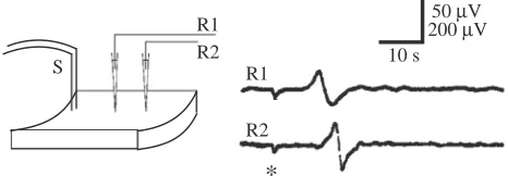
In previous experiments (Leys and Mackie, 1997), measurements of impulse conduction velocity were obtained with a single recording electrode on the assumption that the impulse initiation time was negligible and that the time taken for the impulse to arrive at the recording site was entirely taken up in conduction. To check this, we measured the conduction delay at two recording sites placed at different distances in a linear arrangement with the stimulating electrode (Fig. 2). Impulses were found to propagate at the same velocity across all parts of the array, showing that the impulse was initiated without significant delay following a shock and quickly reached its maximum velocity. The conduction velocity was measured at 0.27±0.1 cm s−1(mean ±S.D., N=10). This is close to the value
of 0.26±0.07 cm s−1given previously for spread of water current
arrests as measured with a flow meter (Mackie et al., 1983).
Absolute and relative refractory periods
When two shocks were given less than 30 s apart, the second
S
R1 R2
R1
R2
10 s 50 µV 200 µV
[image:3.609.324.557.578.659.2]*
Fig. 2. Conduction time: an impulse initiated at the stimulating site (S) propagates past recording electrodes (R1, R2). Stimulating and recording electrodes were arranged in a linear fashion as shown; S to R1, 3.45 cm; S to R2, 5.00 cm. The impulse crossed the 1.55 cm distance between R1 and R2 in approximately 5.5 s; calculated conduction velocity approximately 0.28 cm s−1. Asterisks in this and
shock usually failed to evoke a propagated impulse regardless of shock strength (Fig. 3). The absolute refractory period in several experiments was 29 s. With a 30 s inter-shock interval, the response to the second shock showed a lower amplitude and a reduced conduction velocity (<0.2 cm s−1). In an experiment in
which conduction times were measured simultaneously at two recording sites, 36 mm and 50 mm respectively, from the stimulating site, the conduction time for the second of the two shocks decreased progressively with increasing inter-shock intervals above 30 s. Fig. 4 shows that the recovery of the action potential conduction velocity at the two sites can be fitted with a simple exponential with a time constant of 22 s. The conduction velocity can be seen to reach control levels after approximately 150 s, which was therefore the relative refractory period.
Correlation of impulses and arrests
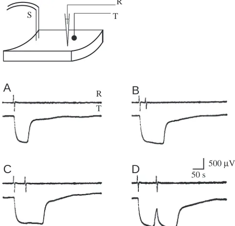
In a preparation where a flow meter probe and a recording suction electrode were placed side by side equidistant from the stimulating site (Fig. 5), changes in flow rate could be directly correlated with impulse arrival. It is clear that arrest of the water current ensues immediately upon arrival of an impulse and that a single impulse is sufficient to produce complete arrest of water flow (Fig. 5A). A second impulse arriving 33 s after the first (Fig. 5B) or 60 s after it (Fig. 5C) prolonged the state of arrest. When a second impulse arrived 90 s after the first, after pumping had started again, a second, more prolonged arrest occurred (Fig. 5D). These recordings show an important feature of the preparation: the independence of the conduction system and the effector system. Impulses were exhibited during periods when the current was completely arrested.
Sensitivity and spontaneity
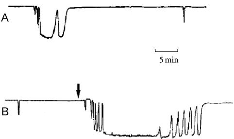
Although specimens of R. dawsoni sometimes stopped pumping without apparent cause (Fig. 6A), when maintained in clean flowing water they were generally found to pump continuously and for hours on end. We have confirmed their sensitivity to mechanical stimuli. Pinching or tapping the sponge itself or tapping the walls of the perfusion chamber set off arrests indistinguishable from those induced by electrical shocks. The sponges showed no sensitivity to changes in ambient light intensity and no evidence of diurnal rhythmicity, but were clearly sensitive to water temperature and quality. They stopped pumping and responding to stimuli when the water temperature
*
* *
200µV
[image:4.609.74.270.73.178.2]10 s 25 30
Fig. 3. Superimposed traces showing the effect of the refractory period. With two shocks 30 s apart, the second shock elicits an impulse with a reduced conduction velocity and amplitude (sponge shows relative refractoriness), but with shocks 25 s apart the preparation is absolutely refractory and no impulse is generated. The asterisk shows the shock artefact.
0 0.2 0.4 0.6 0.8 1 1.2
0 20 40 60 80 100 120 140 Inter-shock interval (s)
Normalised conduction velocity
Fig. 4. Effect of inter-shock interval on conduction velocity. In a preparation like the one shown in Fig. 2, the two recording electrodes were 3.6 cm (R1) and 5.0 cm (R2) from the stimulating site. Shock pairs with inter-shock intervals ranging from 30 to 120 s evoked pairs of propagated impulses recorded at each site. The conduction velocity of the slower second impulse was expressed as a proportion of that of the first. The values plotted are calculated for each recording site (filled circles, R1; open circles, R2) against inter-shock interval. The dotted line represents the exponential time course of a recovery process with a time constant of 22 s. The relative refractoriness lasted approximately 150 s.
T R
50 s 500 µV
D
C
A
B
S
R
[image:4.609.315.557.432.665.2]T
[image:4.609.43.291.459.614.2]rose above 12 °C, recalling the report of Annandale (1907) of a pumping ‘siesta’ in Spongilla sp. Increasing the amount of suspended particles in the incurrent water was found to evoke arrests. In the experiment shown in Fig. 6B, the sponge stopped pumping briefly following the addition of particles, started to pump again before water flow had completely stopped, stopped pumping again four more times for increasingly long periods and finally ceased pumping for 30 min. Return to the full pumping level was likewise accomplished in a series of tentative ‘attempts’ of progressively longer duration, as if the sponge were testing the water. These arrests resembled the responses to mechanical stimulation and were probably caused by contact between particles and the tissue of the sponge. The similarity between this response to particulates and the ‘spontaneous’ arrest sequence shown in Fig. 6A suggests that the latter may also have been evoked by chance entry of particulates with the sea water flowing through the preparation chamber.
A second effect of particulates in the water is to reduce the flow rate during normal pumping. In one set of tests, three sponges approximately 15 cm long had flow rates of 0.85, 0.85 and 1.19 cm s−1prior to addition of sedimentary particles. After
addition of sediment, the sponge stopped pumping, then started again and achieved a steady level of flow, but the flow rates were now 0.76, 0.5 and 0.96 cm s−1, respectively, a mean
reduction of 32 %.
Ionic basis of the action potential Effect of changes in external Na+concentration
When the superfusate consisted of 50 % normal sea water and 50 % Na+-free artificial sea water, action potentials continued
to be propagated, albeit with a slight increase in the time to peak of the externally recorded signal (see Fig. 7A, top trace). There was virtually no change in amplitude. Upon return to normal sea water, there was full recovery of the time to peak. The similar change that occurred when the Na+content was reduced
to 25 % (Fig. 7A, bottom trace) was associated with a small reduction in amplitude. However, in this case, recovery of the
action potential amplitude was incomplete even after prolonged washing. In one experiment, the normal sea water in the bath was exchanged over a period of 8 min with an artificial bathing solution containing dextrose in place of sodium chloride. The preparation continued to generate spikes in the dextrose-containing sea water but was temporarily blocked when the superfusate was returned to normal [Na+]. Spikes finally
recovered when the dextrose solution had been fully exchanged for sea water once more (i.e. after 8 min of perfusion).
Effect of divalent cations: Co2+, Mn2+, Ni2+
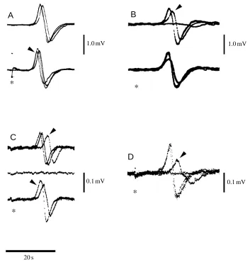
Although sea water containing 1 mmol l−1Co2+had no effect
on spike propagation, an increase in the superfusate Co2+
concentration to 2.5 mmol l−1 reduced the amplitude of the
externally recorded action potential by 33 % after 23 min. Upon washing with sea water, the action potential was restored to 95 % of its control amplitude within 14 min. Exposure to 5 mmol l−1 Co2+ reduced the amplitude by 45 % over 8 min,
with full recovery after a 13 min wash (Fig. 7B, bottom trace). The action potential was fully blocked after 8.6 min of exposure to 10 mmol l−1Co2+(Fig. 7B, top trace). Recovery in
normal sea water followed an approximately exponential time course with a time constant of approximately 40 min. Of the other divalent ions tested, 10 or 1 mmol l−1Mn2+blocked the
spike fully, whereas 2 mmol l−1Ni2+was without effect.
Effect of Ca2+channel antagonists
Superfusion of verapamil for 17 min to a final concentration of approximately 100µmol l−1 had no effect on either spike
propagation or the amplitude of the externally recorded action potential. Similarly, 1µmol l−1 nicardipine (pH 8) had no
effect. However 12µmol l−1nimodipine did extend the time to
peak, and when the concentration was increased to 24µmol l−1,
it blocked propagation; this was followed by a full recovery after 8–9 min superfusion with normal sea water (Fig. 7C).
Effect of ω-Conotoxin
The preparation was first preconditioned by perfusion with 0.01 % bovine serum albumin and then ω-Conotoxin MVIIA was added directly to the bath to make a final concentration of 5×10⁻⁷mol l−1. There was no effect on the spike at this concentration (results not shown).
Effect of TEA+
Low concentrations of tetraethylammonium ions (TEA+) were very effective at blocking spike propagation. Action potentials were blocked after 10 min superfusion with 1–5 mmol l−1TEA+(Fig. 7D). The effect was fully reversible after superfusion with normal sea water for 6–7 min.
Discussion The action potential
The sponge action potential elicited by electrical stimulation is an all-or-nothing propagating event that lasts approximately 30 s. Attempts at intracellular recording having been
A
B
[image:5.609.54.295.69.213.2]5 min
unsuccessful in the past, grafts were used as attachment points for suction pipettes. Amplification of the external signal using a differentiating circuit (time constant approximately 1 s) gave a triphasic waveform (Fig. 8A) with an amplitude that depended on the amount of sponge tissue drawn into the suction pipette and/or the extent to which the tissue had adhered to the pipette wall. Integration of this waveform revealed the diphasic external current associated with propagation (Fig. 8B). A further integration produced a
monophasic signal which, following standard cable theory, we take to be the waveform of the membrane action potential (Fig. 8C). The action potential duration was consistent with the 30 s absolute refractory period shown in Fig. 3.
Propagation in R. dawsoni although slow (0.27±0.1 cm s−1;
mean ± S.D., N=10) had features in common with the rapid
spread of excitation in nerve axons. Although supernormal responses (Adrian and Lucas, 1912; Bullock, 1951) have been reported in many nerve and muscle preparations, a more
10 30 50 10 30 50 -0.8
0 0 . 8
10 30 50
A
Time (s)
B
C
Action potential amplitude (mV)
Fig. 8. (A) The externally recorded sponge action potential amplified using a differentiating circuit with a time constant of approximately 1 s, i.e. approximately one-tenth of the recorded event. (B) Integrated version of A. (C) Integrated version of B. The vertical scale applies to record A only.
*
*
*
*
A
B
C
D
1.0 mV
0.1 mV 0.1 mV
20 s
[image:6.609.209.562.72.442.2]1.0 mV Fig. 7. Effect of different bathing solutions
on the externally recorded action potential. (A) Top: superimposed traces to show effect of a 50 % reduction in [Na+]. After a 10 min
perfusion, to allow control and test solutions to exchange, there was a delay in the time to peak but no change in amplitude. The time to peak recovered fully when the bath was returned to normal sea water (10 min of perfusion) so that the rising phase of the control traces were identical. Bottom: effect of saline containing a 75 % reduction in [Na+]; control and test traces shown
superimposed as above. After a 10 min perfusion, time to peak was delayed as above and spike amplitude was reduced. Recovery of spike amplitude was incomplete even after prolonged washing (arrowhead). In each case, low-[Na+] solutions contained
normal sea water and choline-chloride-containing saline in appropriate proportions. (B) Top: effect of sea water plus 10 mmol l−1
Co2+. Superimposed traces show that action
potential amplitude decreased progressively (arrowhead; 5 min) and was eventually blocked (8.6 min). Controls before and after test solution fully superimpose. Bottom: as top, but with 5 mmol l−1Co2+in the bathing
medium. (C) Effect of 12µmol l−1
nimodipine. Superimposed traces collected at 200 s intervals (top) until complete block (centre). Note the reduced conduction velocity prior to block (arrowhead).
Recovery (bottom traces) shown as superimposed traces 300 and 500 s after starting to perfuse with normal sea water. Amplitude recovered at 300 s, but time to peak remained delayed; after 500 s, amplitude increased and time to peak shortened (arrowhead). (D) Effect of 1 mmol l−1
tetraethyammonium ions (TEA+). Superimposed recordings show that the action potential was blocked after a 10 min perfusion with TEA+
[image:6.609.216.561.644.740.2]general feature in nerve axons is a reduction in conduction velocity during the relative refractory period (Gasser and Erlanger, 1925). Hodgkin and Huxley (1952) were the first to explain the refractory period in terms of inward current inactivation and a prolonged increase in K+conductance, but
did not attempt a correlation with changes in conduction velocity. More recently, ion accumulation within the tissue has been proposed as another possible factor (Bliss and Rosenberg, 1979).
In conducting epithelia, repetitive stimulation decreases conduction velocity in many species (Proboscidactyla
flavicirrata, Spencer, 1974, 1975; Stomotoca atra, Mackie,
1975; Polyorchis penicillatus, Spencer, 1978; Euphysa
japonica, Schwab and Josephson, 1982). In such multicellular
preparations, additional factors such as the delay at each cell junction, an increase in the number of refractory cells and/or a more tortuous conduction path may become significant (Shelton, 1975; Spencer, 1975). In Polyorchis penicillatus, the mechanism appears to be Ca2+-dependent (Grigoriev and
Spencer, 1996) but, as Schwab and Josephson (1982) point out, the field is hindered by the lack of a theoretical analysis of active propagation in two-dimensional sheets of tissue.
Whatever its basis in R. dawsoni, the effect of the relative refractory period is to reduce the conduction velocity just as it does in axons and epithelia. This means that the response to the second of two stimuli given 30 s apart (i.e. within the relative refractory period) will become progressively delayed the further the impulses travel from their initiation site. The conduction velocity recovers to its control level with a 22 s time constant (see Fig. 4), so that impulse separation will increase from 30 s to approximately 150 s. If each action potential arrests pumping for 70 s (as in Fig. 5), there will be a prolonged pause in the inward water flow near the stimulus site, but at greater distances pumping will be able to restart before the second action potential arrives.
The nature of the ionic currents
The extended period of absolute refractoriness following excitation suggests that the ion channels carrying inward current undergo a prolonged period of inactivation and that this could account for the repolarising phase of the action potential. Whether a slowly developing outward current also contributes is not clear. Although it delayed and blocked propagation, low levels of TEA+ did not affect the duration of the waveform
(Fig. 7D, arrowhead) as would be expected from K+channel
participation. One explanation for the block is that TEA+
reduced the resting K+conductance, depolarised the membrane
potential and inactivated the excitatory conductance.
We attempted to identify the excitatory conductance by testing the effect of different bathing solutions on impulse propagation. Reducing the Na+ composition to 50 % had no
effect on the impulse amplitude or on the rate of propagation as judged from the point of initial rise (Fig. 7A). A 75 % reduction in [Na+] did reduce the amplitude, but the effect was
small and permanent. Inward current density is one factor that determines the speed of propagation and, had Na+ made a
significant contribution, a delay in the appearance of action potentials at the recording site would be expected. We examined the effect of a Na+-free solution, but the results were
inconclusive because either of the possible outcomes may be plausibly explained. For example, persistence of the action potential might indicate the presence of a protected pool of Na+
within the sponge tissue; block may follow a secondary change in intracellular ion levels in the absence of Na+/Ca2+or Na+/H+
exchange. In the event, the signal persisted for longer than expected if Na+ had made an important contribution to the
action potential, but became blocked during the washing process.
A more clear-cut result was obtained using low concentrations of Co2+ or Mn2+. Propagation of the sponge
action potential was prevented by 10 mmol l−1 Co2+ or
1–10 mmol l−1 Mn2+, both known to block Ca2+ channels at
concentrations up to 20 mmol l−1 (Hagiwara and Takahashi,
1967); 2 mmol l−1Ni2+ was without effect. Co2+reduced the
amplitude of the signal in a dose-dependent fashion (range 1–10 mmol l−1) and also increased its time to peak. The
sensitivity of the sponge to nimodipine, a 1,4-dihydropyridine Ca2+ channel antagonist (Rampe and Triggle 1989), at a
relatively low concentration (12–24µmol l−1), provides further
support for a contribution from Ca2+influx because there was
a clear decrease in conduction velocity (Fig. 7C, arrowhead, top trace). However, the sponge was insensitive to verapamil (100µmol l−1) and nicardipine (1µmol l−1), and the possibility
remains that the divalent ions were blocking an H+current such
as that found in molluscan neurones (Thomas and Meech, 1982; Byerly et al., 1984). Such an explanation would also account for the blocking effect of TEA+ and would be
consistent with the findings of Nawata and Sibaoka (1979) on
Noctiluca miliaris (see below).
Co2+, Mn2+and Cd2+are also known to be effective blockers
of a tetrodotoxin (TTX)-resistant Na+ channel isoform found
in cardiac Purkinje fibres (DiFrancesco et al., 1985; Hanck and Sheets, 1992; Sheets and Hanck, 1992). A similar TTX-resistant, Cd2+-sensitive Na+channel has been recorded from
the epithelium (Grigoriev and Spencer, 1996) and motor neurones (Spafford et al., 1996; Grigoriev et al., 1996) of the hydrozoan jellyfish Polyorchis penicillatus and from the motor nerve net of the scyphomedusan jellyfish Cyanea capillata (Anderson, 1987). To exclude such channels as possible contributors to the action potential, the effect of Na+-free sea
water must be tested under conditions where intracellular management of ions such as Ca2+or H+is known to remain
uncompromised.
As a final caveat, we should note that it is as yet uncertain whether propagation in the sponge is entirely via current flow through local circuits, models that involve the diffusion of intracellular messengers not having been excluded. Nevertheless, the evidence presently available, i.e. the sensitivity of the sponge action potential to nimodipine and different divalent ions together with the virtual absence of an effect of Na+-deficient sea water, strongly suggests that
Mechanism of current arrest
Inhibition of flagellar beating, a process well-documented in both protozoa and invertebrates, has always seemed the most likely cause of water current arrest in R. dawsoni, but as it has still not been possible to observe the flagella themselves during arrests, other possibilities have to be considered. Seemingly normal arrests have been recorded after removal of both atrial and dermal pinacoderm layers, showing that closure of the pores in these membranes cannot be the primary mechanism involved (Mackie et al., 1983). Furthermore, visual observations of the pores in living sponges carried out during the course of the present study strongly suggest that the pores themselves are not contractile. Another possibility is that the prosopyles (perforations in the trabecular reticulum through which water enters the flagellated chambers) are contractile, but electron microscopy has failed to reveal any sphincter-like bundles of microfilaments in these structures. Persuasive evidence that current arrests are due to cessation of flagellar beating can be seen in the suddenness with which arrests stop and start and the fact that arrests are essentially all-or-none events, as shown in the flow meter traces in Fig. 5. In analysing these traces, allowance has to be made for the momentum of the moving water mass following cessation of pumping and for inertial resistance following its recommencement. In some specimens, the arrest is so brief and the inertia of the water mass so great, that pumping restarts before the flow can stop completely. In this case, a second arrest can bring about a further reduction in water flow (see Fig. 1D in Leys and Mackie, 1997).
A calculated arrest/recovery curve derived from velocity distributions for accelerating flow in a smooth-walled pipe, assuming rapid switching on and off of the pumping mechanism (as would be expected with flagella), conforms closely to the actual curves obtained from flow meter recordings (Mackie et al., 1983). If pumping stopped and started more gradually (as would be expected if it were controlled by cytoskeleton-based changes in pore diameter), the falling and rising slopes would be much less steep. The evidence therefore points to sudden, coordinated flagellar arrest as the primary or sole cause of cessation of pumping. The basic mechanism is presumably a simple on/off switch, such as that controlling the cilia in the ascidian branchial sac (Arkett, 1987), rather than a complex system of modulation such as that in lamellibranch gills, where the ciliated cells receive a dual innervation and beat at variable rates (Aiello, 1974). Even in the simple systems of ascidians and sponges, however, the repetition of all-or-none arrests at certain frequencies makes possible a de facto intermediate range of pumping rates.
While it would be premature to speculate about the ionic basis of flagellar arrest in R. dawsoni, the evidence that the action potential is Ca2+-dependent suggests that Ca2+channels
may also mediate the flagellar arrests. There is good evidence that changes in patterns of ciliary beating (arrests or reversals) are triggered by increases in intracellular Ca2+levels in ciliate
and flagellate protozoa (Naitoh and Eckert, 1974), in lamellibranch molluscs (Satir, 1975), in ascidians (Mackie et al., 1974) and in ctenophores (Moss and Tamm, 1987).
Nature of the conducting tissue
It is now generally agreed that sponges lack nerves (Pavans de Ceccatty, 1989), so we must look for a tissue that forms a continuous pathway through the whole sponge and is linked with the flagellated chambers where the effector response takes place. Early structural studies (Ijima, 1904) showed that the only tissue distributed in this manner is the trabecular reticulum. Reiswig (1979b) confirmed earlier reports that this tissue is a syncytium, ‘a thin, flat, layer of homogeneous cytoplasm, 0.4µm in thickness continuous over distances which can be surveyed by light and electron microscopy suggesting that this system may constitute a single, continuous, cytoplasmic network throughout the entire specimen’. There can be little doubt that this is the tissue responsible for impulse conduction. We estimate that the trabecular reticulum constitutes 75 % of the organic matter in the sponge. In R.
dawsoni, its syncytial character has been confirmed by electron
microscopy (Mackie and Singla, 1983a), in vitro studies (Leys and Mackie, 1994; Leys, 1995, 1997) and observation of the movement of ingested particles in sandwich cultures (Wyeth et al., 1996). It is continuous with the pinacoderm on both atrial and dermal sides and its processes enter the flagellated chambers, where they are intimately associated with the flagellated tissue. We envisage impulses generated in the pinacodermal or trabecular portions of the syncytium propagating to all parts of the sponge unimpeded by internal membrane barriers. The syncytium is linked to the choanoderm by cytoplasmic bridges containing dense material (‘perforate septa’ or ‘plugs’), but the latter are not membrane barriers and therefore need not block the forward flow of action currents. Impulses would therefore enter the choanoderm and cause flagellar arrest, presumably by altering the balance of ions in the cytoplasm. In this scenario, the trabecular reticulum would continue to conduct impulses even when the flagella were fully arrested.
There is no evidence for specialized sensory structures associated with the trabecular reticulum. Presumably, mechano-electric transduction is a property of the whole system. In this regard, the system resembles the excitable epithelia of hydromedusae and amphibian tadpoles (Anderson, 1980).
Functional significance of the response
of their filtration systems. Crabs and other interlopers walking on or near a sponge tend to dislodge clouds of sediment, so current arrest in response to mechanical stimulation would seem adaptive. As shown here for R. dawsoni, arrests also occur when particulates are added to the incurrent water. The return to full pumping occurs gradually through a series of brief starts and stops, as the sponge ‘tests’ the water. Assuming that current arrest protects the sponge from taking in unsuitable water, there would be obvious advantages in an early warning system of this sort, because it would enable distant parts of the sponge to stop pumping before the noxious water impinged on them. The limited arrest duration brought about at distant sites by the relative refractory period (see above) raises the possibility that water pumped from a remote site may help to clear particulate matter from non-pumping regions by generating an outward flow through normally ‘incurrent’ pore cells.
Uniqueness of hexactinellids
The question of whether electrical signalling occurs in other members of the Porifera is unresolved. Calcareous sponges and demosponges are cellular organisms, lacking any tissue comparable with the hexactinellid trabecular syncytium. To conduct electrical signals from cell to cell, they would require low-resistance pathways equivalent to gap junctions or plant plasmatodesmata, and it is not clear that they possess such features. Electron microscopy shows close membrane appositions, but connexon-like units have not been resolved despite lanthanum staining (Green and Bergquist, 1982). There is one report of electrical coupling in aggregated sponge cells (Loewenstein, 1967), but the experiments were performed under rather artificial conditions and it is doubtful whether the findings can be extrapolated to cover normal sponge tissue. Slow contractile responses have been described in several species (for a review, see Mackie, 1979), but the cells responsible do not seem to be electrically excitable, and there is no evidence of associated changes in transmembrane potential (Prosser et al., 1962). An ability to arrest the feeding current is found in some tropical demosponges (Reiswig, 1971). In some of these, contraction of the exhalant water channels appears to be responsible, while in others, cessation of flagellar beating is thought to accompany contractile changes, but in no case has it been demonstrated that the response is spread by a conduction system rather than simply representing summed local responses. Only in Hymedesmia sp. have rapid (<1 s latency) responses been documented. When touched, the sponge apparently immediately drops flaps that are usually raised up over the ostial fields (inhalant openings) (Reiswig, 1979a). What distinguishes R. dawsoni is that a purely local stimulus, such as plucking a single spicule projecting from the outer surface, can send an electrical signal throughout the entire sponge that causes an effector response in all parts. Nothing comparable has been reported in other groups of sponges.
Electrogenesis and architecture
Recent molecular data (Schütze et al., 1999) suggest that the metazoa are a monophyletic group, that they include the sponges, and that the hexactinellids are the most ancient sponges and therefore the most ancient metazoans. Along with other multicellular organisms (fungi, plants), the metazoans trace their origin to unicellular eukaryotes. This encourages comparisons between electrogenesis in sponges and protozoa. Multiple forms of Ca2+-selective ion channel provide the basis
for electrogenesis in Paramecium tetraurelia (Oertel et al., 1977) and Stylonychia mytilus (Deitmer, 1984), while the action potential in the heliozoan Actinocoryne contractilis is Na+-dependent (Febvre-Chevalier et al., 1986). In
Paramecium, the action potentials are not truly all-or-none
because their amplitude depends on the size of the stimulating pulse. Simulations of such graded spikes using the NEURON Simulation Environment (Hines and Carnevale, 1997) suggest that membranes with these characteristics do not produce propagating action potentials when introduced into an axon-like architecture. In Actinocoryne contractilis, simultaneous intracellular recordings show that the Na+-dependent action
potential travels rapidly from the 50µm diameter head to the flattened base but, as yet, it is not clear whether it propagates along the 250µm connecting stalk in an all-or-none fashion (Febvre-Chevalier et al., 1986).
The best example of spike propagation in the protozoa is in the dinoflagellate Noctiluca miliaris, which ranges in diameter from 300 to 600µm and has two functionally distinct excitable systems. In one, electrically elicited impulses propagate around the surface of the sphere (6 cm s−1; Eckert, 1965a,b) and trigger
luminescence. The system can be treated as a thin sheet of electrogenic tissue, albeit linked to a central mass of cytoplasm by a network of fine cytoplasmic threads. In two-dimensional structures of this kind, a high current density is necessary for spike propagation because of the large increase in membrane area with distance from the initiation site. In Noctiluca, a potentially toxic increase in [H+] brought about by such a large
ionic current density is likely to be managed by the high-capacity pH buffers normally present in cell cytoplasm. However, in other examples of this type of architecture, such as epithelial conduction in the cnidarians Hippopodius hippopus, Chelophyes
appendiculata and Polyorchis penicillatus, Na+is established as
the basis of the action potential. Just as with fast axonal conduction (Hille, 1992) a lower toxicity may confer some advantage on a Na+-channel-based system
through a set of interconnecting one-dimensional cables, and an apparently slow conduction velocity at the sponge surface might reflect the circuitous route taken by some contributing elements. If the branch length was short compared with its space constant, the system should behave like a three-dimensional mass. In such a case, the voltage decline from a point source of current would be even steeper than in the two-dimensional sheet (Jack et al., 1975) because of the even larger increase in membrane area with distance from the current source. In R. dawsoni, the low conduction velocity reduces the maximum current needed to raise this membrane area to threshold and minimises the demands on intracellular Ca2+
management. With the evolution of tubular axons in the cnidarian nervous system, both Na+- and Ca2+-dependent
propagating signals appear (Mackie and Meech, 1985), although here too the Ca2+spike operates with a relatively low
current amplitude. A Ca2+-current-based solution is also
responsible for signalling in nematodes such as Caenorhabditis
elegans (Goodman et al., 1998) in which voltage-gated Na+
channel genes are apparently absent (Bargmann, 1998).
Non-nervous conduction in other multicellular organisms
The conduction system described here in a sponge shows many parallels with non-nervous pathways carrying action potentials in other multicellular organisms. With some notable exceptions, such as carnivorous plants like Drosera capensis,
Dionaea muscipula and Aldrovanda vesiculosa, these systems
mediate responses best explained in terms of protection or defence.
Electrical signalling in plants has been reviewed by Sibaoka (1966), Pickard (1973) and Simons (1992). In the ‘sensitive plant’ Mimosa pudica, for example, the folding of the leaflets following contact or injury is probably a protective response making the plant less vulnerable to phytophagous insects. Similarly, a caterpillar attacking a tomato leaf evokes a propagated action potential (Wildon et al., 1992; Stankovic´ and Davies, 1997) that travels to neighbouring leaves, triggering synthesis of proteinase inhibitors, which are toxic to caterpillars (Green and Ryan, 1972).
In animals (Mackie, 1970; Spencer, 1974; Anderson, 1980), excitable epithelia play an important part in defensive responses, for example in the protective involution or ‘crumpling’ of many hydromedusae and the escape swimming behaviour of certain siphonophores. These responses are sometimes accompanied by dramatic visual displays such as bioluminescence or loss of transparency. In salps, excitable epithelia are part of a relay system transmitting changes in the direction and speed of escape locomotion along the chain of blastozooids, enabling the colony to respond as a single unit. In amphibian tadpoles, the excitable skin gives the tadpole the ability to respond to touch with escape behaviour prior to the outgrowth of the sensory innervation. In general, excitable epithelia serve as extensions of neurosensory fields and as ways of spreading motor excitation to effectors where there is no need for each effector cell to be independently innervated. The responses tend to be of a simple, overall kind, involving
synchronous activation of effectors over a wide area, as in R.
dawsoni.
Non-nervous conduction in plants involves low-resistance pathways (plasmatodesmata) between the phloem cells, comparable with the gap junctions coupling cells in excitable epithelia and muscles in animals. Syncytiality (the sponge solution) is rarely employed. The ionic mechanisms underlying impulse propagation vary widely. In animals, the action potentials are primarily Na+-dependent events, but Ca2+
-dependent action potentials are found in some higher plants (Iijima and Sibaoka,1985; Hodick and Sievers, 1986), as in R.
dawsoni. Again, as in R. dawsoni, conduction velocities are
often very slow: 0.1–0.4 cm s−1in tomato seedlings (Wildon et
al., 1992), 1.0–3.5 cm s−1 in colonial hydroids (Josephson,
1961) and 1.5–2.0 cm s−1 in colonial tunicates (Mackie and
Singla, 1983b, 1987).
Should non-nervous conducting tissues in animals be seen as antiquated systems, significant only as relics of pre-nervous evolution? The fact that they co-exist with nerves in many animals today makes this seem unlikely. We take the view that they have arisen repeatedly in evolution and for good reasons, serving efficiently as adjuncts or alternatives to nerves rather than being merely the vestiges of an earlier creation. At the same time, R. dawsoni shows that electrical impulse conduction is feasible in an animal lacking a nervous system and may therefore have characterized some of the very earliest lines of metazoa to evolve.
S.P.L. and G.O.M. gratefully acknowledge the support of the Natural Sciences and Engineering Research Council of Canada. R.W.M. thanks the Wellcome Trust for travel funds. We thank the staff of the Bamfield Marine Station, where this work was carried out, for providing excellent facilities. In particular, we thank Nikita Grigoriev and Andy Spencer for many useful discussions and Joelle Bélanger for much practical help. We also thank the anonymous referees for their comments and suggestions.
References
Adrian, E. D. and Lucas, K. (1912). On the summation of propagated disturbances in nerve and muscle. J. Physiol., Lond. 44, 68–124.
Aiello, E. L. (1974). Control of ciliary activity in Metazoa. In Cilia and Flagella (ed. M. A. Sleigh), pp. 353–376. London: Academic Press.
Allen, R. D., Metuzals, J., Tasaki, I., Brady, S. T. and Gilbert, S. P. (1982). Fast axonal transport in squid giant axon. Science 218, 1127–1129.
Anderson, P. A. V. (1980). Epithelial conduction: its properties and functions. Prog. Neurobiol. 15, 161–203.
Anderson, P. A. V. (1987). Properties and pharmacology of a TTX-insensitive Na+ current in neurones of the jellyfish Cyanea
capillata. J. Exp. Biol. 133, 231–248.
neurons in a urochordate (Ascidiacea). J. Comp. Physiol. A 161, 837–847.
Bargmann, C. I. (1998). Neurobiology of the Caenorhabditis elegans genome. Science 282, 2028–2033.
Bliss, T. V. P. and Rosenberg, M. E. (1979). Activity-dependent changes in conduction velocity in the olfactory nerve of the tortoise. Pflügers Arch. 381, 209–216.
Bullock, T. H. (1951). Facilitation of conduction rate in nerve fibres. J. Physiol., Lond. 114, 89–97.
Byerly, L., Meech, R. and Moody, W., Jr (1984). Rapidly activating hydrogen ion currents in perfused neurones of the snail, Lymnaea stagnalis. J. Physiol., Lond. 351, 199–216.
Deitmer, J. W. (1984). Evidence for two voltage-dependent calcium currents in the membrane of the ciliate Stylonychia. J. Physiol., Lond. 355, 137–159.
DiFrancesco, D., Ferroni, A., Visentin, S. and Zaza, A. (1985). Cadmium-induced blockade of the cardiac fast Na channels in calf Purkinje fibres. Proc. R. Soc. Lond. B 223, 475–484.
Eckert, R. (1965a). Bioelectric control of bioluminescence in the dinoflagellate Noctiluca. I. Specific nature of triggering events. Science 147, 1140–1142.
Eckert, R. (1965b). Bioelectric control of bioluminescence in the dinoflagellate Noctiluca. II. Asynchronous flash initiation by a propagated triggering potential. Science 147, 1142–1145. Febvre-Chevalier, C., Bilbaut, A., Bone, Q. and Febvre, J.
(1986). Sodium–calcium action potential associated with contraction in the heliozoan Actinocoryne contractilis. J. Exp. Biol. 122, 177–192.
Gasser, H. S. and Erlanger, J. (1925). The nature of conduction of an impulse in the relatively refractory period. Am. J. Physiol. 73, 613–635.
Goodman, M. B., Hall, D. H., Avery, L. and Lockery, S. R. (1998). Active currents regulate sensitivity and dynamic range in C. elegans neurons. Neuron 20, 763–772.
Green, C. R. and Bergquist, P. R. (1982). Phylogenetic relationships within the invertebrata in relation to the structure of septate junctions and the development of ‘occluding’ junctional types. J. Cell Sci. 53, 279–305.
Green, T. R. and Ryan, C. A. (1972). Wound-induced proteinase inhibitor in plant leaves: a possible defense mechanism against insects. Science 175, 776–777.
Grigoriev, N. and Spencer, A. N. (1996). A mechanism for fatigue of epithelial action potentials in the hydromedusa, Polyorchis penicillatus: a case of non-neuronal habituation. In Zooplankton: Sensory Ecology and Physiology (ed. P. H. Lenz, D. K. Hartline, J. E. Purcell and D. L. Macmillan), pp. 461–473. Amsterdam: Gordon and Breach.
Grigoriev, N. G., Spafford, J. D., Przysiezniak, J. and Spencer, A. N. (1996). A cardiac-like sodium current in motor neurons of a jellyfish. J. Neurophysiol. 76, 2240–2249.
Hagiwara, S. and Takahashi, K. (1967). Surface density of calcium ions and calcium spikes in the barnacle muscle fiber membrane. J. Gen. Physiol. 50, 583–601.
Hanck, D. A. and Sheets, M. F. (1992). Extracellular divalent and trivalent cation effects on sodium current kinetics in single canine cardiac Purkinje cells. J. Physiol., Lond. 454, 267–298.
Hille, B. (1992). Ionic Channels of Excitable Membranes. Sinauer Associates Inc., Sunderland, Mass.
Hines, M. L. and Carnevale, N. T. (1997). The NEURON simulation environment. Neural Comput. 9, 1179–1209.
Hodgkin, A. L. and Huxley, A. F. (1952). A quantitative description
of membrane current and its application to conduction and excitation in nerve. J. Physiol., Lond. 117, 500–544.
Hodick, D. and Sievers, A. (1986). The influence of Ca2+on the
action potential in mesophyll cells of Dionaea muscipula Ellis. Protoplasma 133, 83–84.
Iijima, T. and Sibaoka, T. (1985). Membrane potentials in excitable cells of Aldrovanda vesiculosa trap-lobes. Plant Cell Physiol. 26, 1–13.
Ijima, I. (1904). Studies on the Hexactinellida. Contribution IV. (Rossellidae). J. Coll. Sci. Imp. Univ. Tokyo 28, 13–307.
Jack, J. J. B., Noble, D. and Tsien, R. W. (1975). Electric Current Flow in Excitable Cells. Oxford: Clarendon Press.
Josephson, R. K. (1961). Colonial responses of hydroid polyps. J. Exp. Biol. 38, 559–577.
Lawn, I. D., Mackie, G. O. and Silver, G. (1981). Conduction system in a sponge. Science 211, 1169–1171.
Leys, S. P. (1995). Cytoskeletal architecture and organelle transport in giant syncytia formed by fusion of hexactinellid sponge tissues. Biol. Bull. 188, 241–254.
Leys, S. P. (1997). Sponge cell culture: a comparative evaluation of adhesion to a native tissue extract and other culture substrates. Tissue & Cell 29, 77–87.
Leys, S. P. and Lauzon, N. R. J. (1998). Hexactinellid sponge ecology: growth rates and seasonality in deep water sponges. J. Exp. Mar. Biol. Ecol. 230, 111–129.
Leys, S. P. and Mackie, G. O. (1994). Cytoplasmic streaming in the hexactinellid sponge Rhabdocalyptus dawsoni (Lambe, 1873). In Sponges in Time and Space. Proceedings of the Fourth International Porifera Congress (ed. R. W. M. van Soest, T. M. G. van Kempen and J.-C. Braekman), pp. 417–423. Rotterdam: Balkema.
Leys, S. P. and Mackie, G. O. (1997). Electrical recording from a glass sponge. Nature 387, 29–30.
Loewenstein, W. R. (1967). On the genesis of cellular communication. Devl. Biol. 15, 503–520.
MacGinitie, G. E. (1939). The method of feeding of tunicates. Biol. Bull. 77, 443–447.
Mackie, G. O. (1970). Neuroid conduction and the evolution of conducting tissues. Q. Rev. Biol. 45, 319–332.
Mackie, G. O. (1975). Neurobiology of Stomatoca. II Pacemakers and conduction pathways. J. Neurobiol. 6, 357–378.
Mackie, G. O. (1979). Is there a nervous system in sponges? Colloques Internationaux du C.N.R.S. 291 Biologie des Spongiaires (ed. C. Lèvi and N. Boury-Esnault), pp. 145–151. Mackie, G. O., Lawn, I. D. and Pavans de Ceccatty, M. (1983).
Studies on hexactinellid sponges. II. Excitability, conduction and coordination of responses in Rhabdocalyptus dawsoni (Lambe, 1873). Phil. Trans. R. Soc. Lond. B 301, 401–418.
Mackie, G. O. and Meech, R. W. (1985). Separate sodium and calcium spikes in the same axon. Nature 313, 791–793.
Mackie, G. O., Paul, D. H., Singla, C. L., Sleigh, M. A. and Williams, D. E. (1974). Branchial innervation and ciliary control in the ascidian Corella. Proc. R. Soc. Lond. B 187, 1–35. Mackie, G. O. and Singla, C. L. (1983a). Studies on hexactinellid
sponges. I. Histology of Rhabdocalyptus dawsoni (Lambe, 1873). Phil. Trans. R. Soc. Lond. B 301, 365–400.
Mackie, G. O. and Singla, C. L. (1983b). Coordination of compound ascidians by epithelial conduction in the colonial blood vessels. Biol. Bull. 165, 209–220.
Moss, A. G. and Tamm, S. L. (1987). A calcium regenerative potential controlling ciliary reversal is propagated along the length of ctenophore comb plates. Proc. Natl. Acad. Sci. USA 84, 6476–6480.
Naitoh, Y. and Eckert, R. (1974). The control of ciliary activity in protozoa. In Cilia and Flagella (ed. M. A. Sleigh), pp. 305–352. London: Academic Press.
Nawata, T. and Sibaoka, T. (1979). Coupling between action potential and bioluminescence in Noctiluca: effects of inorganic ions and pH in vacuolar sap. J. comp. Physiol. 134, 137–149. Nedergaard, M. (1994). Direct signaling from astrocytes to neurons
in cultures of mammalian brain cells. Science 263, 1768–1771. Oertel, D., Schein, S. J. and Kung, C. (1977). Separation of
membrane currents using a Paramecium mutant. Nature 268, 120–124.
Pavans de Ceccatty, M. (1989). Les éponges, à l’aube des communications cellulaires. Pour la Science 142, 64–72.
Pickard, B. G. (1973). Action potentials in higher plants. Bot. Rev. 39, 172–201.
Prosser, C. L., Nagai, T. and Nystrom, R. A. (1962). Oscular contractions in sponges. Comp. Biochem. Physiol. 6, 69–74. Rampe, D. and Triggle, D. J. (1989). New advances in molecular
pharmacology of Ca2+ channels. Trends Pharmac. Sci. 10,
388–389.
Reiswig, H. M. (1971). In situ pumping activities of tropical Demospongiae. Mar. Biol. 9, 38–50.
Reiswig, H. M. (1979a). A new sponge with rapid contraction systems. Programme Abstracts, Annual Meeting of the Canadian Society for Zoology, May, 1979, Laval University, Quebec, p. 83.
Reiswig, H. M. (1979b). Histology of Hexactinellida (Porifera). Colloques Internationaux du C.N.R.S., Paris 291 Biologie des Spongiaires (ed. C. Lèvi and N. Boury-Esnault), pp. 173–180. Satir, P. (1975). Ionophore-mediated calcium entry induces mussel
gill ciliary arrest. Science 190, 586–588.
Schütze, J., Krasko, A., Custodio, M. R., Efremova, S. M., Müller, I. M. and Müller, W. E. G. (1999). Evolutionary relationships of Metazoa within the eukaryotes based on molecular data from Porifera. Proc. R. Soc. Lond. B 266, 63–73.
Schwab, W. E. and Josephson, R. K. (1992). Lability of conduction
velocity during repetitive activation of an excitable epithelium. J. Exp. Biol. 98, 175–193.
Sheets, M. F. and Hanck, D. A. (1992). Mechanisms of extracellular divalent and trivalent cation block of the sodium current in canine cardiac Purkinje cells. J. Physiol., Lond. 454, 299–320.
Shelton, G. A. B. (1975). The transmission of impulses in the ectodermal slow conduction system of the sea anemone Calliactis parasitica (Couch). J. Exp. Biol. 62, 421–432.
Sibaoka, T. (1966). Action potentials in plant organs. Symp. Soc. Exp. Biol. 20, 49–74.
Simons, P. (1992). The Action Plant: Movement and Nervous Behaviour in Plants. Oxford: Blackwell.
Spafford, J. D., Grigoriev, N. G. and Spencer, A. N. (1996). Pharmacological properties of voltage-gated Na+currents in motor
neurones from a hydrozoan jellyfish Polyorchis penicillatus. J. Exp. Biol. 199, 941–948.
Spencer, A. N. (1974). Non-nervous conduction in invertebrates and embryos. Am. Zool. 14, 917–929.
Spencer, A. N. (1975). Behavior and electrical activity in the hydrozoan Proboscidactyla flavicirrata (Brandt). II. The medusa. Biol. Bull. 149, 236–250.
Spencer, A. N. (1978). Neurobiology of Polyorchis. I. The function of effector systems. J. Neurobiol. 9, 143–157.
Stankovic´, B. and Davies, E. (1997). Intercellular communication in plants: electrical stimulation of proteinase inhibitor gene expression in tomato. Planta 202, 402–406.
Takahashi, K., Baba, S. A. and Murakami, A. (1973). The ‘excitable’ cilia of the tunicate, Ciona intestinalis. J. Fac. Sci. Univ. Tokyo 13, 123–137.
Thomas, R. C. and Meech, R. W. (1982). Hydrogen ion current and intracellular pH in depolarised voltage-clamped snail neurones. Nature 299, 826–828.
Wildon, D. C., Thain, J. F., Minchin, P. E. H., Gubb, I. R., Reilly, A. J., Skipper, Y. D., Doherty, H. M., O’Donnell, P. J. and Bowles, D. J. (1992). Electrical signalling and systemic proteinase inhibitor induction in the wounded plant. Nature 360, 62–65. Wyeth, R. C., Leys, S. P. and Mackie, G. O. (1996). Use of