Arteriovenous Fistula
T. Terada, Y. Kinoshita, H. Yokote, M. Tsuura, Y. Tanaka, T. Itakura, Y. Ryujin, S. Hayashi, and J. Minamikawa
Summary: Seven dural arteriovenous fistulas were successfully embolized with mechanical detachable coils. Two lesions were located in the transverse-sigmoid sinus, four in the cavernous sinus, and one in the marginal sinus. All lesions were completely occluded on postembolization angiography. No recurrent symp-toms appeared during a mean follow-up period of 11 months. Owing to the length and retrievability of the mechanical detach-able coils, embolization was quicker and safer, and coils were packed more densely, than is possible with conventional coils. Coil migration was avoided because coils of the appropriate size were chosen before they were placed.
Index terms: Fistula, arteriovenous; Interventional instruments, coils
Microcoils are now commonly used for the treatment of intracranial aneurysms and dural arteriovenous fistulas (AVFs) (1). However, conventional microcoils lack retrievability, which sometimes results in coil migration or insufficient coil packing of the involved dural sinuses (2). Guglielmi and coworkers devel-oped an electrolytically detachable coil (the GDC) that is retrievable (3, 4). GDCs have made embolization of intracranial aneurysms safer, but these coils still have two drawbacks. One is the time required for coil detachment (up to 30 minutes [5]), and the other is the possibil-ity of embolic debris resulting from electrolytic coil detachment (Halbach VV, Dowd CF, Hi-gashida RT, et al, “Metallic Fragment Emboli Resulting from Treatment with Electrolytically Detachable Coils [GDC],” presented at the An-nual Meeting of the American Society of Neu-roradiology, Nashville, Tenn, 1994). Newly de-veloped mechanical detachable coils (MDCs) (Target Therapeutics, Fremont, Calif), which are investigational devices and not approved by the Food and Drug Administration, have several
advantages over conventional microcoils and GDCs: namely, retrievability, quick detach-ment, and no possibility of metallic embolic de-bris being created during detachment (6). How-ever, reports concerning the use of MDCs for transvenous embolization of dural AVFs are few (6). We present our experience with the use of MDCs for the embolization of seven dural AVFs.
Methods and Representative Cases
Embolization was performed with MDCs in seven pa-tients with dural AVFs. Five lesions were initially treated by transarterial embolization with polyvinyl alcohol particles (ITC, South San Francisco, Calif) before the transvenous coil embolization. Two dural AVFs were located in the transverse-sigmoid sinus, four in the cavernous sinus, and one in the marginal sinus. One transverse-sigmoid dural AVF had previously been embolized with conventional mi-crocoils (Hilal coils, Cook Inc, Bloomington, Ind), but this resulted in incomplete fistula embolization and migration of the one microcoil to the heart. The patient had repeat embolization with MDCs. A profile of each case is pre-sented in the Table.
All procedures were performed by means of fluoros-copy and digital road-mapping functions. Five AVFs were treated by transarterial embolization (before the trans-venous embolization) with polyvinyl alcohol particles, which ranged in diameter from 150 to 500 mm. Trans-femoral or transjugular approaches were used for trans-venous embolization. A Tracker-18 two-tip marker cathe-ter (Target Therapeutics, Fremont, Calif) was navigated into the affected sinus coaxially through a larger guiding catheter. Various sizes of MDCs, ranging from 2 mm34 cm to 8 mm 3 20 cm, were used for embolization. The retrieval function was examined for all MDCs before they were introduced into the catheter. A total of 113 MDCs were used in our seven cases. The range of applied coils was from 2 mm34 cm to 8 mm320 cm. The average number of coils used for each embolization was 16.
Received July 24, 1995; accepted after revision November 1.
From the Departments of Neurological Surgery at Wakayama Medical College, (T.T., Y.K., M.T., Y.T., H.Y., T.I.), Kokuritsu Minami Wakayama Hospital (Y.R., S.H.), and Kishiwada Municipal Hospital (J.M.), Wakayama City, Japan.
Address reprint requests to Tomoaki Terada, MD, Department of Neurological Surgery, Wakayama Medical College, 7 ban-cho 27, Wakayama City, 640 Japan.
AJNR 17:1343–1348, Aug 1996 0195-6108/96/1707–1343qAmerican Society of Neuroradiology
Coil embolization was finished when transarterial an-giography documented the disappearance of the AVF or when insertion of additional MDCs into the fistulous com-partment became impossible.
Case 1
A 65-year-old woman with a left transverse-sigmoid dural AVF with proximal left sigmoid sinus occlusion was admitted to our hospital for treatment (Fig 1A). The AVF was treated by transvenous embolization with conven-tional microcoils, but the coil migrated to the heart (Fig 1B). A pulmonary embolism occurred on the seventh day, after which the patient was given anticoagulants for 4 months. Subsequent transarterial embolization of the left occipital artery was attempted by using polyvinyl alcohol particles, which resulted in temporary improvement of the patient’s tinnitus. The second transvenous embolization was tried by using MDCs and resulted in a complete cure (Fig 1C and D).
Case 4
A 60-year-old woman with pulsatile tinnitus had an-giography that showed a left transverse-sigmoid dural AVF with left distal transverse sinus occlusion and stenosis of the distal left internal jugular vein (Fig 2A and B). Transar-terial embolization with polyvinyl alcohol particles (diam-eter range, 500 to 710mm) was performed to decrease the shunt flow for the left occipital artery, which is a main feeding artery and safely accessible. A 5F guiding catheter was introduced into the left internal jugular vein and a Tracker-18 microcatheter was introduced into the in-volved dural sinus segment. The fistulous portion was lo-cated at the transverse-sigmoid junction, forming a small venous pouch. This portion was embolized with MDCs that were 2 mm34 cm and 4 mm38 cm in size. An 8-mm3 20-cm MDC was inserted into the proximal sigmoid sinus before insertion of a 4-mm38-cm coil to prevent migra-tion of the smaller coil. Five 2-mm 3 4-cm coils, one 4-mm38-cm coil, and three 8-mm320-cm coils were
used, resulting in complete embolization of the dural AVF (Fig 2C and D).
Case 6
A 60-year-old woman with a carotid cavernous fistula presented with tinnitus. Bilateral periorbital and posterior auricular bruits were audible. Bilateral cavernous sinus fistulas were present at angiography (Fig 3A and B). A Tracker-18 two-tip marker catheter was navigated into the left cavernous sinus via the right transfemoral and trans-petrosal routes. Both cavernous sinuses were packed with variously sized MDCs from left to right. The fistula ap-peared occluded on angiograms obtained immediately af-ter embolization (Fig 3C and D). One 6-mm320-cm coil unraveled during the retrieval procedure, but was success-fully withdrawn.
Results
All dural AVFs were embolized successfully with MDCs. Two lesions were completely oc-cluded immediately after embolization. Com-plete occlusion of the remaining five AVFs was confirmed by follow-up angiography. Four MDCs unraveled during attempted coil retrieval. Three of these coils were easily retrieved, but one prematurely detached inside the catheter. A snare wire was necessary to retrieve this coil. The particulars for all cases are given in the Table.
Deliverability of MDCs
All MDCs were easily delivered into the fistu-lous portion of the affected dural sinuses. If the volume of coils was larger than the fistulous space, it was difficult to extrude the entire coil from the catheter. In such cases, coils were retrieved and exchanged for smaller ones. Seven patients treated with MDCs for treatment of dural AVFs
Patient Age, y/Sex Location
of AVF Embolization Route
No. of MDCs
used Complications
1 65/F L TS-SS Transarterial and transvenous 13 . . .
2 59/F L CS Transvenous 18 Unraveling and
premature detachment 3 63/M L CS Transarterial and transvenous 15 Unraveling 4 60/F L TS-SS Transarterial and transvenous 9 . . .
5 63/M L CS Transvenous 23 Unraveling
6 60/F Bilateral CS
Transarterial and transvenous 27 Unraveling
7 60/F R MS Transarterial and transvenous 8 . . .
Total 113
When coils were detached, the distal end (inter-locking portion) of the pusher sometimes hooked previously detached coils. In these in-stances, the pusher was easily retrieved by gen-tly pushing and rotating this device.
Coil Positioning
Each coil was positioned accurately to oblit-erate the involved dural sinus segment fully. Because of their retrievability, coils were repo-sitioned until they were in the optimal location. In cases of dural AVFs of the cavernous sinus, venous drainage channels—including the sphe-noparietal sinus, the ophthalmic vein, and the entire cavernous sinus—were packed with MDCs. These procedures were finished more quickly than those involving conventional mi-crocoils, owing to the longer length of the MDCs.
Coil Stability
None of the MDCs changed their position, and all demonstrated good stability at the fistu-lous sites. In two cases of transverse-sigmoid dural AVFs, MDCs of an appropriate size were first partially deployed and then examined for stability. We judged a coil to be stable if its diameter was slightly larger than the sinus and it did not move after a few minutes’ observation before detachment. If a coil was stable in an appropriate portion of the fistula, it was de-tached. Larger coils served as a nest for smaller MDCs, allowing denser coil packing of the in-volved dural sinus.
Complications
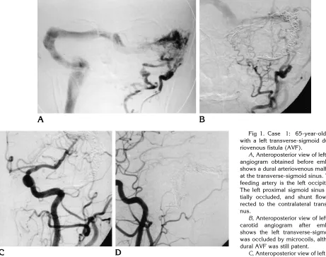
[image:3.612.64.525.84.448.2]Four MDCs unraveled during repeated inser-tion and retrieval. Three of them were easily Fig 1. Case 1: 65-year-old woman with a left transverse-sigmoid dural arte-riovenous fistula (AVF).
A, Anteroposterior view of left occipital angiogram obtained before embolization shows a dural arteriovenous malformation at the transverse-sigmoid sinus. The main feeding artery is the left occipital artery. The left proximal sigmoid sinus was par-tially occluded, and shunt flow was di-rected to the contralateral transverse si-nus.
B, Anteroposterior view of left external carotid angiogram after embolization shows the left transverse-sigmoid sinus was occluded by microcoils, although the dural AVF was still patent.
C, Anteroposterior view of left common carotid angiogram after embolization with mechanical detachable coils (MDCs) show that the dural AVF has completely disappeared.
retrieved, but one detached inside the catheter. A snare wire was necessary to retrieve this pre-maturely detached coil.
Follow-up Results
The average follow-up period for the seven patients was 11 months (range, 5 to 15 months). Angiographic disappearance of an AVF was confirmed immediately after emboli-zation in two patients, 1 week after emboliemboli-zation in three patients, 7 months after embolization in one patient, and 5 months after embolization in one patient. No recurrence of symptoms was noted in any of these cases.
Discussion
The transvenous approach for the treatment of dural AVFs was described by Halbach et al (7); it is now accepted as the standard
treat-ment for these lesions. Coils are normally used for the occlusion of venous outflow, and micro-coils with or without fibers are used for the transvenous embolization of dural AVFs. Ordi-narily, these coils work well to occlude AVFs (7). However, one of the pitfalls of these devices is coil migration (2), especially in the case of high-flow dural AVFs, as in our case 1. Another pitfall is inaccurate coil positioning distant from the fistulous site. In this regard, MDCs have several advantages over conventional micro-coils (6), the most important being their retriev-ability. This feature allows accurate and tight coil packing with less chance of coil migration. Also, because of the longer length of MDCs, complete occlusion of a target venous space can be achieved more rapidly.
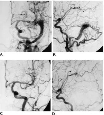
[image:4.612.226.562.81.453.2]Fibered platinum coils are believed to be more thrombogenic than bare platinum coils (5). And although MDCs are composed of bare platinum coils and may be less thrombogenic, Fig 2. Case 4: 60-year-old woman
with a left transverse-sigmoid dural arte-riovenous fistula (AVF).
A, Anteroposterior view of left com-mon carotid angiogram shows a left transverse-sigmoid dural AVF. The left distal transverse sinus is occluded and the distal internal jugular vein is ste-nosed.
B, Lateral view of left common carotid angiogram.
C, Anteroposterior view of left com-mon carotid angiogram after emboliza-tion shows that the dural AVF has com-pletely disappeared.
accurate and compact packing allows complete obliteration of fistulas, as demonstrated in our cases, which were all completely occluded after embolization.
As compared with GDCs, MDCs have both advantages and disadvantages. The advan-tages are their rapid detachment and lack of generation of metal emboli during detachment (Halbach VV et al, “Metallic Fragment Emboli”). The disadvantages are their possible premature detachment and unraveling during deployment (although the latter complication also occurs with GDCs). In our cases, unraveling of MDCs occurred when coils were already inserted into the sinus, when a coil was resistant to being introduced into the lesion, when a coil was re-sistant to retrieval, and when large coils (6 mm
3 20 cm to 8 mm 3 20 cm) were used. We
think that newly introduced coils may become tangled with other coils, making them difficult to retrieve and susceptible to unraveling. The
longer coils are more likely to become tangled and to unravel. If coils do unravel, fixation of their interlocking portion becomes unstable. Premature detachment is thought to happen when unraveled MDCs pass through a sharp vascular bend. It is important to change to shorter coils to prevent unraveling when coil insertion becomes difficult and always to be prepared for this eventuality. Another disadvan-tage of MDCs is the hard interlocking joint lo-cated at the end of the coil that may potentially injure a vessel wall during coil detachment. This is a major drawback in the use of MDCs for the treatment of cerebral aneurysms.
[image:5.612.63.395.82.461.2]We treated seven cases of dural AVFs using only MDCs except for one case previously em-bolized with Hilal coils. For cases of carotid cavernous fistulas, MDCs do not seem to be more effective than conventional microcoils, except for reducing the duration of the emboli-zation procedure by virtue of the longer length Fig 3. Case 6: 60-year-old woman with a carotid cavernous fistula.
A, Anteroposterior view of left exter-nal carotid angiogram obtained before embolization shows the left cavernous si-nus is opacified in the arterial phase.
B, Anteroposterior view of right exter-nal carotid angiogram before emboliza-tion shows the right cavernous sinus is opacified in the arterial phase.
C, Anteroposterior view of left com-mon carotid angiogram after emboliza-tion shows the carotid cavernous fistula has completely disappeared.
of the MDCs. However, for cases of high-flow fistulas with large sinus spaces, such as dural AVFs of the transverse-sigmoid or marginal si-nus, MDCs, because of their retrievability, were effective for stabilizing the initial coil and for ascertaining correct coil size.
References
1. Hodes JE, Aymard A, Gobin YP, et al. Endovascular occlusion of intracranial vessels for curative treatment of unclippable aneu-rysms: report of 16 cases.J Neurosurg1992;75:694 –701 2. Numaguchi Y, Pevsner PH, Rigamonti D, Ragheb J. Platinum coil
treatment of complex aneurysms of the vertebrobasilar circulation.
Neuroradiology1992;34:252–255
3. Guglielmi G, Vinuela F, Duckwiler G, et al. Endovascular treatment of posterior circulation aneurysms by electrothrombosis using elec-trically detachable coils.J Neurosurg1992;77:515–524
4. Sadato A, Taki W, Nishi S, Yamashita K, Kikuchi H, Ikada Y. Treatment of spontaneous carotid cavernous fistula using an elec-trodetachable microcoil.AJNR Am J Neuroradiol1993;14:334 – 336
5. Graves VB, Strother CM, Rappe AH. Treatment of experimental canine carotid aneurysms with platinum coils.AJNR Am J Neuro-radiol1993;14:787–793
6. Yoshimura S, Hashimoto N, Kazekawa K, Nishi S, Sampei K. Em-bolization of dural arteriovenous fistulas with interlocking detach-able coils.AJNR Am J Neuroradiol1995;16:322–324