Neurosonographic Abnormalities Associated with Maternal History
of Cocaine Use in Neonates of Appropriate
Size for
Their Gestational Age
Vikram Singh Dogra, Jaye M. Shyken, P. A. Menon, Jesse Poblete, Dina Lewis, and James S. Smeltzer
PURPOSE: To determine whether increased incidence of neurosonographic abnormalities (pre-dominantly of the basal ganglia and thalamus) in cocaine-exposed neonates who are small for their gestational age is attributable to the cocaine or to neonatal size. METHODS: Neonates whose sizes were appropriate for their gestational age with no evidence of hypoxia or respiratory distress were identified prospectively by a maternal history of cocaine use. Scans were performed within 72 hours of birth using a 7.5-MHz transducer following a standard protocol. The images were analyzed without access to patient information. Forty study neonates were compared with 34 control subjects who were appropriate in size for their gestational age, scanned using the same
protocol. Comparisons were made using Fisher Exact Test, t test, and logistic regression.
RESULTS: No control infant had neurosonographic abnormalities. In the study group, gestational age ranged from 27 to 41 weeks. Of the 40 study neonates, 14 (35%) had one neurosonographic abnormality; two had two abnormalities. The predominant lesion was focal echolucencies, mainly in the area of the basal ganglia (10 of 40, 25%). Other findings were caudate echogenicity (3 of
40, 7.5%), ventricular dilation (2 of 40, 5%) and one "moth-eaten" appearance of the thalamus.
Lesions were more likely approaching term and were not related to prematurity or alcohol use. CONCLUSION: Apparently normal neonates with a maternal history of cocaine use are likely to have degenerative changes or focal infarctions in their basal ganglia attributable to cocaine.
Neurosonography should be used to evaluate these neonates. The long-term significance of these
lesions needs further evaluation.
Index terms: Infants, newborn; Ultrasound, in infants and children; Cocaine
AJNR Am J Neuroradio/15:697-702, Apr 1994
We (Dogra VS et al, Abnormal Neurosono-graphic Findings Associated with Maternal His-tory of Cocaine Use in Appropriate-for-Gesta-tional-Age Neonates, presented at the 31st An -nual Meeting of the American Society of Neuro-radiology, Vancouver, 1993) and others ( 1, 2) have found an increased incidence of
echolucen-Received February 24, 1993; accepted pending revision April 26;
revision received July 22.
Presented at the annual meeting of the American Society of Neuro-radiology, Vancouver, BC, May 1993.
From the Departments of Radiology (V.S.D., J.P.), Neonatology (P.A.M., D.L.), and Obstetrics and Gynecology (J.M.S., J.S.S.), St Louis Regional Medical Center, and Department of Obstetrics and Gynecology (J.M.S., J.S.S.), Washington University, StLouis.
Address reprint requests to Vikram S. Dogra, MD, Department of Radiology, StLouis Regional Medical Center, 5535 Delmar Blvd, StLouis, MO 63112.
AJNR 15:697-702, Apr 1994 0195-6108/94/1504-0697 © American Society of Neuroradiology
697
cies of the basal ganglia in neonates who are small for their gestational age and have been exposed to cocaine. In 1988 about 11% of the US population were regular cocaine users. About 6% of these used cocaine during pregnancy (3). Cocaine use during pregnancy adversely affects the development of the fetus (4). Are the echol-ucencies observed in neonates who are small for their gestational age attributable to cocaine use or to size of the neonates? To resolve this ques-tion we studied the neurosonographic abnormal-ities found in neonates of size appropriate for their gestational age with a maternal history of cocaine use.
Materials and Methods
698 DOGRA AJNR: 15, April1994
TABLE I: Abnormal neurosonographic findings associated with maternal history of cocaine use in neonates appropriate for their gestational age
Cocaine metabolites Significance versus
Control Group Category
Absent
Number Normal Abnormal
Focal echolucencies Caudate echogenicity Ventricular dilation "Moth-eaten" appearance of
thalamus
Note: P value by Fisher Exact Test. • Two had two abnormalities.
10 6 4 2
2
0
no evidence of hypoxia or respiratory distress at birth, who were born in our institution to women with a history of cocaine use.
Gestational ages were assigned using the New Ballard Score, a maturational assessment of the gestational age based on neuromuscular and physical maturity (5), ex-panded to include extremely premature infants. Size appro-priate for gestation age was defined as weight, head circum -ference, and length all within the 1Oth to 90th percentile range using Lubchenco norms (6). Neonatal urine toxicol-ogy by immunoassay was performed for the metabolites of cocaine, marijuana, opiates, and phencyclidine within 48 hours of birth. All positive specimens were confirmed by thin layer chromatography (7). Umbilical arterial and ve-nous pH and pC02 were normal at birth. Apgar score at
five minutes was seven or greater.
Real-time sonography was performed within 72 hours of birth, with a 7 .5-MHz sector transducer, through the anterior fontanelle at a constant scan depth of 100 mm and constant gain settings. Six coronal (two anterior, two mid, and two posterior), two sagittal, and 10 parasagittal views (five each to right and left) were recorded.
The maternal history of alcohol use was recorded. All neonates in the study group had prenatal care with a minimum of six visits. The urine results were not known to the neurosonographer. The images were analyzed with-out access to information on patient status. The studies were interpreted twice by the same observers and scored as positive when the second interpretation concurred with the first. This study group was compared with a control group of neonates of size appropriate for their gestational age who had no maternal history of cocaine use who otherwise met study criteria and who had negative urine
screening for cocaine metabolites, using Fisher Exact Test,
logistic regression, and unpaired t tests.
Results
In the study group of 42, two were excluded.
One was not of a size appropriate for gestational
age on review, and one had no urine toxicology
results. The remaining 40 of the study group
Total Present
30 20 10
8 2 0
40 26
14' P<.OOI
10 P< .002
3 P< .24 2 Not significant
Not significant
were compared with the 34 control subjects ap-propriate for gestational age whose urine
toxicol-ogy for cocaine metabolites returned negative.
Infants included in the study ranged from 27 weeks to 41 weeks of age. In the study group,
mean head circumference was 32. 1
±
2. 1 em(mean
±
SD, range 24.5 to 37 em); meangesta-tional age was 37.6
±
3 weeks (range 27 to 41weeks), and mean birth weight was 2643
±
594g (range 940 to 3780 g). Seventy-five percent (30
of 40) of the subjects were positive for cocaine metabolites, and a positive history of alcohol use was obtained from the mothers of 19 of the 40
subjects. None of the 34 control subjects was
positive for maternal history of cocaine or cocaine
metabolites by design. No other drugs tested were
detected in either group.
No control infant had a neurosonographic
ab-normality. Thirty-five percent of the study group
(14 of 40, P
<
.001) had neurosonographicab-normalities. Neurosonographic findings are
sum-marized in Table 1. Twenty-five percent of the study group (10 of 40, P < .01) had focal echo-lucencies measuring 2 to 4 mm in size and with-out evidence of hemorrhage. These were often multiple and distributed in the caudate, periven-tricular (inferolateral to the body of the lateral ventricle), and choroidal regions of the brain (Figs 1A and 18).
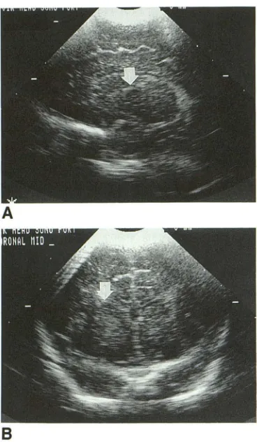
Three of 40 in the study group (7%, P = .23) had caudate echogenicity in the area of the head of the caudate nucleus. This echogenicity did not
change in echo texture on three follow-up scans,
[image:2.612.119.486.101.220.2]hypoechogenici-AJNR: 15, April 1994
A
8
A 8
ties and hyperechogenicities with blurred margins (Figs 3A and 38), a "moth-eaten" appearance.
In the study group of 40 neonates of size appropriate for their gestational age, 11 were preterm and 29 were term infants. The mean age of the neonates with normal scans was less than the mean age of those whose scans were abnor-mal (P
<
.05). A neurosonographic abnormality was more likely at term. Distribution of neuroson-ographic abnormalities in relation to gestational age is given in Table 2. The likelihood of a lesion was somewhat reduced with prematurity (P =.15) and unrelated to history of alcohol use (P =
.81 ), but these two were positively related to each other (9 of 11 preterm subjects versus 1 0 of 19 at term had alcohol history, P< .06, Fisher Exact Test).
Within the study group who all had a history of cocaine use, neurosonographic abnormalities were not associated with cocaine metabolites in the urine (P = .71).
Discussion
Cocaine is the most potent naturally occurring central nervous system stimulant, sharing many pharmacologic and adverse effects with synthetic stimulants such as amphetamines and
phenme-NEUROSONOGRAPHIC ABNORMALITIES 699
Fig. 1. A, Left parasagittal section sh ow-ing a focal echolucency in the region of the caudate (arrowhead).
B, Coronal section corresponding to A.
Fig. 2. A, Right parasagittal section showing echogenicity of the caudate (ar -row).
B, Coronal section corresponding to A (arrowhead pointing to caudate).
trazine (8). More than 100 000 babies born in the United States annually are believed to have been exposed to cocaine or other drugs during the critical period of fetal brain development (9). The immediate perinatal effects can be abruptio pla-centae, prematurity, microcephaly, and symmet-rical growth retardation.
Of potentially greater long-term significance are the neurodevelopmental problems of cogni-tive, emotional, and social development found in infants exposed to cocaine in utero (1 0), which become more apparent later in infancy (11). To identify the structural effects of cocaine that are attributable directly to cocaine exposure and not to its indirect effects on fetal growth we examined a cohort of infants who were of size appropriate for their gestational age with cocaine exposure but without perinatal and neonatal complications. This baseline structural data will be helpful in correlating neurodevelopmental abnormalities observed with structural lesions of the brain.
[image:3.612.54.410.78.373.2]700 DOGRA
A
8
Fig. 3. A, Parasagittal section showing thalamus with multiple small hypoechogenicities and hyperechogenicities with blurred margins (moth-eaten appearance, arrow).
8, Coronal section corresponding to A.
TABLE 2: Distribution of neurosonographic lesions in relation to gestational age
Category
Number Abnormal
Focal echolucencies Caudate echogenicity Ventricular dilation "Moth-eaten" appearance
of thalamus
Term (>37 weeks) Preterm (<37 weeks)
29
12'
9 2
11 2'
1 0
' One in each group with two abnormalities.
our series are consistent with those shown by others in primates and humans in association with various forms of asphyxia (12-16). There is one case of moth-eaten appearance of the thala-mus in our present series and three cases in our study of infants who are small for their gestational age. The normal thalamus has medium echoes; normal variation could have given this appear-ance, despite the control of technique, depth, and gain. We sought histopathologic correlation of this observation by reviewing 157 neonatal au-topsies done at our center in the past 5 years and
AJNR: 15, April 1994
identified nine reports mentioning a maternal his-tory of cocaine use. One of these reported a specific thalamic lesion on autopsy. It showed aggregates of microcalcification, neuronal degen-eration, and edema (Fig 4). Microcalcifications were not present in other parts of the brain. Ultrasound findings before death were reported to suggest thalamic hemorrhage. The moth-eaten sonographic appearance may correspond to these pathologic changes. The thalamus has the second highest uptake of cocaine in the brain (17) and a high metabolic rate, which may make this structure more susceptible to the actions of cocaine.
Focal echolucencies were the predominant findings in the study group. It is important to distinguish these echolucencies from other cystic lesions in the brain. To differentiate them from periventricular leukomalacia and porencephalic cysts we have called them focal echo/ucencies. Periventricular leukomalacia and the focal echo-lucencies we observed are parts of the spectrum of hypoxic-ischemic encephalopathy but are es-sentially different from each other. Periventricular leukomalacia has been mainly described in rela-tion to immaturity and acute perinatal distress ( 18) and refers to predominant involvement of white matter. Various morphologic forms of it have been described ( 19). In our study the echo-lucencies and echogenicities were not related to prematurity and were mainly of the gray matter of the caudate nucleus and area of the basal ganglia. None of the neonates had evidence of intrapartum asphyxia, as measured by umbilical pC02 and Apgar score. The location of lesions observed corresponds to the patterns of status
[image:4.612.86.270.74.390.2] [image:4.612.52.296.475.568.2] [image:4.612.313.557.535.700.2]AJNR: 15, April 1994
marmoratus associated with chronic partial an-oxia as shown by Myers et al (20-22) in primates and Volpe in humans (23). This could be related to the binge pattern of cocaine use. Porencephalic cysts are associated with intraparenchymal hem-orrhage; we found no evidence of hemorrhage in any of these lesions.
The particular distribution of cerebrovascular lesions observed in our study seems to be related to the state of maturation of the cerebral vessels and the resulting responsiveness to placentally transferred cocaine (2). Furthermore, these
le-sions correspond to the patterns of 11C-labeled
cocaine uptake shown by Fowler et al in a posi-tron emission tomography study of healthy adult volunteers (17), which was maximal in the corpus
striatum, then the thalamus. The
neurosono-graphic abnormalities found probably represent ischemic changes secondary to cocaine-induced vasospasm or hypoxic changes from maternal placental vasoconstriction.
The neurosonographic appearance of 26 neo-nates with a maternal history of cocaine use is normal either because of low sensitivity of this technique in detecting subtle lesions or, more
likely, because of variations in fetal response,
dosage, duration, maternal hemodynamic effects, or timing of exposure to cocaine. The lack of difference in lesions found between those with and those without cocaine metabolites within the study group indicates these lesions antedate de-livery by an uncertain time period. The urine cocaine metabolite results in neonates are posi-tive only in cases with maternal cocaine use within approximately 7 days before delivery (7).
Neurosonographic lesions have been reported
by Dixon and Bejar in 28 cocaine-exposed infants.
Unlike our subjects, 39% of their study group showed hemorrhagic cerebral infarcts by
ultra-sound at birth (1). This study confirms the
echo-lucencies and ventricular dilation found in theirs, but we did not find the ventricular hemorrhages they reported, and our findings of caudate echo-genicity and moth-eaten appearance of the thal-amus, to our knowledge, are new. Our selection criteria for this appropriate-for-gestational-age se-ries, based on maternal history of cocaine use, differs from the small-for-gestational-age series (Dogra VS et al, Abnormal Neurosonographic Findings Associated with Maternal History of Co-caine Use in Appropriate-for-Gestational-Age Neonates, presented at the 31st Annual Meeting
of the American Society of Neuroradiology,
Van-couver, May 1993), which was based on cocaine
NEUROSONOGRAPHIC ABNORMALITIES 701
metabolite detection in the urine. The findings
are qualitatively identical but lower in incidence.
Schellinger et al have shown that echoencephal-ography is more sensitive than computed tomog-raphy (CT) for detection of small cystic lesions in
neonates (24). The one infant in our study eval
-uated by CT confirms this. The echolucency shown in Figures 1 A and 1 B was not seen on CT scan.
Of two magnetic resonance (MR) studies of
infants exposed to cocaine, that of Link et al (25)
and that of Bandstra et al (Abnormal Neuroson-ographic Findings Associated with Maternal His-tory of Cocaine Use in Appropriate-for-Gesta-tional-Age Neonates, presented at the 31st An-nual Meeting of the American Society of Neuroradiology, Vancouver, May 1993), neither found lesions in the brain with cocaine exposure. The first study had a small number of subjects (only nine with MR), and the median age of the neonate at MR was 3.6 months. It is likely that small cystic lesions could have healed by gliosis and would be impossible to detect at this age. The MR study of Bandstra et al showed an in-crease of the subarachnoid space in six of 21 neonates with cocaine exposure and reported no other central nervous system abnormality. We did not find a case of enlarged subarachnoid
space except two with ventricular dilation. The
enlargement of the subarachnoid space makes us wonder what structures have been diminished to create this space. Ultrasound is not the modality
of choice to visualize the subarachnoid space, so
we may have missed this finding. A prospective contemporaneous study of MR and neurosonog-raphy in the same cocaine-exposed neonates will be helpful in explaining these differences.
A frequent problem of studies attempting to identify the effects of cocaine is the potential
confounding with effects of malnutrition, alcohol,
and other drugs. In this series infants were in the appropriate growth range by standard norms (as a part of selection criteria). It is impossible to control strictly for malnutrition, because cocaine affects appetite. Control for maternal history of alcohol use did not alter the results. No other
drugs were used by history or drug screening.
Limitations of the present study are the lack of
evaluation by other modalities, lack of a
702 DOGRA
Strengths of the study are the clear uniform selection criteria, consecutive examination of an entire cohort of infants meeting these criteria,
standardized examination technique applied by a
single sonographer in all cases, and review and
confirmation of all positive and negative findings
using a uniform classification system based on prior experience. This permits an estimation of the true incidence of lesions in this population. Neurosonography is the most operator dependent of all the neuroimaging modalities. We believe that correct identification of neonatal brain lesions requires standardization of sonographic technique used (depth and gain settings) and images ob-tained. This will prevent variability of settings and
anatomy shown from interfering with
sana-graphic interpretations.
Neurosonographic abnormalities were ob-served in 35% of the neonates of size appropriate for their gestational age in the control group with maternal history of cocaine use. The most com-mon lesion was focal echolucency in the area of the basal ganglia, followed by caudate
echogen-icity, ventricular dilation, and thalamic
heterogen-icity. These lesions were more likely in term
neonates (P
<
.05). They probably represent small infarctions of the structures involved, but the clinical significance of these findings needs to be defined. These findings will be helpful in study-ing the neuroanatomic correlates in the follow-up of cocaine-exposed infants. Apparently normal neonates of size appropriate for their gestational age born to mothers with a history of cocaine use have a high incidence of neurosonographicab-normalities attributable to cocaine. This study
justifies routine neurosonographic evaluation of neonates with cocaine exposure.
References
I. Dixon SD, Bejar R. Echoencephalographic findings in neonates
as-sociated with maternal cocaine and methamphetamine use: Incidence
and clinical correlates. J Pediatr 1989; II :770-778
2. Volpe JJ. Effects of cocaine use on the fetus. N Eng/ J Med
1992;327:339-407
3. Lindberg CS, Alexander EM, Gendrop SC, Nencioli M, Williams DG. A review of the literature on cocaine abuse in pregnancy. Nurs Res 1991;40:69-75
4. Hadeed AJ, Siegel SR. Maternal cocaine use during pregnancy: effect on the newborn infant. Pediatrics 1989;84:205-210
AJNR: 15, April 1994
5. Ballard JL, Khoury JC, Wedig K, Wang L, Elerswalsman BL, Lipp R.
New Ballard Score, expanded to include extremely premature infants.
J Pediatr 1991; 119:417-423
6. Lubchenco LO, Hansman C, Boyd E. Intrauterine growth in length and head circumference as estimated from live birth at gestational
ages from 26 to 42 weeks. Pediatrics 1966;37:403-408
7. Ostrea EM Jr, Welch RA. Detection of prenatal drug exposure in the pregnant woman and her new born infant. C/in Perinatal
1991;18:629-645
8. Bejerot N. A comparison of effects of cocaine and synthetic central
stimulants. Br J Addict 1970;75:35-37
9. Chasnoff IJ. Drug use and woman: establishing a standard of care.
Ann NY A cad Sci 1989;562:208-21 0
10. Rodning C, Beckwith L, Howard J. Prenatal exposure to drugs:
behavioral distortions reflecting CNS impairment? Neurotoxico/ogy
1989; 10:629-634
II. Coles C, Platzman K, Smith I, James M, Falek A. Effects of cocaine, alcohol and other drugs used in pregnancy on neonatal growth and
neurobehavioral status. Nurotoxico/ Terato/1991;13:1-11
12. Myers RE. Four patterns of perinatal brain damage and their
condi-tions of occurrence in primates. Adv Neural 1975; I 0:223-234
13. Fulling KH. Hypoxic ischemic encephalopathy in term infants. Diag-nosis and prognosis evaluated by ultrasound. Radiology
1984; 152:395-399
14. Kreusser KL, Schmidt RE, Shackelford GD, Volpe JJ. Value of
ultrasound for identification of acute hemorrhagic necrosis of
thala-mus and basal ganglia in an asphyxiated term infant. Ann Neural
1984;16:361-363
15. Shen EY, Huang CC, Chyou SC, Huang HY, Hsu CH, Huang FY.
Sonographic findings of the bright thalamus. Arch Dis Child
1986;61: I 096-1099
16. Colamaria V, Curatolo P, Cusmai R, Bernardina BD. Symmetrical
bithalamic hyper densities in asphyxiated full-term newborns: an
early indicator of status marmoratus. Brain Dev 1988;10:57-59
17. Fowler JS, Volkow ND, Wolf AP, et al. Mapping cocaine binding ~tes in human and baboon brains in vivo. Synapse 1989;4:371-377 18. Grant EG. Neurosonography: periventricular leukomalacia. In:
Text-book of neurosonography of the pre-term neonate. New York: Sprin-ger Verlag, 1986:69-84
19. Leech RW, Alvord EC Jr. Morphologic variations in periventricular leukomalacia. Am J Patho/1974;74:591-602
20. Myers RE. Two patterns of perinatal brain damage and their
condi-tions of occurrence. Am J Obstet Gyneco/1972; 112:246-276 21. Myers RE, Beard R, Adamsons K. Brain swelling in the newborn
rhesus monkey following prolonged partial asphyxia. Neurology
1969; 19: I 012-1018
22. Seltzer ME, Myers RE, Holstein SF. Prolonged partial asphyxia: effects of fetal brain water and electrolytes. Neurology 1972;22:732-737
23. Volpe JJ. Neurology of the newborn. Philadelphia: Saunders,
1987:215-217
24. Schellinger D, Grant EG, Richardson JD. Cystic periventricular
leu-komalacia: sonographic and CT findings. AJNR Am J Neuroradio/
1984;5:439-445