Cerebral Palsy: MR Findings
in
40 Patients
Charles L. Truwit, 1'2 A. James Barkovich,2'3 Thomas K. Koch,3 and Donna M. Ferriero3.4
Purpose:, We used MR to retrospectively analyze the brains of patients suffering from cerebral
palsy, our aim being to determine MR's role in the assessment of brain damage and the relationship of pre-, peri-, and post-natal events to cerebral palsy. Methods: Forty patients (aged 1 month to 41 years) underwent MR scanning and findings were correlated with clinical histories in all cases.
Results: Review of MR scans of 11 patients who had been born prematurely revealed findings of
periventricular white matter damage, indicative of hypoxic-ischemic brain injury (82%), the
chronology of which was difficult to determine. Among 29 patiel")ts who had been born at term,
three major patterns emerged: (1), gyral anomalies, suggestive of polymicrogyria, consistent with
mid-second trim'ester injury; (2), isolated periventricular leukomalacia reflecting late second- or
early third-trimester injury; and (3), watershed cortical or deep gray nuclear damage, consistent
with late third-trimester, perinatal or postnatal injury. In 16 (55%) of 29 patients born at term, MR findings of intrauterine brain damage were observed; in over half of these cases MR revealed developmental anomalies, which is nearly twice the rate reported in prior studies employing CT.
Conclusion: Our results support a growing consensus that cerebral palsy in term infants is often
the result of prenatal factors, and less commonly related to the perinatal period.
Index term: Cerebral palsy
AJNR 13:67-78, January /February 1992
Cerebral palsy (CP) has been the subject of numerous clinical, neuropathologic, and neuro-radiologic studies to assess the relationship of
pre-, peri-, and postnatal events to CP (1-9).
Clinical studies have been limited by the availa-bility of historical information regarding prenatal care and possible prenatal insults. Neuropatho-logic studies have been limited by the small
Received February 20, 1991; accepted and revision requested April
15, revision received May 22; final acceptance May 29.
The views expressed in this article are those of the authors and do not reflect the official policy or position of the Department of the Army, the Department of Defense, or the United States Government.
1
U.S. Army Medical Department, Academy of Health Sciences,
HSHA-GPS Fort Sam Houston, TX 78234-6100. Address reprint requests to C.L.
Truwit, Department of Radiology, Fitzsimons Army Medical Center,
Au-rora, CO 80045-5000. 2
Department of Radiology, Neuroradiology Section, University of
California, San Francisco, 505 Parnassus Ave., Box 0628, San Francisco,
CA 94143-0628.
3 Department of Pediatric Neurology, University of California, San
Francisco, San Francisco, CA 94143. 4
Department of Pediatric Neurology, San Francisco General Hospital, San Francisco, CA 94110.
AJNR 13:67-78, Jan/Feb 1992 0195-6108/92/1301-0067 © American Society of Neuroradiology
67
numbers of cases that have gone to autopsy. Neuroradiologic studies have been limited in their
usefulness, primarily because of the limited
ca-pabilities of neuroimaging in the era before
com-puted tomography (CT) and magnetic resonance
(MR) imaging. With the development of CT,
ra-diographic correlation with clinical data regarding
prenatal, perinatal, and postnatal asphyxia be-came possible, particularly with respect to mor-phologic changes of the cerebral white matter (1,
7-1 0). The sensitivity of CT, however, is limited
in many cases of cerebral palsy. In particular,
while state-of-the-art CT is sensitive to dramatic
anomalies of brain development, such as
schizen-cephaly, MR is more sensitive than CT in the
detection of both subtle brain malformations,
such as callosal hypogenesis, polymicrogyria, and
mild degrees of white matter damage. Therefore,
we studied MR images to retrospectively analyze
the brains of patients with cerebral palsy.
Materials and Methods
Forty patients, aged 1 month to 41 years, underwent
68
strength. Three patients were initially scanned at less than 1 year of age. They are now 4 years, 6 months (case 15),
3 years, 4 months (case 24) and 2 years, 6 months (case 27) and have spastic, hypotonic and spastic CP, respec-tively. As the study was undertaken at a major referral
center, the study population was limited by selecting only those patients with the referral diagnosis of CP, and only those for whom MR studies were included. The MR studies were retrospectively reviewed by two of the authors with knowledge of the preceding clinical diagnosis of CP, but without knowledge of the specific clinical manifestations
of each case. Thus, the MR review was only partially
blinded, since the diagnosis of CP was known. Nevertheless,
because we set out to assess what abnormalities appeared
on MR scans of patients with CP, not the efficacy of MR in diagnosing CP, we believe this methodology was appropri-ate.
All MR studies included sagittal and axial T1-weighted
images, 400-800/20-35/1-2 (TR/TE/excitations), and
ax-ial T2-weighted images, 2000-3000/30-120/1-2. The MR
studies were evaluated for developmental or acquired
ab-normalities of the corpus callosum, deep and peripheral white matter, cerebral cortex, basal ganglia/thalami, and brainstem, and for the presence of parenchymal cysts, hydrocephalus, and ventricular contour defects. The corpus
callosum, brainstem, cerebral cortex, and basal ganglia/
thalami were subjectively assessed as thinned or normal. As the radiologic manifestations of preterm and term inju-ries differ, patients born prematurely were grouped se pa-rately.
Classification of cerebral anomalies was based on typical MR findings. Polymicrogyria was diagnosed by the
pres-ence of abnormally thick cortex, diminished and absent arborization of subjacent white matter, and mild ventricu-lomegaly beneath the anomalous parenchyma ( 11, 12).
Hypoplasia of the ipsilateral thalamus, cerebral peduncle,
and pons was commonly associated. The cortical mantle
typically demonstrated multiple sulci, in contradistinction to the broad, shallow gyri of pachygyria, and in
contradis-tinction to the mushroom-shaped, small gyri of ulegyria.
We recognize that this definition is broader than the strict pathologic definition, which requires an abnormal four-layered cortex. Nevertheless, although it is possible that
the MR findings reflect another, unspecified migration
anomaly, in our experience, this MR pattern has repeatedly
correlated with pathologic specimens of polymicrogyria.
The diagnosis of schizencephaly required MR definition
of an abnormal gray matter cleft extending all the way to
the lateral ventricle. A cleft that reached toward, but that
was clearly separated from, the ventricle was defined as a cleft of polymicrogyria (13). Although others have consid -ered this to be type I schizencephaly (14), we consider the
presence of white matter between the gray matter cleft
and the lateral ventricle to distinguish the two entities, since
a pial-ependymal seam, the hallmark of schinzencephaly,
is impossible in this situation.
MR findings were correlated with clinical histories, which
were available for all patients. In one patient (case 4), no
birth history was available other than delivery at 28 weeks
AJNR: 13, January /February 1992
gestation. Specifically, pre-, peri- and postnatal histories were assessed. Eleven patients (27%) were born prema-turely, by the criterion of a gestational age (by dates) of 36 weeks or less at the time of birth. Twenty-nine patients (73%) were delivered at or near term. Although included in Table 1, we did not segregate the patients by type of CP (e.g., diplegic, quadriplegic), as we commonly encountered clinical discrepancies, such as one clinician labeling the patient diplegic only to find the next clinician labeling the same patient quadriplegic or hemiplegic.
For purposes of this study, we defined the perinatal period as that time starting 2 weeks before birth and continuing through the first 2 weeks of life. Perinatal asphyxia was defined to include fetal distress and respira-tory distress (endotracheal oxygenation, meconium aspi-ration, arrest, or near-arrest). We did not try to separate the types and degrees of perinatal asphyxia. Periventricular leukomalacia (PVL), isolated periventricular white matter damage without cortical, subcortical, or deep gray matter lesions, was considered to be representative of brain dam-age that occurred prior to the 36th week of gestation. This concept is supported by recent reports on the MR assess-ment of perinatal asphyxia (15, 16). As a result, isolated PVL in the term infant was considered to be consequent to intrauterine brain injury, even in the face of perinatal insults.
Results
As seen in Table 1, the MR scans revealed
readily apparent abnormalities in every patient except three patients born at term. One of these patients (case 16) had choreoathetoid CP; her mother had experienced third-trimester pre eclampsia. Another patient (case 22) had spasti-city and was cortically blind. The third patient (case 29) underwent meconium aspiration and had spasticity.
MR Findings in Patients Born Prematurely
Among the 11 patients born prematurely, sev-eral radiographic features were apparent. MR scans in all 11 revealed deep white matter loss, especially in the peritrigonal regions. In nine of the 11, scans demonstrated thinning of the cor-pus callosum, especially in the posterior body
(Fig. 1 ). In case 8, the corpus callosum was
severely hypogenetic. The one patient with a normal corpus callosum (case 5) had hypotonic CP and had diminished deep, and to a lesser
extent peripheral, white matter. The ventricular
trigones were slightly rounded. All had abnormal
ventricles, usually manifested as irregular contour
of the trigones, consistent with prior incorpora-tion of peri ventricular cysts. One patient (case 9) had discrete periventricular cysts. In three
TABLE 1: Clinical and MR Findings in Patients with Cerebral Palsy G r oup/ Ges tat io n a l Age Case Age at MR Sex at Bi r th : Peri-, Dominant Clinica l Co rpu s Ca ll osum Deep White Matter P eri p heral Cys t s Lat era l Ven tricl es Cortex Basal Brai nste m Other Find ings Postnatal Findings White Matter Ganglia No. Hi story Born prematurely I 9 yr, 8 mo M 27 wk: Grade 4 Spastic di pl egia Dif f u se l y thinn ed, D i mini s h e d, especia ll y Normal No Irr eg ular co nt o ur , espec iall y No rmal No rmal N o rmal intraventricular especia ll y pos-p eri trigonal midb ody and trigones h emorr h age, t e r io rl y sh unt , RDS 2 I yr, 6 mo F 28 wk: Grade 3 Spastic diplegia Thinned p oster i or Dimin i shed peritr i gonal & Normal No Dilat ed trig o n es No rmal No rmal Nor mal i nt raven t ricu l ar body long T 2 hemorrhage, RDS 3 I yr, 2 mo M 28 wk: RDS, Spas t ic it y Diffusely t hinn ed, Diminished p e ritri go n a l N or mal No Minimally irregular co nt o ur No rm al No rmal Small Cerebe llar hem is pher ic i ntrave ntr icu l ar poste ri or > a n -hypoplasia vs atro-hemorrhage terior ph y 4 41 y r F 28 wk Spastic it y, blindness Thinned body Diminished & l o n g T 2 Norma l No Ab n orma l contour No rm al No rm a l No rmal 5 2 y r , 6 mo F 30 wk: S u rviving H ypoton i c, athetoi d , No rm a l Diminished peritrigonal & S li ghtly di-No S li g htl y r ounded tr igo n es Norma l No rmal No rmal twin; endot r achea l paraparesis occipita l mini shed intubatio n for 2 wk 6 7 yr, 5 mo M 30 wk: Severe RDS, Spast i c it y, LE Thinned pos t e ri o r Diminished peritrigonal Scatte r ed foci No Irregular contou r poste-No rm al No rmal No rmal i ntr aventricu l a r body & splen-of pro-r i o rl y hemorrhage , s hun t ium longed T2 7 2 yr, 8 mo F 30-31 wk: 5 days' Spast i c diplegia Th i nn ed p oste ri or Diminished perit r igonal, Normal No D il ated trigones & occipital Normal No rmal No rmal endot r acheal i ntu -body & s pl en-long T 2 h o rn s w ith i r regular co n-bation , ductus li ga-ium t our tion 8 10 yr M 32 wk: NSVD, hy-Spastic d i p l eg i a, Severe h ypo-Markedly diminished Mildy dimin-No Markedly irregular co n tour Normal Norma l No rmal Chiasmatic hypoplasia drocephalu s n oted me nt al retarda-genesis ished pos- thick-at birth; s hunt at 3 tion teriorly ness, wk
dis- torted 20 sh
TABLE 1: Clinical and MR Findi n gs in Patients with Ce r eb r al Palsy-Continued Group / Gestational Age Case Age at MR Sex at Birth: Peri-, Dominant Clinica l Corpus Callosum Deep Wh ite Matter Peripheral Cysts Lateral Ventricles Cortex Basal Brainstem Other Findi ngs No. P os tnatal Finding s White M atter Ga ngli a H isto r y 25 3 yr, 6 mo M T e rm : Normal until Spasticity , mental Thinned posterior Very delayed myel i nat ion Ve r y delayed No Bu l bous f ro ntal tips & Norma l N or m al S ma ll 6 m o retardati on, b ody & s pl en -myel ina-trigones developmenta l ium ti o n delay 26 2 yr, 1 mo F T erm : Apgars , 9 / 9 Spastic dipl egia, S lightl y thinned Delayed myelination D e l ayed m ye-No Nor mal Normal N o rmal Normal truncalataxia , ver-diffusely, but lination ; tical n ys tagmu s, n or mal f or de-s li g htly L side l ayed m yelina-prolonged tion T 2, peritri-go nal 27 1 0 m o M T e rm : Spotting since Spas ti c d iplegia, L Thinn ed s pl e n -L o ng T1 & T2 , trig o nal , Dimin is hed No Enlarg ed temporal & occip-Diffuse Small R D iff us e l y fir s t month , p re-hemiparesis ium temporal , occ ipit a l on R, ita! h orns, trigones poly-thalamus small , R mature r upture of bilateral micro-side membranes >36 s ub co rti cal gy ri a, R hr , nu cha l X2, e n cep ha l a-frontal, home birth malacia , pa r ie t a l
temporal tip
-o
c:
"'
-.;
u
:
s
0
U.J ...J
a:l <:
I-"'
Ol
c '6 c
[i:
"'
.<:; 0
E
.!!l
"' c '§ til
~~
"' c til (J "'
)(
.!!l 0 u
"' "'
u
~
"'
>
"iii
:;;
.§
E iil .2
e
"'
:J a. 0 u
)( "' (fJ
"
"' Ol <"' §
"' > "iii
E
(fJ
"iii E
0
z:
"iii
E 0 z:
:J .9
c 8
"iii
E 0
..5
<
0 z:
"iii E
0
z:
E
~ ~
0 ·-" ,~ c
"' c a. "'
:_§lD
1-0 E
"'
"iii .<:; a. "' u
c "' c "'
-u
"' :r:
0 E
"iii E
0
z:
.: 8 "iii
E 0
c
.0
"'
~L-~ :J
"' 0
c ~
w
0 z:
0
E
..:
"'
"'
"iii
E
0
z:
c
8 "iii
E 0 c
.0 "'
1
~-
:J"' 0 c ~
w
0 z:
1JN
.<:
1-:~ g>
E .2
i5
"iii E
0
z:
0
>
(fJ z:
0
E
__J :c Ol ·;:
"'
I .. "'
.!!l E 0 0
z: ~
" c ~
gestation, peripheral white matter damage was
seen on MR. This is not typically observed prior
to 34 weeks of gestation (16). Two of these three
patients (cases 6 and 8) had been previously
shunted for hydrocephalus; it is possible that the
peripheral white matter damage was related to
the hydrocephalus and/or shunting (17). The
other patient was a twin, and it is possible that
the gestational age was underestimated.
No abnormalities were noted in the basal
gan-glia or thalami. Three patients (27%) (cases 10
and 11) had diminished caliber of the brainstem.
Three patients (27%) (cases 1, 6, and 8) had been
shunted for hydrocephalus. Developmental
anomalies were noted in one patient with severe hypogenesis of the corpus callosum and optic chiasmatic hypoplasia (case 8). There were no
apparent significant morphologic differences
between the spastic and hypotonic CP cases.
Clinical Findings in Patients Born Prematurely
Of the 11, nine (82%) had varying degrees of
spastic CP (cases 1-4, 6-9, and 11) and two
(18%) had hypotonic CP (cases 5 and 10). Nine
patients (82%) had perinatal asphyxia (cases
1-3 and 5-1 0). One patient (case 11) had elevated
serum titers of toxoplasmosis antibodies, as did
the mother, which suggested intrauterine insult.
MR Findings in Patients Born at Term
The MR results in the 29 patients born at term were not as uniform as in those in the subgroup of patients born prematurely. Nineteen patients
born at term (66%) had diminished deep white
matter (cases 13-15, 17-21, 28, 30-37, 39, and
40) (Figs. 2 and 3), seven of whom also had prolonged T2 relaxation of the white matter
(cases 18, 28, 31, 34, 36, 39, and 40) and six of
whom had overlying anomalies of neuronal
mi-gration (cases 21, 30, 32, 33, 35, and 40) (Fig.
4). Three patients had abnormal signal of the
white matter but normal white matter quantity: two had delayed myelination (cases 25 and 26)
and one had foci of T2 prolongation (case 27).
Only six of the 22 patients with abnormal white
matter had perinatal asphyxia (cases 18-19, 24,
27, 37, and 39), although one of the six (case 27)
had clinical and MR findings of both intrauterine damage (neuronal migration anomaly) and
peri-natal damage (premature rupture of membranes
and nuchal cord, abnormal peripheral white
mat-ter of the contralateral hemisphere) (Fig. 4). Five
of the 22 had findings typical of PVL, manifested
AJNR: 13, January/February 1992 73
A B
Fig. Periventricular leukomalacia in patient born prematurely (case 7).
A, Sagittal T1-weighted image (600/20/1) reveals thinning of posterior callosal body (arrows) where peritrigonal fibers cross. Band C, Axial proton-density- (2800/30/1) (B) and T2-weighted (2800/80/1) (C) images show diminished peritrigonal (deep) white
matter and spared peripheral white matter (C, arrows). Note rounded contour of ventricular trigones and adjacent gliosis (B, arrows).
A B
c
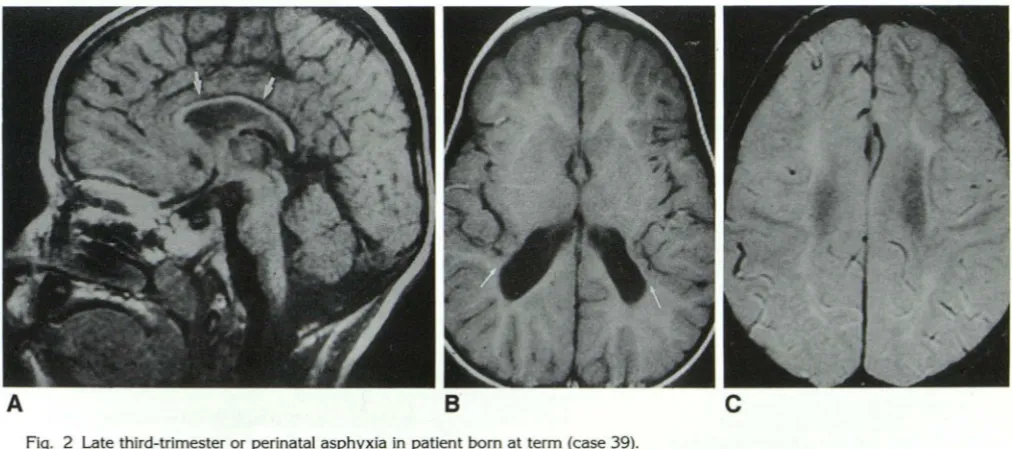
Fig. 2 Late third-trimester or perinatal asphyxia in patient born at term (case 39).
A, Sagittal T1-weighted image (600/20/1) shows diffuse callosal thinning (arrows).
B, Axial T1-weighted image (600/20/1).reveals apposition of posterior sylvian cortex to enlarged ventricular trigone, where marked
loss of peritrigonal white matter is apparent (arrows). Ventricle has abnormal contour where periventricular cysts have been incorporated.
C, Axial proton-density image (2800/30/1) at most cephalad portion of lateral ventricles shows markedly diminished deep and peripheral white matter with abnormal T2 prolongation consistent with gliosis. Relatively normal peripheral white matter is seen in frontal lobes.
white matter, and ventricular-parenchymal
inter-face. Of these five patients, one had clinical
evidence of intrauterine insult (case 36), one had
a two-vessel umbilical cord and perinatal as·
-phyxia (case 37), and two had unremarkable
prenatal and perinatal histories (cases 13 and 14).
The corpus callosum was abnormal in 16
pa-tients born at term (55%) (cases 13-15, 17, 21,
25-28, 30, 36, 37, and 39) (Figs. 2 and 3) and
[image:7.614.58.558.77.313.2] [image:7.614.55.561.368.592.2]74 AJNR: 13, January/February 1992
A
8
c
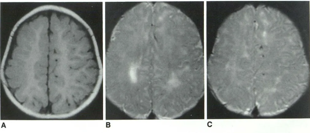
Fig. 3 Diffuse atrophy consistent with perinatal asphyxic event in patient born at term (case 20). A, Sagittal T1-weighted image (600/30/1) reveals diffuse callosal thinning and prominent CSF spaces.
B, Axial image shows marked atrophy. Thinned cortex is well seen in sylvian region (arrows). Ventricles are enlarged ex vacuo.
C, T2-weighted image (2800/80/1) confirms atrophy and reveals markedly diminished deep and peripheral white matter.
A
8
Fig. 4 Prenatal and perinatal insults in patient born at term (case 27).
A, Axial T1-weighted image reveals abnormal gyral pattern of right hemisphere and nearly complete absence of peripheral white matter on right. This image is typical of diffuse polymicrogyria, evidence of intrauterine insult. More caudal images (not shown) showed
small right thalamus and cerebral peduncle and underdeveloped sylvian fissure. These findings are consistent with intrauterine brain damage.
B and C, Axial T2-weighted images (2800/80/1) show abnormal T2-prolongation of cerebral white matter. In particular, note
abnormal signal on left side, uninvolved by polymicrogyria. Both deep and peripheral white matter are involved, consistent with clinical history of perinatal asphyxia in term infant.
callosum was thinned either focally (involving the posterior body and/or splenium) or diffusely. In
one patient (case 30), the corpus callosum
ap-peared foreshortened, although normal in shape and thickness.
Ventricular abnormalities were apparent in 20
patients born at term, including 12 (41 %) with
an irregular ventricular contour and ventricular enlargement, reflecting incorporation of paren-chymal cysts or white matter hypoplasia (cases
13-15, 18, 19, 21, 23, 33, 34, 37, 39 and 40),
[image:8.614.59.561.82.295.2] [image:8.614.57.562.348.565.2]AJNR: 13, January/February 1992
Fig. 5 Hydranencephaly in patient born at term (case 38).
Sagittal Tl-weighted image (600/30/1) reveals minimal subfrontal
brain and otherwise absent supratentorial brain. Posterior fossa is
normal.
(cases 17, 20, 24, 25, 27, 30, and 31), and one with hydranencephaly (case 38). Cortical thinning was seen in three term patients (10%) (cases 18-20), one of whom had findings diagnostic of multicystic encephalomalacia (case 19).
Six patients born at term had abnormalities of the basal ganglia and/or thalami (cases 15, 17, 24, 27, 30, and 39). Foci of marked T1 and T2 shortening in the basal nuclei, possibly represent-ing hemorrhage, were apparent on the scan of one patient who had had perinatal asphyxia (case
24). Another patient (case 15), whose gestation
had involved a 24-hr period of decreased fetal movements at 30 weeks, had a cyst involving the left thalamus and retrolenticular internal cap-sule with a small ipsilateral left cerebral peduncle. Clinically, there was spasticity of the right upper extremity. The third patient (case 17), born at 43 weeks with meconium staining, had bilaterally small thalami and slightly diminished peripheral white matter in addition to callosal and deep white matter thinning and bulbous ventricular trigones.
Two patients (cases 27 and 30) had small right
thalami, right frontoparietal anomalies of neu-ronal migration, and hypoplasia of the right
brain-stem. The gestation and birth in one of these
patients (case 27) were complicated by vaginal spotting from the first month, premature rupture of membranes, a nuchal cord, and delivery at home. Clinically, the patient had spastic diplegia and superimposed left hemiparesis. The sixth patient (case 39) had postnatal apneic spells at 2 weeks, requiring endotracheal oxygenation. MR revealed srriall basal ganglia and thalami, as well as a diffusely thinned corpus callosum and dif-fusely diminished deep white matter, a
constel-75
lation of findings most consistent with anoxic
brain damage (16).
Scans in 10 patients born at term (cases 21,
23, 27, 30, 32-35, 38, and 40) revealed
devel-opmental brain anomalies. Gyral anomalies
suggestive of polymicrogyria were evident in
eight (Fig. 4); one of these also demonstrated
full-thickness porencephaly (schizencephaly) (13, 18).
The scan in the ninth patient revealed gray matter
heterotopias of the ventricular trigones (13, 18).
Clinically, these nine patients had seizure
disor-ders, spasticity or hemipareses, hypotonia, or a
combination thereof. Birth histories were
unre-markable in all, except for the patient who was
born at home with a nuchal cord following
pre-mature rupture of membranes for greater than
36 hr (case 27). Finally, in a 5-year-old child with spastic quadriparesis, seizures, and global
devel-opmental delay, MR revealed hydranencephaly
(Fig. 5).
Clinical Findings in Patients Born at Term
The clinical data of the 29 patients born at term revealed 21 patients (72%) with varying
degrees of spastic CP; four (14%) with hypotonic
CP, five (17%) with hemiparesis, either isolated
or superimposed on spasticity; and one (3%) with
choreoathetotic CP. Additionally, many patients
were developmentally delayed (21 %), had
sei-zures (28%), or were cortically blind (7%). Two
patients (cases 15 and 36) clinically had
intra-uterine insult. In both, MR findings suggested prenatal brain damage. Histories in six patients
(cases 18, 19, 24, 27, 29, and 37) revealed
peri-natal asphyxia. Of these six, three (cases 18, 19,
and ~4) had MR findings suggestive of perinatal
insult, one (case 38) had findings more suggestive
of an intrauterine event, one (case 29) had a
normal MR scan, and one (case 27) had findings
of both intrauterine and perinatal asphyxia. Four
more patients had clinical histories that may have
been compatible with perinatal injury, although
the clinical data did not meet our criteria for
perinatal asphyxia. They included two with
me-conium staining (cases 16 and 17), one with a
nuchal cord (case 28), and one born at 37 weeks
who was cyanotic at birth and required 24 hr of
oxygen therapy (case 12). Two patients had
clin-ical histories suggesting postnatal injury,
includ-ing one hospitalized for dehydration at 3 weeks
(case 31) and one who had several apneic spells
that required endotracheal oxygenation at 2
[image:9.614.60.284.85.228.2]76
born at term, pregnancies and deliveries had been unremarkable.
Discussion
CP refers to a clinical set of static encephalop-athies comprising a broad array of neuropathol-ogies that are linked by their expression of vari-able disabilities of movement and posture. CP can be defined as a disorder of movement and posture consequent to a nonprogressive lesion of the immature brain (7, 19, 20). Several types of CP have been classified, of which spastic CP is the dominant type (21). In addition, choreoathe-totic, hypotonic, and ataxic varieties have been described (21). Not infrequently, children with CP manifest overlap or evolution from one variety to another.
The first major study of children with CP dates
to 1862, when Little reported 47 cases of
persist-ent spastic rigidity in which abnormal circum-stances attended the act of parturition (22). In particular, he focused on asphyxia neonatorum as the primary contributing factor. Since Little's work, the terminology has changed, such that persistent spastic rigidity is referred to as spastic CP, and asphyxia neonatorum has become peri-natal asphyxia. What has endured, however, is the general perception in the medical, legal, and Jay communities that CP is oftentimes conse-quent to perinatal asphyxia (19). (Note.-While Little's was the first large study, earlier reports by Pinel and Cazauvieilh had already addressed what would later be called CP. In addition, Little's lectures in 1843 formed the foundations of his later work ( 1, 2, 16).)
A neuropathologic classification of CP was first proposed by Freud, who distanced himself from Little by recognizing the etiologic significance of prenatal, perinatal, and postnatal brain insults (21,
23). Since Freud's contribution, abundant
litera-ture has focused on CP (2-7, 9, 15, 19, 20,
23-28). Whether studied from a neuropathologic,
clinical, or even radiologic vantage, the underly-ing thematic questions have been: "What causes
CP?" "How much of CP can be attributed to
perinatal asphyxia?" and, in particular, the ques-tion of Hall ( 19), "How may the cases that are caused by 'asphyxia' be identified?" Recently, major epidemiologic studies have been published that contradict Little's hypothesis. In the Collab-orative Perinatal Project of the National Institute of Neurological and Communicative Disorders
AJNR: 13, January /February 1992
and Stroke, Nelson and Ellenberg (6, 28) con-cluded that "the proportion of CP associated with intrapartum asphyxia .. . was in the range of 3%
to 13% and did not exceed 21 %." In a study of the Western Australia Cerebral Palsy Register, Blair and Stanley (2) estimated intrapartum brain injury to be possible or likely in 4.9-8.2%.
In the present study, we sought to establish a role for MR in assessing brain damage in patients with CP. We started with the premise that all cases referred for study with the clinical diagnosis of CP qualified as case material. It became evident very early that cases were being included that by neurologic and neuropathologic criteria would have been excluded from earlier studies. Three patients with developmental delay or seizures, but Jacking motor impairments, were excluded from the present study, despite referral diagnoses of CP. On the other hand, several cases with undeniable clinical manifestations of CP proved to have anomalies of brain development. We justify the inclusion of these cases on the grounds that, clinically, these patients cannot be differen-tiated: all have static, usually spastic encephalop-athies with variable developmental delay and cog-nitive dysfunction. Despite the valuable contri-butions of earlier studies by Benda, Nelson and Ellenberg (6), and Adsett et al ( 1 ), patients with malformations of the central nervous system (CNS) were excluded from their populations of patients with CP (1, 6, 23). By such exclusion, a considerable portion of static encephalopathies are neglected, and attribution of CP to factors other than prenatal injury increases.
Patients were segregated into two subgroups'
based on gestational age at birth. The first group'
included 11 patients born prematurely (27% ) ..
This is consistent with the findings of earlier: studies, such as that by Eastman and Deleon
(4), which found 35% of CP cases in preterrrf
deliveries. Nine of the 11 patients in this group manifested typical MR findings of PVL (15, 16,
29). Eight of these nine cases had clinical evi.:
dence of perinatal asphyxia; in one patient no_
history was available. In only one of the 11 cases was a congenital malformation of the CNS (cal-losal hypogenesis) or extra-CNS organs noted.
Because the pattern of brain injury is similar in fetuses and neonates of 27-34 gestational weeks
( 16), it is not possible, on the basis of MR findings,
AJNR: 13, January/February 1992
the nine patients born prematurely with PVL had intrauterine events that predisposed the infants to prematurity. If so, perinatal asphyxia may not
have been the sole insult resulting in brain injury.
In the second subset of 29 patients born at term (73%), a wide variety of abnormalities were
observed. Most striking was the finding that, in
16 (55%) of the 29 patients, the MR scans
re-vealed changes compatible with intrauterine in-sult to the developing brain. This included nine patients with anomalies of neuronal migration, one with hydranencephaly, five with PVL similar to that seen in premature infants, and one with
findings of PVL and small infarcts of the left
retrolenticular internal capsule and thalamus. Of these 16 patients, 10 had unremarkable gesta-tional and birth histories, two had documented intrauterine insults, and three had histories perti-nent to the perinatal period, although only two of these 16 patients are likely to have suffered perinatal asphyxia.
In contradistinction to the high percentage of intrauterine injuries in patients born at term, MR
findings suggested perinatal brain injury in only
seven (24% ). This subset included the patients
with focal deep gray matter injury, diffuse white matter diminution and parietal cortical thinning, multicystic encephalomalacia, diffuse parasagit-tal cortical and white matter injury, hemorrhage within the lentiform nuclei, small basal ganglia and/ or thalami with callosal thinning and white matter loss, and the one with MR features of both intrauterine damage (polymicrogyria) and peri-natal asphyxia (prolonged T2 relaxation of the deep and peripheral white matter bilaterally). In five of these seven patients, a history consistent with significant perinatal injury was present.
By cross-referencing the clinical data and MR findings, in only seven (24%) of the 29 patients
born at term were we able to identify CP
associ-ated with perinatal asphyxia. Moreover, two of
the seven had evidence of both intrauterine and
perinatal brain damage. In most cases, therefore,
neither clinical evidence nor MR findings of
peri-natal asphyxia could be identified. In 16 (55%) of
29 patients born at term, clinical and/or MR
evidence of prenatal brain damage was present.
In two cases (7% ), brain damage most likely
occurred in the postnatal period.
The high percentage of cases with intrauterine brain damage (55% of term patients, 9% of preterm, 43% of both term and preterm patients
combined) was quite remarkable. Even excluding
the cases of PVL in term infants, which we believe
77
should be classified as intrauterine insults, 10
term and one preterm cases of congenital mal
-formations (28%) must be accounted for. Most
of these patients had a gyral pattern suggestive
of polymicrogyria, an anomaly of neuronal
mi-gration that occurs at approximately 20-22
weeks' gestation (13). In the postmortem study
of Malamud et al. (3), 35% of patients with CP
had CNS malformations: 10% had specific anom
-alies and 25% had microscopic aberrations of the
CNS. A more recent clinical study of diplegic CP
found seven (14%) of 49 term and one (2%) of
4 7 preterm patients to have CNS malformations,
although no specific mention was made as to
how these malformations were discovered (20).
Several recent radiologic studies also found CP
patients with developmental brain anomalies (7,
15). In aCT study of CP patients, Kolawole et al
(9) found 15.8% of their patients had underlying
prenatal (developmental) factors. Similarly, Wik
-lund et al (7) observed anomalies of neuronal
migration on CT in 16% of their cases of
congen-ital hemiplegia. Interestingly, they also found
peri-ventricular atrophy (roughly equivalent to our
definition of PVL) in 42%. In an MR study, Koeda
et al ( 15) found two (29%) of seven patients born
at term with spastic diplegia had brain anomalies,
one of whom had schizencephaly and
contralat-eral polymicrogyria. The findings of our study
and that of Koeda et al are not surprising because
MR is known to be more sensitive than CT in the
detection of subtle gyral anomalies.
In conclusion, although our study is limited by
its retrospective nature and selected population,
our results support an enlarging consensus that
the group of patients with clinical CP consequent to intrauterine brain injury has been
underem-phasized in the past (7). With the increased
sen-sitivity of MR in the detection of subtle brain
injuries, a significantly higher proportion of CP
patients may be determined to have suffered
intrauterine brain damage. In light of these
find-ings, CP should not be assumed to be consequent
to a perinatal event; rather, consideration of
pre-natal, and occasionally postpre-natal, causes may be
productive in determining the time of the brain
injury. In furtherance of this search, MR imaging
may be very helpful in cases of CP.
References
78
2. Blair E, Stanley FJ. Intrapartum asphyxia: a rare cause of cerebral palsy. J Pediatr 1988;112:515-519
3. Malamud ~. ltabashi HH, Castor J, Messinger HB. An etiologic and
diagnostic study of cerebral palsy. J Pediatr 1964;65(2):270-293 4. Eastman NJ, Deleon M. The etiology of cerebral palsy. Am J Obstet
Gyneco/1955;69:950-961
5. Hagberg B, Hagberg G. Prenatal and perinatal risk factors in a survey of 681 Swedish cases. Clin Dev Med 1984;87:126-134
6. Nelson KB, Ellenberg JH. Antecedents of cerebral palsy: multivariate analysis of risk. N Eng/ J Med 1986;315:81-86
7. Wiklund L-M, Uvebrant P, Flodmark 0. Morphology of cerebral
lesions in children with congenital hemiplegia. Neuroradiology
1990;32:179-186
8. Kulakowski S, Larroche J-C. Cranial computerized tomography in
cerebral palsy. An attempt at anatomo-clinical and radiological cor-relation. Neuropediatrics 1980; 11:339-353
9. Kolawole TM, Patel PJ, Mahdi AH. Computed tomographic (CT)
scans in cerebral palsy (CP). Pediatr Radio/ 1989;20:23-27 10. Ando Y, Eda I, Nakano C, et al. Cranial computerized tomography of
prematurely born children with cerebral palsy. Acta Neonatal Jpn
1985;21:281-287
11. Osborn RE, Byrd SE, Naidich TP, Bohan TP, Friedman H. MR imaging
of neuronal migrational disorders. AJNR 1988;9:1101-1106 12. Smith AS, Blaser Sl, Ross JS, Weinstein MA. Magnetic resonance
imaging of disturbances in neuronal migration: illustration of an
embryologic process. RadioGraphies 1989;9(3):509-522
13. Barth PG. Disorders of neuronal migration. Can J Neural Sci
1987;14:1-16
14. Bird CR, Gilles FH. Type I schizencephaly: CT and neuropathologic
findings. AJNR 1987;8:451-454
15. Koeda T, Suganuma I, Kohno Y, Takamatsu T, Takeshita K. MR imaging of spastic diplegia. Neuroradiology 1990;32: 187-190 16. Barkovich AJ, Truwit CL. Brain damage from perinatal asphyxia:
AJNR: 13, January/February 1992
correlation of MR findings with gestational age. AJNR 1990;11: 1087-1096
17. Weller RO, Williams BN. Cerebral biopsy and assessment of brain damage in hydrocephalus. Arch Dis Child 1975;50:763-768
18. Barkovich AJ. Pediatricneuroimaging. New York: Raven, 1990 19. Hall DMB. Birth asphyxia and cerebral palsy. BMJ 1989;299:
279-282
20. Veelken N, Hagberg B, Hagberg G, Olow I. Diplegic cerebral palsy in Swedish term and preterm children: differences in reduced optimality,
relations to neurology and pathogenetic factors. Neuropediatrics
1983;14:20-28
21. Ingram TTS. A historical review of the definition and classification of the cerebral palsies. Clin Dev Med 1984;87:1-11
22. Little WJ. On the influence of abnormal parturition, difficult labour,
premature birth and asphyxia neonatorum on mental and physical
conditions of the child, especially in relation to deformities. Trans
Obstet Soc London 1862;3:293-344
23. Christensen E, Melchior JC. Cerebral palsy: a clinical and
neuropath-ological study. Clin Dev Med 1967;25:1-14
24. Lilienfeld AM, Parkhurst EA. A study of the association of factors of pregnancy and parturition with the development of cerebral palsy.
Am J Epidemio/1951 ;53:262-282
25. Jarvis SN, Holloway JS, Hey EN. Increase in cerebral palsy in normal birthweight babies. Arch Dis Child 1985;60:1113-1121
26. Stanley FJ, Watson LD. The methodology of a cerebral palsy register: the Western Australian experience. Neuroepidemiology 1985;4: 146-160
27. Stanley FJ. The changing face of cerebral palsy? Dev Med Child Neuro/1987;29:263-265
28. Nelson KB. What proportion of cerebral palsy is related to birth asphyxia? J Pediatr 1988;112:572-573
29. Flodmark 0, Lupton B, Li D, et al. MR imaging of periventricular
leukomalacia in childhood. AJR 1989; 152:583-590