107
Follow-up of Conservatively Managed Epidural
Hematomas: Implications for Timing of Repeat CT
Thomas P. Sullivan, Jeffrey G. Jarvik, and Wendy A. Cohen
BACKGROUND AND PURPOSE: Small asymptomatic epidural hematomas (EDHs) are fre-quently managed nonoperatively with good neurologic outcome. Our goals were to determine the frequency and timing of enlargement of acute traumatic EDHs that are not immediately surgically evacuated as well as to identify factors associated with rehemorrhage.
METHODS: Of 252 consecutive patients with acute traumatic EDH who were treated over a 5-year period, 160 were managed nonoperatively. Their CT scans, imaging reports, and medical records were reviewed retrospectively. Parameters analyzed as possible predictors of rehemorrhage during nonoperative management were size of the EDH, presence of an associ-ated fracture, contralateral brain injury, midline shift, coagulopathy, and neurologic and phys-iological injury as measured by the Revised Trauma Score. We compared discharge disposition as a proxy for neurologic condition at discharge.
RESULTS: The EDH enlarged in 37 (23%) of the 160 patients during conservative manage-ment. Mean enlargement was 7 mm, and the mean time to enlargement was 8 hours after injury and 5.3 hours after CT diagnosis. EDH enlargement occurred within 36 hours after injury in all cases. Of the parameters analyzed, only a high Revised Trauma Score correlated significantly with EDH rehemorrhage, suggesting that intubation and chemical paralysis may prevent rehemorrhage through the restriction of head movement and the control of blood pressure. The subgroup of patients with rehemorrhage experienced no difference in neurologic outcome despite a higher rate of clinical deterioration.
CONCLUSION: EDH enlargement occurs frequently, but early. Repeat imaging with CT is most appropriate within 36 hours after injury.
Nonoperative management of small asymptomatic epidural hematomas (EDHs) is increasingly ac-cepted for patients with convexity lesions without associated midline shift or significant mass effect. However, no firm consensus in the literature exists regarding the required length of conservative ob-servation (1), and it is unclear whether immediate surgery is not more cost-effective than serial CT scanning, intracranial pressure (ICP) monitoring, and neuropsychological testing performed to deter-mine the presence of delayed deterioration (2).
Delayed deterioration after EDH is typically the result of progressive cerebral swelling and ischemia (3). Rehemorrhage or continued hemorrhage
re-Received March 23, 1998; accepted after revision August 20. Supported by a grant from the GE-Radiology Research Ac-ademic Fellowship (J.G.J.).
Presented at the annual meeting of the American Society of Neuroradiology, Toronto, Canada, May 1997.
From the Department of Radiology, University of Washing-ton Medical Center, 1959 NE Pacific St, 357115, Seattle, WA 98195.
Address reprint requests to Jeffrey Jarvik, MD.
qAmerican Society of Neuroradiology
mains a concern as a cause of delayed deterioration in nonoperatively managed patients. In small series, the frequency of EDH enlargement has been re-ported to range from 5.5% to 65% (1, 4–7).
The primary goals of this study were to deter-mine the frequency of enlargement of acute EDHs that are conservatively managed, to determine when that enlargement is most likely to occur, to identify the clinical factors at presentation and ini-tial CT findings that might predict subsequent EDH enlargement, and, finally, to compare clinical out-comes between patients with conservatively man-aged EDHs that enlarged and those that did not.
Methods
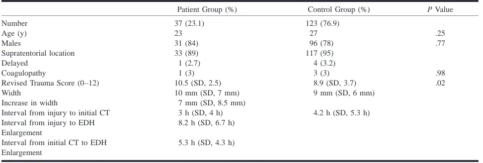
TABLE 1: Comparison of clinical data for patients with enlarging epidural hematoma (EDH) (patient group) and those with stable EDH (control group)
Patient Group (%) Control Group (%) P Value
Number Age (y) Males
Supratentorial location Delayed
Coagulopathy
Revised Trauma Score (0–12)
37 (23.1) 23 31 (84) 33 (89) 1 (2.7) 1 (3) 10.5 (SD, 2.5)
123 (76.9) 27 96 (78) 117 (95) 4 (3.2) 3 (3) 8.9 (SD, 3.7)
.25 .77
.98 .02 Width
Increase in width
Interval from injury to initial CT Interval from injury to EDH Enlargement
Interval from initial CT to EDH Enlargement
10 mm (SD, 7 mm) 7 mm (SD, 8.5 mm) 3 h (SD, 4 h) 8.2 h (SD, 6.7 h)
5.3 h (SD, 4.3 h)
9 mm (SD, 6 mm)
4.2 h (SD, 5.3 h)
Of the 252 patients with acute traumatic EDH, 216 initial diagnostic CT scans were obtained at the time of admission to the trauma center, and 36 were obtained at outside institutions before transfer. All follow-up CT scans were obtained at the trauma center, except for one patient, whose EDH was iden-tified as enlarging on a follow-up scan at the outside institution to which he was admitted before his transfer to our center. Scans were obtained without contrast in 5-mm axial sections at 7-mm intervals from vertex to foramen magnum and dis-played in window settings for brain parenchyma (width, 80 HU; level, 30 HU), extraaxial hemorrhage (width, 150 HU; level, 40 HU), and bone (width, 4000 HU; level, 400 HU) and filmed in 16-on-1 format until 1994 and subsequently in 20-on-1 format. Follow-up scans were obtained emergently if neu-rologic status deteriorated or headache was reported as wors-ening. Routine follow-up scans were generally obtained within 24 hours in neurologically stable, conservatively managed pa-tients. Those patients with a nonoperatively managed EDH that did not enlarge on at least one follow-up CT scan were used as control subjects for comparison against those with an EDH that did enlarge under observation.
Two observers rated the presence and time interval of en-largement (measured with calipers as an increase in width) of the EDH, the presence of skull fracture, contralateral brain in-jury, or extraaxial hemorrhage, and the presence and degree of midline shift. Scans showed no or mild midline shift if less than 5 mm, moderate midline shift if 5 to 10 mm, and severe shift if more than 10 mm. The only disagreements between the readers were whether a mild versus no midline shift had oc-curred in two patients. We resolved one of these disagreements by consensus; the other required a third neuroradiologist’s opinion.
We reviewed medical records to determine the interval be-tween injury and initial CT scan, the clinical factors at the time of presentation (including coagulation parameters [pro-thrombin time/partial thromboplastin time]), and the Revised Trauma Score (RTS), a physiological indicator of injury se-verity that combines respiratory rate, systolic blood pressure, and the Glasgow Coma Scale (GCS) score, with 0 to 4 points for each category (8). A higher RTS indicates a patient with less severe injuries, and an intubated and chemically paralyzed patient receives a score of 4 (0 points for a GCS score of 3, 0 points for respiratory rate, and 4 points for normal systolic blood pressure).
Disposition at discharge was measured as a proxy for clin-ical outcome. Disposition to home, to a rehabilitation facility, or to a skilled nursing facility corresponded to progressively more severe neurologic impairment.
x2and Fisher’s exact tests were performed to compare
cat-egorical variables and independent samples t-tests were used
for continuous variables. We also performed a multivariate lo-gistic regression, with rehemorrhage as the dependent variable and the imaging parameters (fracture, shift, contralateral inju-ry) and clinical parameters (coagulopathy and RTS) as inde-pendent variables.
Results
In the 5-year period studied, 252 patients were admitted with acute traumatic EDH, 92 (36.5%) of whom underwent immediate surgical evacuation and were not included in the study group. The re-maining 160 patients (63.5%) were examined with at least one follow-up CT scan, either as part of conservative observation or until definitive surgery could be performed. Medical records were avail-able for all patients whose EDH enlarged and for all but seven of the patients whose EDH did not enlarge.
The EDHs enlarged in 37 patients (23%) (Table 1). In all 37 patients, enlargement was detected on the first follow-up scan after diagnosis. One EDH enlarged 17 hours after injury and again 11 hours later, without subsequent enlargement thereafter. It was located in the posterior fossa, was presumed venous in origin, was 9 mm in width, and did not require surgery. All but four EDHs were supraten-torial. Only one enlarging EDH was delayed in ini-tial appearance (not present on the iniini-tial postinjury CT scan, but seen on a 3-hour follow-up scan). The mean age of patients with an enlarged EDH was 23 years (range, 1 to 87 years), and 84% were male. The average size of the initial EDH was 10 mm (SD, 7 mm), and the average increase in width was 7 mm (SD, 8.5 mm).
TABLE 2: Comparison of imaging features for patients with en-larging epidural hematoma (EDH) (patient group) and those with stable EDH (control group)
Patient Group (%)
Control
Group (%) P Value
Fracture
Contralateral injury No midline shift Mild shift Moderate shift Severe shift
32 (86) 14 (38) 22 (60) 10 (27) 3 (8) 2 (5)
101 (82) 53 (53) 85 (69) 25 (20) 11 (9)
2 (2)
[image:3.612.310.544.227.450.2].41 .43 .44 .54 .76 .23
TABLE 3: Comparison of neurologic status at discharge as mea-sured by disposition
Discharge Disposition Patient Group (%) Control Group (%)
Home
Rehabilitation facility Skilled nursing facility Died
23 (62) 8 (22) 2 (5) 4 (11)
71 (62) 27 (24) 12 (11) 3 (3)
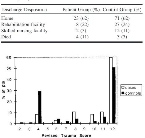
FIG1. Graph of RTS subgroups in patients (enlarging EDH
un-der conservative management) and control subjects (stable EDH). Note that a higher RTS indicates less severe injury, and an RTS of 4 indicates an intubated and paralyzed patient.
patient was discharged home within 48 hours with no neurologic deficit.
The mean interval between injury and EDH en-largement was 8.2 hours (SD, 6.7 hours). All 37 EDHs enlarged within 24 hours after the initial CT scan: 35 (95%) of 37 enlarged within 12 hours, one (3%) enlarged after 14 hours, and one (3%) en-larged 24 hours after the initial diagnostic scan.
Because of the retrospective nature of this study and the physical constraints of routine CT scanning in patients with multiple injuries in a busy trauma center, these data points represent times at which EDH enlargement was detected, not necessarily when it occurred. In all but one of the 37 patients, EDH enlargement was seen on the first follow-up scan. In one patient, the EDH was not present on the admission scan but was detected on a follow-up scan at 3 hours. Careful evaluation of interme-diate window settings (width, 150 HU; level, 40 HU) excluded an isodense EDH on the admission scan. It an enlarged 4 hours later and remained sta-ble on a subsequent scan. The patient did not re-quire surgery and was discharged home without neurologic deficit.
Our control group consisted of 123 patients in whom the EDH remained stable under observation (Table 1). The mean age of the control subjects was 27 years, and 78% were male. The average width of the EDH was 9 mm (SD, 6 mm). All but six were supratentorial, and four were delayed in ap-pearance (not present on the initial CT scans but present on follow-up scans, at a mean interval of 9.7 hours [SD, 6.3 hours]). Delayed EDHs are a somewhat controversial entity. With delayed EDHs, a vascular injury presumably has occurred by the time the initial CT scan with negative findings is obtained, but the extraaxial hemorrhage is identi-fied later, when factors such as increasing venous or arterial blood pressure overcome the tamponade effect of elevated ICP (9–11). These subjects were not included in the patient group because our goal was to identify risk factors for EDH enlargement in patients with known EDH. Patients without an identified EDH (even if it was present but too small to detect by CT) were treated differently, clinically, from those with an identified EDH and, as such, for our primary analyses, were not included in our patient group. The single delayed EDH in the pa-tient group and the four delayed EDHs in the con-trol group all had fractures on the initial scan, which showed no EDH.
No statistically significant difference in the ini-tial CT findings was found between the patient and control groups when examined for the size of the EDH, fractures associated with the EDH, contra-lateral brain injury or extraaxial hemorrhage, or the presence or degree of midline shift, quantitated as mild, moderate, or severe (Table 2). We did not have the data available to examine ICP values in this series.
Clinical data, including coagulation parameters (prothrombin time/partial thromboplastin time) and
FIG 2. Unenhanced CT scans of a 36-year-old male assault victim (initial CT scan 2 hours after injury).
A and B (at a level 5 mm contiguously higher thanA), Scans show a left temporal EDH, 2 cm in width. The patient was neu-rologically normal at the time of the initial CT scan.
C and D (at a level 5 mm contiguously higher thanC), Follow-up scans 10 hours later, after decline in neurologic status prompted repeat scanning. The EDH en-larged to 3 cm in width, associated with early ipsilateral uncal herniation. The pa-tient underwent surgical evacuation and was discharged to a skilled nursing facility after a 17-day hospitalization.
We repeated the logistic regression twice, switching the four control subjects with delayed EDH into the group of 37 patients as well as ex-cluding delayed EDH cases from both the patient and the control groups. The significance of the RTS ( P 5 .03) remained essentially unchanged when we excluded patients with delayed EDH from both the patient and the control groups, nor did it change the resulting odds ratio (exponent of coefficient b
51.15). However, switching the four subjects with delayed EDH from the control group to the patient group changed the significance of the RTS as a predictor of rehemorrhage ( P 5 .07), making it borderline insignificant. This P value is still much smaller than any of the other factors analyzed, and the apparent lack of stability of the significance of the RTS is most likely attributable to our relatively small sample size.
Slightly less than half (16 of 37) of the patients with EDH enlargement subsequently underwent surgery. All but two of these patients had deterio-rated clinically, manifested by worsening headache or a decreasing level of consciousness. At our in-stitution, worsening neurologic status, in addition to EDH enlargement, is an indication for surgical EDH evacuation. Five of the 37 patients had EDH enlargement greater than 10 mm, four of whom de-teriorated clinically. Five of nine patients with EDH enlargement of 5 to 10 mm deteriorated clin-ically, as did five of the 23 patients with EDH en-largement of less than 5 mm. In contrast, only 22
(18%) of 123 patients with a stable EDH under-went surgery, of whom 17 (77%) had deteriorated clinically. This was a statistically significant differ-ence ( P5.001). No statistically significant differ-ence ( P 5 .18) was found between patients and control subjects regarding discharge disposition (Table 3).
Discussion
EDH enlargement was found on follow-up CT scans in nearly one fourth (23%) of patients in our series with conservatively managed EDH (Figs 2 and 3). EDH enlargement occurred early, detected on average within 8 hours of injury and within 5 hours of initial diagnosis. A higher RTS was pre-dictive of rehemorrhage, but no imaging features were found to correlate significantly with EDH en-largement. Patients with EDH rehemorrhage re-ported a worsening of their clinical condition more frequently and underwent subsequent surgical evacuation twice as often as those with stable EDH, but they did not experience a worse neurologic out-come, as measured by disposition at discharge.
FIG 3. Unenhanced CT scans of an 87-year-old woman struck by a car (initial CT scan 2 hours after injury).
A and B (at a level 15 mm higher than A), Scans show a 12-mm-wide high-right-convexity EDH with subarachnoid hemor-rhage in the right parietal lobe inferiorly. The patient had a neurologic deficit, but surgery was not performed because of the patient’s religious preferences. A further decline in neurologic status prompted a fol-low-up scan 9 hours later.
C and D (at a level 10 mm higher than C), Scans show an increase in width of the high-right-convexity EDH to 18 mm, with ipsilateral parenchymal hemorrhage and severe right to left midline shift. The patient did not survive the injury.
true frequency of enlargement in their series is higher, since they reported but did not enumerate other EDHs that increased by 3 mm or less. The highest incidence was reported by Sakai et al (6) (in what is also the largest series to date), who found 24 of 37 conservatively managed patients with EDH enlargement, all within 5 hours. While the incidence is higher than that reported here, the time frame is similar to ours (the mean time to EDH enlargement after injury was 8 hours).
We found, as expected, that the majority of EDHs are associated with underlying fractures, in-cluding all five cases of delayed EDH. The pres-ence of a fracture, however, did not correlate with a greater tendency to rehemorrhage. A contralateral subdural hematoma located at the countercoup site is frequently found with EDH and may tamponade an EDH at the coup site (13). We found no lesser tendency for EDH enlargement in the presence of a contralateral subdural hematoma. Abnormal co-agulation factors do not seem to play a role in re-hemorrhage, with similar numbers in both groups. Sakai et al also reported no increased occurrence of bleeding diathesis in their 24 patients with an enlarging EDH (6). Clinical deterioration in our pa-tient group corresponded in general to the degree of enlargement. This may not be entirely attribut-able to mass effect, since associated parenchymal shear injury is most likely a contributing factor.
Our data controverts previous assertions that EDH attains maximum size within minutes of
within 6 hours of injury) is crucial to improving outcome (12), but the longer an EDH is asympto-matic, the greater is the likelihood that it will re-main so (20).
The RTS (8) at presentation correlated signifi-cantly with rehemorrhage. We found that patients with a higher RTS, apparently less severely injured, had a statistically greater likelihood of EDH en-largement. A bimodal distribution to the RTS was found in most patients, either with a score of 4 (because of intubation and chemical paralysis) or a score of 12 (because of being physiologically and neurologically normal). This is not unexpected, since any patient with head injury and a lower GCS score or more severe signs of physiological injury (hypotension or tachypnea) would be treated with immediate surgical evacuation. It is possible that blood pressure and ICP were better controlled in patients with an RTS of 4 (chemically paralyzed patients with appropriate mechanical ventilation). Conversely, in awake patients with head injuries and no physiological injury according to trauma score (RTS 5 12), agitation may lead to blood pressure elevation and hyperventilation with a low-ering of the ICP, both of which may promote bleed-ing into an existbleed-ing EDH. However, in the series of Sakai et al (6), five patients with EDH enlarge-ment were monitored for ICP: two had ICP increas-es and three were unchanged. We lacked the data to examine ICP values in our series.
Our initial analysis included delayed EDH in pa-tients and control subjects. An argument can be made that delayed EDH represents enlargement of an EDH from invisible to visible (assuming high-quality CT scans at diagnosis and follow-up) and should, therefore, be considered an enlarging le-sion. A counter argument is that patients without EDH on initial CT scans are treated differently from those with EDH, and thus these groups should not be lumped together. One solution to this dilem-ma is to remove all patients with delayed EDH (in both the patient and the control group) from the analysis. When we did this, our results remained essentially unchanged. Another approach is to con-sider the appearance of delayed EDH as an enlarg-ing lesion and include these patients with cases of rehemorrhage. This analysis changed the RTS to a borderline insignificant predictor, increasing the P value from .02 to .07. This lack of stability most likely reflects our small sample size, although other explanations, such as misclassification, are possible as well. Other variables in the logistic regression analysis remained insignificant on both repeat analyses.
Nonoperative management has been advocated for EDHs that are less than 1.5 cm in width, as-sociated with minimal or no midline shift, and lo-cated in the convexities (1, 19, 20). The majority of EDHs occur in the temporal region (21) and, when large, cause the most pronounced symptoms associated with temporal lobe herniation and brain stem mass effect. EDHs in the posterior fossa are
usually venous in origin but are generally less fa-vorable lesions for nonoperative management be-cause of the small volume of the posterior fossa (22). Our cohort of nonoperatively managed pa-tients with EDH generally conformed to these cri-teria, with a mean EDH width of 9 to 10 mm, with 94% being supratentorial.
EDH enlargement, while common in our series, was not found to cause any difference in immediate clinical outcome, as measured by discharge dispo-sition. This is most likely attributable in great part to close clinical monitoring and prompt surgical evacuation of clinically significant EDH enlarge-ment, as evidenced by the 43% rate of subsequent surgical treatment of enlarging EDHs. We acknowl-edge that our measure of outcome is approximate, and perhaps its insensitivity accounts for the lack of difference between patients and control subjects. However, this was the only variable available in the database. Those who caution against nonoperative management of EDHs cite the safety of EDH sur-gery, the cost of hospitalization and monitoring, and the devastating effects of late swelling and her-niation (2, 20, 23). We found a higher rate of clin-ical deterioration in patients with EDH enlarge-ment, which occurred more frequently in patients with an enlargement of 1 cm or greater. Although mass effect may play a role in clinical decline, it is likely that associated brain injury, such as diffuse shear, may be more common in patients with an enlarging EDH. Prompt surgical evacuation once EDH enlargement or clinical decline was detected no doubt accounts for the observation that outcome was unchanged as compared with patients with sta-ble a EDH managed conservatively.
Conclusion
Our findings suggest that CT monitoring of con-servatively managed EDH for rehemorrhage is most appropriately timed in the first 36 hours after injury, with most cases of EDH enlargement oc-curring by 8 hours after injury. Moreover, intuba-tion and chemical paralysis seem to have protective effects against EDH enlargement, perhaps by the control of head movement, blood pressure, and possibly ICP. Finally, EDH rehemorrhage does not appear to result in worse neurologic status at dis-charge in conservatively managed patients.
References
1. Hamilton M, Wallace C. Nonoperative management of acute epidural hematoma diagnosed by CT: the neuroradiologist’s role. AJNR Am J Neuroradiol 1992;13:853–859
2. Marshall LF. Nonoperative management of extradural hema-toma (letter). Neurosurgery 1985;16:–606
3. Langfitt TW, Gennarelli TA. Can the outcome from head injury be improved? J Neurosurg 1982;56:19–25
6. Sakai H, Takagi H, Ohtaka H, Tanabe T, Ohwada T, Yada K. Serial changes in acute extradural hematoma size and asso-ciated changes in level of consciousness and intracranial pres-sure. J Neurosurg 1988;8:566–570
7. Servadei F, Faccani G, Roccella P, et al. Asymptomatic extra-dural haematomas: results of a multicenter study of 158 cases in minor head injury. Acta Neurochir (Wien) 1989;96:39–45 8. Champion HR, Sacco WJ, Copes WS, Gann DS, Gennarelli TA,
Flanagan ME. A revision of the Trauma Score. J Trauma 1989; 29:623–629
9. Domenicucci M, Signorini P, Strzelki J, Delfini R. Delayed post-traumatic epidural hematoma: a review. Neurosurg Rev 1995; 18:109–122
10. DiRocco A, Ellis SJ, Landes C. Delayed epidural hematoma. Neuroradiology 1991;33:253–254
11. Smith HK, Miller JD. The danger of an ultra-early computed tomographic scan in a patient with an evolving acute epidural hematoma. Neurosurgery 1991;29:258–260
12. Knuckey NW, Gelbard S, Epstein MH. The management of ‘‘asymptomatic’’ epidural hematomas: a prospective study. J Neurosurg 1989;70:392–396
13. Maurer JJ, Mayfield FH. Acute bilateral extradural hematomas. J Neurosurg 1965;23:63–66
14. Ford LE, McLaurin RI. Mechanisms of extradural hematomas. J Neurosurg 1963;20:760–769
15. Lofgren J. Traumatic intracranial hematomas: pathophysiolog-ical aspects on their course and treatment. Acta Neurochir Suppl (Wien) 1986;36:151–154
16. McLaurin RL, Ford LE. Extradural hematoma: statistical sur-vey of 47 cases./TITLE J Neurosurg 1964;21:364–371 17. Gean AD. Imaging of Head Trauma. New York: Raven; 1994;
107–124
18. Bender MB, Christoff N. Non-surgical treatment of subdural hematomas. Arch Neurol 1974;31:73–79
19. Pozzati E, Tognetti F. Spontaneous healing of acute extradural hematomas: study of twenty-two cases. Neurosurgery 1986;18: 696–700
20. Sagher O, Ribas GC, Jane JA. In discussion of: Hamilton M, Wallace C. Nonoperative management of acute epidural he-matoma diagnosed by CT: the neuroradiologist’s role. AJNR Am J Neuroradiol 1992;13:853–862
21. Zimmerman RA, Bilaniuk LT, Gennarelli T, Bruce D, Dolinskas C, Uzzell B. Cranial computed tomography in diagnosis and management of acute head trauma. AJR Am J Roentgenol 1978; 131:27–34
22. Kawakami Y, Tamiya T, Tanimoto T, et al. Nonsurgical treatment of posterior fossa epidural hematoma. Pediatr Neurol 1990;6: 112–118