SWELLING BEHAVIOUR OF THE CATCH CONNECTIVE TISSUE IN HOLOTHURIAN BODY WALL
BY JOHN P. EYLERS*
Biology Department, Mercer University, Macon, GA, USA
AND ALAN R. GREENBERG
Department of Mechanical Engineering, University of Colorado, Boulder, CO, USA
Accepted 6 October 1988
Summary
Swelling tests in a series of isotonic and isoionic solutions of varying calcium-to-sodium ratios were conducted on isolated dermal connective tissue of the holothurian Thyonella gemmata Verrill. The tissue swelled rapidly and attained a maximum volume increase of approximately 40 % when transferred from distilled water to NaCl solution; however, the volume did not change significantly in isotonic CaCl2 solution. At Ca2 +/Na+ ratios =S0-04 the tissue swelled at its
maximum rate. The rate decreased with increasing calcium concentration, until at Ca2 +/Na+ ^0-40 no detectable swelling occurred. Similar results were obtained for Pentacta pygmaea Goldfuss. When tissues previously swollen in NaCl were placed in CaC^, the volume decreased significantly. Uniaxial tensile tests indicated that the elastic modulus of the tissue was much greater in Ca2+ solutions than in Na+ solutions. We hypothesize that dermal stiffness in holothurians is regulated by cation-sensitive crosslinks.
Introduction
Since Takahashi (1967) first drew attention to the echinoderm connective tissue 'catch' mechanism, the body of mechanical, chemical and morphological data on these unique tissues has been growing steadily. Motokawa (1984c) and Wilkie (1984) catalogue the instances in which animals of this phylum appear to possess the ability to alter the stiffness of their connective tissues in response to signals from the nervous system. This voluntary modulation of connective tissue mechanics has been invoked to explain autotomy in sea cucumbers (Byrne, 1985) and brittle stars (Wilkie etal. 1984), variable rigidity in starfish rays (Eylers, 1976), compliance changes in ligaments of the sea urchin spine (Smith et al. 1981; Hidaka & Takahashi, 1983; Hidaka, 1983) and sea lily cirrus (Wilkie, 1983), as well as the * Present address, to which requests for reprints should be directed: Department of Pharmacology, Box 3813, Duke University Medical Center, Durham, NC 27710, USA.
defensive stiffening of sea cucumber body wall (Stott et al. 1974; Eylers, 1976, 1982; Motokawa, 1981, 1982, 19Wa-d; Hayashi & Motokawa, 1986). A common finding is that in the presence of Ca2+ and other divalent cations these tissues become stiffer when subjected to instantaneously applied forces while showing less viscous flow in their time-dependent response to constant stress or strain. Greenberg & Eylers (1984) showed that the stress relaxation time of holothurian body wall connective tissue (HBW) increases significantly when it is transferred from a bathing solution of NaCl to an isotonic CaCl2 solution. This is in accord with the observation that creep compliance of the tissue is reduced in an environment of Ca2+ (Eylers, 1982). Although in many of the instances cited above the presence of viable cells in the tissue preparations somewhat complicates the analysis, the results suggest the presence of calcium-dependent crosslinks within the proteoglycan and collagen matrix. When these crosslinks are activated they increase the stiffness of the matrix and restrict the relative motion of the polymer molecules. They can be disrupted by sodium and other monovalent cations, which results in decreased stiffness and increased compliance of the tissue. During the analysis of the HBW stress relaxation experiments (Greenberg & Eylers, 1984), it was noted that tissue left for several hours in sodium chloride solution swelled whereas tissue remaining in calcium chloride solutions did not change significantly in size. This behaviour was attributed to osmotic swelling of the weakly crosslinked matrix present in the former solvent and resistance to such swelling in the latter solution owing to the formation of calcium-induced crosslinks. This suggested two simple tests of the calcium crosslink hypothesis: (1) tissue swollen in sodium solution should shrink when transferred to calcium solution; and (2) the rate of swelling should be a function of the Ca2 +/Na+ ratio in the solution. Consequently, a series of swelling experiments was carried out in isotonic and isoionic solutions of varying Ca2 +/Na+ ratio using two species of holothurians. The results of these experiments support the existence of calcium-mediated crosslinks and also give some insight into the physiological boundary conditions of the system.
Materials and methods
Tissue preparation
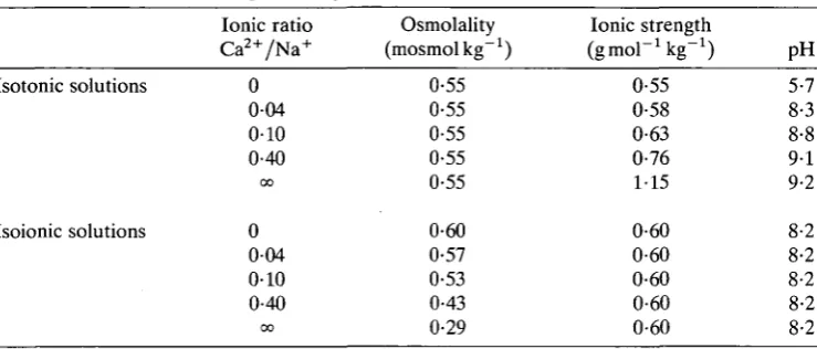
Table 1. Properties of isotonic and isoionic test solutions
Isotonic solutions
Isoionic solutions
Ionic ratio Ca2 +/Na+
0 0 0 4 0-10 0-40 0 0 0 0-04 0-10 0-40 0 0 Osmolality (mosmolkg"1)
0-55 0-55 0-55 0-55 0-55 0-60 0-57 0-53 0-43 0-29 Ionic strength (gmol^kg"1)
0-55 0-58 0-63 0-76 1-15 0-60 0-60 0-60 0-60 0-60 pH 5-7 8-3 8-8 9-1 9-2 8-2 8-2 8-2 8-2 8-2
removed by scraping with a razor blade, and the circumoral tentacles and nerve ring were cut out. During this procedure the animal contracted maximally and the body wall became noticeably stiffer. The carcass was split open lengthwise and the viscera and muscle layer were separated from the inner surface of the body wall with a blunt probe. The preparation (HBW), consisting only of body wall connective tissue and its embedded cell population, was washed and soaked in distilled water for 1 h. Previous experience has shown that HBW can be held in distilled water at room temperature for up to a week or at 4°C for more than a year without showing any signs of structural breakdown (unpublished results).
Solutions
Test solutions consisted of aqueous mixtures of NaCl and CaCl2 (Table 1) in which the Ca2 +/Na+ molar ratios varied from 0 (pure NaCl) to infinity (pure CaCli). Two types of solutions were prepared: isotonic, in which the osmolality of the solutions was held constant at seawater strength but the ionic strength was allowed to vary; and isoionic, in which the ionic strength was held equal to sea water and the osmolality varied. A buffer was added to the isoionic solutions to maintain pH8-2, but the pH of the isotonic solutions was not regulated.
Swelling
All swelling experiments were conducted at a temperature of 24-26°C. Tissue samples were taken from the preparations using a 5 mm punch which produced a discoid sample about 1 mm in thickness and 20-40 (A in volume. Five samples were taken from the interambulacral regions of each preparation so that every animal was tested in each of the test solutions. The samples were held in distilled water until they were transferred to the test solutions at the beginning of the experiment.
(W2). Having previously weighed the pycnometer filled with distilled water only (Wi), the sample volume could be calculated using the equation:
V = ( W1- W2 + W3) / D , (1)
where D is the density of water. Samples were then transferred to the assigned test solutions. Periodically each sample was surface-dried, weighed and examined for signs of physical deterioration. Volume change was calculated from the change in wet mass. At the end of the experiment, sample volumes were again determined by pycnometry. After accounting for the mass of the salts in the test solutions, no significant change in W2 between the beginning and the end of the experiment was observed in any sample. From this it was inferred that none of the tissue dissolved during the experiment and that the changes in sample masses reflected only the osmotic uptake of water. Finally, the samples were dried, and the dry mass, wet mass and density were used to calculate the volume fraction of water in the tissue. Swelling rate constants (q) were calculated by pooling the data for all samples in a given solution and fitting them to an exponential regression of the form: Vt/V0 = Qexp(qt), where V, is the volume at time t, Vo is the initial volume, and Q is a constant, ideally equal to 1.
Reversibility
Since HBW swelled rapidly in sodium chloride solutions but not at all in calcium chloride solutions, an experiment was devised to test the reversibility of this effect. Two sets of three samples each were prepared and their volumes measured by pycnometry. At the beginning of the experiment, one set was placed in a 0-55 mol P1 NaCl solution and the other in an isotonic 0-38 mol I"1 CaCl2 solution. After 10 h the volumes were measured and the solutions reversed, i.e. those in sodium chloride were placed in calcium chloride and vice versa. Volume changes were then monitored at hourly intervals for 5h, and the experiment was terminated with a final volume measurement at 26 h.
Tensile properties
A
Load cell •
Grip-Metal rod Glass chamber -O silk suture —
Specimen
Metal ring Rubber
[image:5.451.111.347.78.265.2]stopper-Crosshead -
J
Fig. 1. Holothurian body wall preparation for tensile test. (A) The specimen, with metal rings attached, is mounted on the Instron mechanical tester before the glass chamber is filled with test solution. (B) The appearance of the specimen just before failure. Note the region of localized necking where failure will occur.
from the photographs, and engineering strains (e) were calculated according to e = A L / LO. The cross-sectional area of the tissue was estimated from the initial
length and the volume of the sample. Recorded forces were divided by the initial cross-sectional area to generate values of engineering stress (s). Ignoring a small low-modulus toe region often observed in connective tissue (Silver, 1987), the elastic modulus (E = s/e) was taken as the slope of the initial straight-line portion of the stress-strain curve. The point at which the line deviated was identified as the proportional limit (sp), and the ultimate properties (su and eu) were taken at the maximum load.
Results Swelling
Representative results of the swelling experiment carried out on tissue samples from Pentacta pygmaea in isotonic solutions are shown in Fig. 2. As C a2 +/ N a+ increased, the swelling rate constant (q) decreased. When Ca2 +/Na+ increased, the swelling rate constant (q) decreased. When Ca2 +/Na+ equalled or exceeded 0-40 no significant swelling was observed over 66 h. Table 2 presents the statistical analysis of the results in Fig. 2 as well as those for Thyonella gemmata in isotonic and isoionic solutions. Negative values of q at high ionic ratios indicate deswelling or decrease in volume, but this result is significant in only one instance.
Reversibility
Fig. 2. The swelling of Pentacta pygmaea tissue in isotonic solutions of varying Ca2 +/Na+ ratios. V,/Vo: volume at time t relative to initial volume. ( • ) Ca2 +/
Na+ = oo; (©) C a2 +/ N a+= 0-40; (3) Ca2 +/Na+=0-10; (C) C a2 +/ N a+= 0-04;
( O ) C a2 +/ N a+= 0 .
monovalent salt solutions are exchanged is demonstrated in Fig. 3. In Fig. 3A
Thyonella gemmata tissue which had been exposed to 0-55 moll"1 NaCl solution for 10h was switched to an isotonic 0-38moll"1 CaCl2 solution. An exponential curve fitted to the data by the method of least squares illustrates deswelling over the next 16 h. Conversely, when similar tissue was exposed first to the CaCl2 solution and then switched to isotonic NaCl, the tissue immediately began to swell (see Table 3). The rates at which swelling occurred in the CaCl2- and NaCl-substituted solutions are in reasonable agreement with the values obtained in the initial swelling experiments (see Table 2).
Tensile properties
Table 2. Statistical analysis of swelling data* Species
Solution C a2 +/ N a+ Q N
Pentacta pygmaea Isotonic Thyonella gemmata Isotonic Thyonella gemmata Isoionic 0 0-04 010 0-40 00 0 0-04 010 0-40 00 0 0-04 010 0-40 00
* Equation: Vt/V0 = Qexp(qt).
N, number of observations. F, variance ratio: time/residual. P, statistical probability. NS, not significant.
1-07 1-01 105 1-03 0-97 1-26 1-09 1-00 0-98 0-96 1-00 1-04 1-03 1-02 0-99 0-0184 0-0177 0-0079 -0-0003 -0-0003 0-0146 0-0148 0-0102 -0-0005 -0-0009 0-0276 0-0161 00066 0-0012 0-0010 30 30 30 30 30 23 23 30 30 30 25 25 25 25 25 335 356 148 1-1 0-3 28 87 130 0-9 9-3 33 44 18 2-0 1-5 <0-001 <0001 <0-001 NS NS <0001 <0-001 <0-001 NS <0-01 <0-001 <0-001 <0001 NS NS 1-4 1-2 1-0
15 25 25
Fig. 3. Swelling-deswelling experiments. (A) Thyonella gemmata tissue swollen in 0-55moll"1 NaCl for lOh is transferred to 0-38 moll"1 CaCl2. (B) Tissue held in
Table 3. Statistical analysis of swelling reversibility experiments
Sequence Q q 0 0 N
0-55 moll"1 NaClto 0-38 m o i r1 CaCl2
0-38moll"1 CaCl2 to
0-55 m o i r1 NaCl
1-35
0-83
-00088
00289
Equation: V,/Vo = Qexp(qt).
18
18
18
168
<0-001
[image:8.451.140.314.208.468.2]<0001
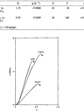
Fig. 4. Stress-strain curves for Thyonella gemmata tissue in distilled water, 0-38moll"1 CaCl2 and 0-55moll"1 NaCl. s, stress; e, strain.
Discussion
Table 4. Tensile behaviour of holothurian body wall connective tissue
E Sp su
Solution (MPa) (MPa) (MPa) eu
Distilled CaCl2
NaCl
H2O 18 ± 7
17 ± 6 6 ± 5
(16) (15) (17) 2-6 ±1-3 2-7 ±0-8 1-5 ±0-8 (14) (14) (10) 4 6 2 •7 •4 •9 ±1 ±2 ±1 •3 •3 •6 (10) (8) (8) 3-3 ± 4-9 ± 4-1 ± 1 3 1 •1 •1 •6 (10) (8) (8)
Sample sizes are given in parentheses.
E, the elastic modulus; Sp, proportional limit; su, stress at maximum load; eu, strain at
maximum load.
dermis. Fibroblasts and wandering coelomocytes are concentrated in the lami-nated dermis and hypodermis. Although numerous, these cells are very small and constitute a negligible portion of the total volume. 80 % of the mass of the tissue is interstitial water, and collagen constitutes about one-third of the dry mass (Stott
et al. 1974). The body wall of holothurians is highly permeable to inorganic ions.
Koizumi (1935) found that potassium, sodium, calcium and magnesium all diffused rapidly through the body wall tissue in either direction.
When a constant stress is applied to a strip of body wall tissue it lengthens at a nearly constant rate until failure (Eylers, 1982; Byrne, 1985). This indicates that although the collagen fibres are closely packed they are not tightly crosslinked but are free to flow past each other as the matrix deforms. Both the histology and the creep behaviour of HBW indicate that the collagen fibres are not tightly bound to each other, and the tissue may be modelled as an acid mucopolysaccharide gel with inclusions. This is in contrast to the usual model of vertebrate connective tissues in which the ground substance is entrapped in a reticulum of collagen fibres (Meyer
etal. 1976).
A gel is composed of a network of randomly coiled macromolecules in thermal motion. It exhibits entropy (or rubber) elasticity, in which stored mechanical energy takes the form of a decrease in the entropy of the deformed network (Wainwright et al. 1976, pp. 54-60). The factors which regulate the entropy elasticity of the gel are related in the classic equation for an ideal rubber:
Em = 3RTN0, (2)
where Em is the elastic modulus of the gel matrix, R is the molar gas constant, Tis absolute temperature, and No is the number of network chains per unit volume (Tanford, 1961; Bueche, 1962). If the average relative molecular mass of polymer between crosslinks is Mc, and the density of the network is P, then equation 2 can be rewritten:
Em = 3RTP/Mc, (3)
(Gosline, 1971). Moreover, the large elastic deformations observed during the tensile tests are indicative of a gel above its glass transition temperature.
Gel swelling and shrinking is governed by the balance between osmotic forces which tend to draw water into the matrix and elastic restoring forces within the network which resist expansion. When the network elastic forces equal the osmotic swelling pressure the gel remains isovolumetric. If the osmotic pressure exceeds the network elastic forces the gel will take on water and swell until a new equilibrium is reached. Conversely, if elastic restoring forces exceed osmotic pressure the gel will shrink. Consequently, a gel at equilibrium volume can be made to swell or shrink by any agent which can alter the elastic modulus of the matrix (see equation 3).
Equation 3 implies that the elastic modulus of a gel may be altered by changing either the density of the network (P) or the mean relative molecular mass (average length of chain) between crosslinks (Mc). The acid mucopolysaccharides of HBW are polyanions which carry fixed negative charges. If there are no mobile cations in the interstitial water, the repulsive forces between fixed charges will cause the network to expand, decreasing the density and consequently the elastic modulus of the gel. If, however, large numbers of mobile cations are present, they will shield the fixed anions and decrease the repulsive forces between them. This will allow the network to shrink, increasing both its density and its elastic modulus. The mean relative molecular mass between crosslinks may be varied by changing the number of crosslinks. Increasing the number of crosslinks leads to shorter network chains and a higher elastic modulus, whereas decreasing crosslink number has the opposite effect.
Although equation 3 emphasizes the importance of the network chain density (No) in characterizing the structure of the network, recent work (Mark, 1970; Oikawa & Murakami, 1987; Erman & Mark, 1987) has indicated that the actual situation is much more complex owing to factors such as chain entanglements and intramolecular crosslinks. Indeed, even basic equilibrium swelling and mechanical modulus experiments usually provide different values for No (Oikawa & Mura-kami, 1987). Moreover, differences may be expected depending upon whether crosslinks are formed in the bulk or solution state (Erman & Mark, 1987). Similarly, the tendency of a network to deswell (syneresis) is also a complex phenomenon involving gel inhomogeneity and the time-dependence of both syneresis pressure and matrix permeability (Van Dijk etal. 1984). Nonetheless, we believe that the simple approach embodied in equation 3 provides a useful first step in understanding the unique behaviour of this tissue.
would decrease and swelling would occur. Therefore a plausible interpretation of the results is that the presence of sodium ions disrupts existing crosslinks and weakens the structure of the gel. This hypothesis is supported by the observation that the tissue can be macerated in a strong sodium chloride solution but remains structurally stable for long periods in distilled water and calcium solutions. That it is sodium specifically, and not just the presence of mobile cations, which disrupts crosslinks is demonstrated by the fact that there is a slight, though not statistically significant, shrinkage of the tissue when it is transferred from distilled water to high-calcium solutions (Table 2), and that swelling occurs upon transfer from a calcium to a sodium solution (Fig. 3).
The second question is why the tissue should shrink when transferred from a sodium chloride solution to an isotonic calcium chloride solution. Since the solutions are isotonic, shrinkage cannot result from osmotic withdrawal of water from the tissue. Also, Donnan equilibrium effects, which could affect interstitial cation concentration, are minimal in a high ionic strength environment. In accordance with the hypothesis presented above, the swelling behaviour suggests that the elastic modulus of the tissue matrix has increased. However, it is not clear whether the modulus increase is caused by additional crosslinking in the presence of calcium ions, ameliorating the disruptive influence of Na+, or by the higher ionic strength of the calcium chloride solution (Table 1). Increased ionic strength promotes contraction of the network by means of more effective shielding of the polyanions.
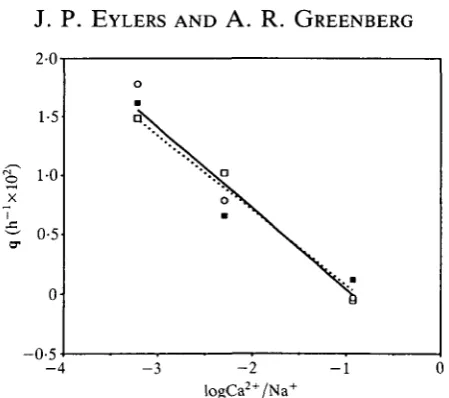
The significance of the latter factor can be evaluated by a more detailed comparison of the swelling results in isotonic and isoionic solutions (see Table 2). For gel samples of similar size and composition the rate of osmotic swelling is inversely proportional to the elastic modulus (Tanaka & Fillmore, 1979). In isotonic solutions the swelling rate decreases steadily with increasing calcium concentration (Fig. 5), as would be expected from the reversibility experiment. However, if the increase in modulus were the result of increasing ionic strength or pH, then swelling rate would not depend upon the ionic ratio in the buffered isoionic solutions. Since analysis indicates that there is no significant difference between the behaviour of the buffered isoionic and the isotonic series, we conclude that the increased value of the modulus is not a result of changes in pH or ionic strength.
2-OT
- 4 - 3 - 2
[image:12.451.113.338.48.247.2]logCa2+/Na+ - 1
Fig. 5. Swelling rate constant (q) vs logCa2+/Na+ ratio. ( • ) Thyonella gemmata isotonic; (O) Pentacta pygmaea isotonic; ( • ) Thyonella gemmata isoionic. Least-squares regression lines are given for all data (solid line) and T. gemmata isotonic (dotted line).
bonds are strong and generally irreversible. In addition, stress relaxation data also demonstrated that the effects of sodium were mimicked by potassium and those of calcium by magnesium, so valence number may be more critical to the mechanism than atomic species.
The difference in tensile elastic properties between tissues in sodium and calcium solutions and distilled water (Table 4; Fig. 4) is also consistent with a model incorporating matrix crosslinking. When the tissue is stretched at a constant rate of strain three of its major components, interstitial water, fibres and the gel matrix, act in parallel to resist lengthening. The interstitial fluid moving about within the tissue as it lengthens generates viscous forces which resist shape change. Viscous forces are dependent on strain rate, however, and since all tensile experiments were carried out at the same strain rate, it is unlikely that interstitial viscosity could account for the observed difference in elasticity. Fibres, although a small fraction of the tissue volume, have a high elastic modulus and therefore significantly increase tissue stiffness during rapid stretching. However, creep tests (Eylers, 1982) indicate that the fibres are not tightly bound together and tend to slide past one another as the tissue deforms. Thus, the contribution of collagen to the tissue modulus is in part controlled by the ability of the intervening matrix to prevent relative movement of the fibres. These factors suggest that the increase in tissue modulus produced in the presence of calcium ions or distilled water results from crosslinking within the gel matrix, and possibly also between the matrix and its fibre inclusions.
creep tests, whereas sodium ions decreased viscosity and caused rapid elongation. Hayashi & Motokawa (1986) also reported that the tissue contracted in distilled water. The effect of potassium ions was variable, and it has been postulated that potassium has a dual effect on living tissue, causing crosslink disruption in the matrix but at the same time stimulating neurosecretory cells to release a substance (such as calcium) that increases crosslinking (Byrne, 1985).
The question of what relationship these in vitro results have to the behaviour of the tissue in vivo is a legitimate concern. In living tissue there are three possible sites where manipulating ionic concentration could affect tissue mechanics: neurosecretory cells that secrete some substance into the matrix, neurones that regulate the neurosecretory cells, and the extracellular matrix itself (Motokawa & Hayashi, 1987). When the tissue is stripped of epithelium and coelomic lining and soaked in distilled water it can be assumed the cellular components of the tissue have been deactivated and that small organic molecules have diffused out of the interstitial spaces. Indeed, the mechanical properties of tissue so treated have been shown to be unaffected by freezing and thawing (Eylers, 1982) and by long-term storage in distilled water (J. P. Eylers & A. R. Greenberg, in preparation). We therefore conclude that in these experiments we are looking at the effects of cations on the matrix itself, unmasked by the activities of cells.
Finally, the close match between the swelling behaviour of T. gemmata and
P. pygmaea implies that the catch mechanism is very similar in these two species.
True mechanical comparison of catch tissues from different echinoderms is difficult because of the lack of standardized methodology. We propose that a quick and simple swelling test in solutions of varying ionic composition might serve to screen tissues for the presence of the catch mechanism and to reveal variations in sensitivity to ionic composition.
Grateful acknowledgement of support for this work goes to Mercer University Faculty Development Fund (JPE).
References
BUECHE, F. (1962). Physical Properties of Polymers. New York: John Wiley & Sons.
BYRNE, M. (1985). The mechanical properties of the autotomy tissues of the holothurian
Eupentacta quinquesemita and the effect of certain physio-chemical agents. J. exp. Biol. 117, 69-86.
ELDER, H. Y. (1973). Distribution and functions of elastic fibers in the invertebrates. Biol. Bull. mar. biol. Lab., Woods Hole 144, 43-63.
ERMAN, B. & MARK, J. E. (1987). Interpretation of stress-strain isotherms for elastomers cross-linked in solution. Macromolecules 20, 2892-2896.
EYLERS, J. P. (1976). Aspects of skeletal mechanics of the starfish Asterias forbesii. J. Morph. 149, 353-368.
EYLERS, J. P. (1982). Ion-dependent viscosity of holothurian body wall and its implications for the functional morphology of echinoderms. J. exp. Biol. 99, 1-8.
GOSLINE, J. M. (1971). Connective tissue mechanics of Metridium senile. II. Visco-elastic
GREENBERG, A. R. & EYLERS, J. P. (1984). Influence of ionic environment on the stress relaxation behaviour of an invertebrate connective tissue. /. Biomechanics 17, 161-166.
HAYASHI, Y. & MOTOKAWA, T. (1986). Effects of ionic environment on the viscosity of catch connective tissue in holothurian body wall. /. exp. Biol. 125, 71-84.
HIDAKA, M. (1983). Effects of certain physio-chemical agents on the mechanical properties of the catch apparatus of the sea-urchin spine. /. exp. Biol. 103, 15-29.
HIDAKA, M. & TAKAHASHI, K. (1983). Fine structure and mechanical properties of the catch apparatus of the sea-urchin spine, a collagenous connective tissue with muscle-like holding capacity. J. exp. Biol. 103, 1-14.
KOIZUMI, T. (1935). Studies on the change and equilibrium of water and electrolytes in a
holothurian, Caudina chilensis (J. Muller). Ilia. On the velocity of permeation of K, Na, Ca and Mg through the isolated body wall of Caudina. Sci. Rep. Tohoku Imp. Univ. Ser. 4, 10, 269-275.
MARK, J. E. (1970). An experimental comparison of the theories of elasticity of polymer networks. J. Am. chem. Soc. 92, 7252-7256.
MENTON, D. N. & EISEN, A. Z. (1970). The structure of the integument of the sea cucumber Thyone briareus. J. Morph. 131, 17-36.
MEYER, F. A., GELMAN, R. A. & SILBERBERG, A. (1976). The role of proteoglycans and collagen in the swelling of connective tissue. In Hydrogels for Medical and Related Applications (ed. J. D. Andrade), pp. 52-59. Washington, DC: American Chemical Society.
MOTOKAWA, T. (1981). The stiffness change of the holothurian dermis caused by chemical and electrical stimulation. Comp. Biochem. Physiol. 70C, 41-48.
MOTOKAWA, T. (1982). Factors regulating the mechanical properties of holothurian dermis.
J. exp. Biol. 99, 29-41.
MOTOKAWA, T. (1984a). The viscosity change of the body-wall dermis of the sea cucumber
Stichopus japonicus caused by mechanical and chemical stimulation. Comp. Biochem. Physiol. 77'A, 419-423.
MOTOKAWA, T. (19846). Viscoelasticity of holothurian body wall. J. exp. Biol. 109, 63-75.
MOTOKAWA, T. (1984C). Connective tissue catch in echinoderms. Biol. Rev. 59, 255-270.
MOTOKAWA, T. (1984<f). Viscosity increase of holothurian body wall in response to photic stimulation. Comp. Biochem. Physiol. 79A, 501-503.
MOTOKAWA, T. (1987). Cholinergic control of the mechanical properties of the catch connective tissue in the holothurian body wall. Comp. Biochem. Physiol. 86C, 333-337.
MOTOKAWA, T. & HAYASHI, Y. (1987). Effects of ionic environment on viscosity of catch connective tissue in holothurian body wall. J. exp. Biol. 103, 15-29.
OIKAWA, H. & MURAKAMI, K. (1987). Some comments on the swelling mechanism of rubber
vulcanizates. Rubber Chem. Tech. 60, 579-590.
SILVER, F. H. (1987). Biological Materials: Structure, Mechanical Properties and Modeling of
Soft Tissues. New York: New York University Press.
SMITH, D. S., WAINWRIGHT, S. A., BAKER, J. & CAYER, M. L. (1981). Structural features associated with movement and 'catch' of sea-urchin spines. Tissue Cell 13, 299-320.
STOTT, R. S. H., HEPBURN, H. R., JOFFE, I. & HEFFRON, J. J. A. (1974). The mechanical defensive mechanism of a sea cucumber. S. Afr. J. Sci. 70, 46-48.
TAKAHASHI, K. (1967). The catch apparatus of the sea urchin spine. II. Response to stimuli. /. Fac. Sci. Tokyo Univ. 11, 121-130.
TANAKA, T. & FILLMORE, D. (1979). Kinetics of swelling of gels. /. chem. Phys. 70, 1214-1218.
TANFORD, C. (1961). Physical Chemistry of Macromolecules. New York: John Wiley & Sons.
VAN DIJK, H. J. M., WALSTRA, P. & SCHENK, J. (1984). Theoretical and experimental study of one-dimensional syneresis of a protein gel. Chem. Eng. J. 28, B43-B50.
WAINWRIGHT, S. A., BIGGS, W. D., CURREY, J. D. & GOSLINE, J. M. (1976). Mechanical Design in Organisms. New York: John Wiley & Sons.
WILKIE, I. C. (1984). Variable tensility in echinoderm collagenous tissues: a review. Mar. Behav. Physiol. 11, 1-34.