Helgol~inder wiss. Meeresunters. 26, 174-216 (1974)
Nutritional potentials in
Zoanthus sociathus
(Coelenterata, Anthozoa)':
R. K. TRENCH
Biology Department, Osborn Memorial Laboratory, Yale University; New Haven, Conn., USA
KURZFASSUNG: Ern~ihrungsm6gliehkeiten bei Zoantbus|sociatus (Coelenterata, Anthozoa). Die Krustenanemone Zoanthus sociatus (ELLIS) lebt in Symbiose mit Dinoflagellaten der Gat-. tung Gymnodiniurn. Feinstrukturelle, physiologische und biochemische Aspekte dieser sym- biotischen Partnerschaft wurden untersucht. Z. sociatus zeichnet sich durch eine autotropbe Ern~ihrungsweise aus, indem Photosyntheseprodukte der intrazellut~ir iebenden Zooxantbellen verwertet werden. Dartiber hinaus ern~brt sich Z. sociatus auch heterotroph dutch die Auf-. nahme yon Detritus, Bakterien und gel&ten organischen Substanzen. Freiland- und Labor- untersuchungen erbrachten jedoch keine Hinweise, dab lebendes Zooplankton verwertet wird, obgleich in bezug auf das Verhalten gegeniiber Nahrungsstoffen, die Ultrastruktur der Mesen- terialfilamente und die Art der Verdauung keine wesentlichen Abweichungen gegeniiber anderen planktonfressenden Coelenteraten festgestellt werden konnten. Z. sociatus nimmt offensicht- lich - ebenso wie andere wirbellose Riffbewohner - eine polytrophe Stellung innerhalb des Ukosystems Korallenriff ein. Die Vielseltigkeit der Ern~ihrungsweise wird unter dem Gesichts- punkt der tSkologisehen Stabilit~it des Lebensraumes Korallenriff er6rtert.
I N T R O D U C T I O N
Nutritional potential in Zoanthus sociatus 175
It is not necessary at this point to query whether primary production by the endo- symbionts satisfies the total energy requirements of the host animals. That movement of photosynthetic products to the animals is accomplished, at least initially, without disruption of the endosymbionts, implies that the association does not represent an herbivore consuming its plant food. Functionally these "plant-animal" associations should be viewed as primary producers.
In the past, a great deal of controversy has been generated over the question of whether coelenterates, traditionally viewed as specialized carnivores (YoNG~ 1940), should be regarded as autotrophs because of the presence of symbiotic algae, or as heterotrophs (for recent review see GOREAtT et al. 1971), since the animals retain their "normal" heterotrophic mode of nutrition. The implication has been that the two modes of nutrition are mutually exclusive. I would like to propose instead that organisms such as corals or other invertebrates with photosynthetic endosymbionts are nutritonally plastic, and may sl,multaneously occupy several trophic levels, deriving nutrients through a variety of mechanisms from several different sources. The relative amounts of the total nutrient requirements of the animals supplied by each different mode of nutrition is at present unresolved, and would probably be very difficult to quantify. The point is that these organisms possess an autotrophic mode of nutrition complementary to heterotrophic modes.
The relationship between Zoanthus sociatus and its flora of zooxanthellae has in the past been regarded as different from that demonstrated by corals (GoRt~AU 1964, GOR~AtT et al. 1971). This view was based on the following observations: (t) it could not be demonstrated that Z. sociatus showed any feeding response when offered homogenates of animal matter, e.g. crab, clam or lobster muscle, (2) the mesenteric filaments appeared to be undifferentiated and (3) the algae were not expelled by
Zoanthus under stressful conditions that caused corals to expel their zooxanthellae (GoREAU 1964). In addition, the nematocysts in Zoanthus appeared to be non-func- tional (Got~rAG personal communication). The concept has been proposed then, that
Zoanthus, like some of the xeniid alcyonarians, e.g. Xenia (AsHwORTrt 1898, 1899, GOHAR 1940, 1948), are nutritionally more dependent on the photosynthetic products of the zooxanthellae than are corals; in the latter case the importance of the algae was thought to be involved in enhancing calcification (GoI~EAU 1961, 1963), or perhaps contributing trace substances or vitamins (Got~rAu & GOREaU 1960).
VON HOLT & YON H O~T (1968a) provided evidence for the transfer of photo- synthetic products from the zooxanthellae in Zoanthus flos-rnarinus (= Z. sociatus)
to the animal, and found (YON HOLT & YON t-IO~T 1968b) that the isolated algae released a wide variety of 14C-labelled meta~bolites when allowed to photosynthesize in vitro in NaHI*CO3. However, it was not clear from their data to what extent the substances detected in the media were released through a selective process or through indiscriminate liberation resulting from cell lysis.
176 R . K . TRENCH
experiments by RelM~ (1971a, b) showed that other species of Zoanthus, previously thought not to feed, demonstrated a characteristic feeding behaviour. Of course, that
Zoanthus engulfed materials of animal origin did not demonstrate that such materials were digested and assimilated.
This study was undertaken in the hope of providing evidence on the possible different modes of obtaining nutrients potentially utilized 'by Z. sociatus. The feeding behaviour, and ability of the animals to digest and assimilate exogenously s~applied protein were investigated, as well as their capacity to utilize exogeno~asly supplied amino acids (SH-leucine) and sugars (3H-glucose). In addition, the movement of photosynthetic products from the algae in vivo and the release of photosynthetic products in vitro were reinvestigated.
The results of laboratory experiments show that Z. sociatus has the ability to derive nutrients in several distinct ways. The animals are able to utilize photosynthetic products from their zooxanthellae, they are able to absorb dissolved organic matter and to digest and assimilate exogenously supplied proteins. However, it remains unclear how much nutrition is procured by the latter two mechanisms under natural conditions, particularly since field and laboratory observations show that Z. sociatus
continues to be totally unresponsive to live zooplankton or other potential living prey. What is apparent is that Z. sociatus, like many other reef-dwelling invertebrates with photosynthetic endosymbionts, possesses the ability to derive nutrion from several different sources, indicating a possible polytrophic status within the ecosystem.
MATERIALS AND METHODS
C o l l e c t i o n a n d m a i n t e n a n c e o f s p e c i m e n s
Zoanthus sociatus (ELLIS 1767; Fig. 1) was collected from the reefs off Discovery Bay, Jamaica and from W.halebone Bay, Bermuda, from depths of 1.5 to 5 meters.
Since a certain degree of uncertainty exists on the taxonomic identity of the animals, a short discussion of the problem is probably justified. Zoanthid taxonomy is currently in a state of chaos. Y. NEUMANN (personal communication) has suggested that Z. sociatus (E.) and Z. flos-marinus (DucHASSAING 8~; MICHELOTTI) are synony- mous. I have found no major distinguishing characters between these two and Z. pro- teus (V~RRILL). I have therefore selected to treat the zoanthids collected from Jamaica and Bermuda as identical. This is justified since results of the same experiments carried out on the two groups of animals were essentially the same.
Specimens used for experimental work were maintained in running sea water aquaria for not more ~han one week. Some specimens which were used for either histology and histochemistry or electron microscopy were sometimes kept for longer periods ,before fixation. Animals used in experiments were always in a healthy state as indicated by their tactile responses. In experiments involving incubations with isotopes, only specimens which remained fully expanded during the "pulse" period of labelling were selected for the continued "chase" experiments.
Nutritional potential in Zoanthus sociatus 177
Fig. 1: Underwater photograph of Zoanthus sociatus taken at 3 meters on the reef at Disco- very Bay, Jamaica. (1.6:1)
minimum of 72 hours before experiments were begun, in closed aquaria under aeration in Millipore-filtered (porosity 0.25 ~m) sea water, to offset the slightest possibility of feeding. I t subsequently became evident that this precaution was unnecessary.
E x p e r i m e n t a l
Pulse-chase experiments with NaH~4CO~ in vivo
178 R . K . TI~NCH
Isolation and incubation of zooxanthellae in vitro
Algal cells were isolated from Zoanthus sociatus by homogenization of the ani- mals (aPcer removal of the cuticle b y dissection) with a Virtis blender, followed by filtration through bolting silk and centrifugation at 500 xg for 20 min,utes (TRENCH 1971b). Suspensions of algae (2 × 10" cells/ml) were prepared in the homogenate of animal tissue recovered a~er centrifugation of the algae (TRENC~ 1971b, c, MUSCA'rlN~ et al. 1972). To 2 ml algal suspension, NaH14COa was added to give an initial activity of 25 #C/mt. Incubations were conducted in stoppered 10 mt erlenmyer flasks at 23-24 ° C, with an illumination of 5 X 10⁴ ergs cm -2 sec-i delivered by Grolux fluorescent tubes.
Suspensions were incubated for 30 minutes to 2 hours. At the end of the incuba- tion period, the cells were centrifuged at 1000 xg for 10 minutes at 10 ° C and the supernatant medium withdrawn and frozen. The pellet was extracted in 2 ml boiling 80 % ethanol, and extract and debris were stored at - 20 ° C until analysed.
Identification of photosynthetic products of zooxanthellae
The media in which zooxanthellae were incubated were evaporated to dryness in a Buchler Evapomix under reduced pressure at 40 ° C. Organic compounds were recovered from the salt by extraction with absolute ethanol followed by chloroform, accounting for about 97 °/0 of the i4C present.
The radioactive compounds in the cell extracts and from the media were separated by two dimensional paper partition radiochromatography. Labelled compounds were then eluted from the paper chromatograms and rechromatographed along with authentic standards either on. Brinkman precoated Silica Gel plates by ascending chro- matography or by one dimensional descending paper chromatography (see TRENCH 1971b for details).
Light microscopic autoradiography
Affer incubating animals in solutions containing the different isotopes, they were fixed in Bouin's solution for 24 hrs. Specimens were dehydrated with methyl cellosolve (ethylene glycol monomethyl ether), cleared in methyl benzoate and benzene and embedded in vacuo in paraplast. Sections were cut at 7 #m in the case of specimens incubated with 14C and at 4 #m in the case of those incubated with 3H.
Radioactive tissues were exposed to Kodak AR-10 stripping film or Kodak NET-2 liquid emulsion for from 10 to 30 days, developed in Kodak D-19 developer and fixed in Kodak rapid fixer (see ROGERS 1969). Tissues were stained through the emulsion with toluidine blue to enhance contrast for photography.
Electron microscopy
Nutritional potential in Zoanthus sociatus 179
for 6 hours, washed overnight in buffered glucose (pH 7.4) and post fixed in 2 0/00sO4 for two hours at 3 ° C. Tissues were dehydrated through serial changes of ethanol and embedded in either araldite or epon (KAY 1965).
Ultrathin sections were prepared on a Porter-Blum MT-2 ultramicrotome, stained with uranyl acetate and lead citrate, viewed and photographed with a JEM-6C elec- tron microscope.
In order to detect histochemically the presence of phosphomonoesterase II (acid phosphatase) in animal tissues, the tissues in question were dissected out under cold fixative and subsequently fixed for 6 hours at 3 ° C, washed overnight in cacodylate buffer (pH 7.2) and then separated into three groups:
G r o u p I. E x p e r i m e n t a 1 : Tissues were incubated in fresh-filtered Gomori (1950) medium at 37 ° C for two to three hours.
G r o u p I I . N a F c o n t r o t : Tissues were incubated as above, but NaF (enzyme inhibitor) was added to a final concentration of 0.5 °/0 (see FANitBONN~I~ 1971).
G r o u p I I I . S u b s t r a t e c o n t r o l : Tissues were incubated as i n l a b o v e but in Gomori medium lacking ~-glycerophosphate.
After incubation, the tissues were washed in acetate buffer (pH 5.0, 30 rnin), acetic acid (5 min) and then cacodylate buffer (pH 7.4, 15 min), followed by post osmication, dehydration and embedding as described above. These tissues were viewed and photographed without staining.
Feeding experiments
Feeding behaviour
In order to investigate the response of Zoanthus sociatus to live prey, suspensions of freshly hatched Artemia salina nauplii or recently captured zooplankton, were pipetted into a finger bowl containing 5 or 10 polyps. The behaviour of the animals was observed with a dissecting microscope, either with a microscope lamp (which tended to concentrate the Artemia or the zooplankters in the path of the beam) or in a room with subdued diffuse lighting. In several cases these experiments were continued for up to 6 hours in order to enhance the possibility that organic compounds diffusing from the zooplankton may influence a feeding response by lowering the threshold for nematocyst discharge.
In addition to intact organisms, experiments were conducted using fresh suspen- sions of tissue from a range of invertebrates. In each case the tissue in question was homogenized in a glass tissue grinder and aliquots of the suspension were pipetted on to the surface of the oral disc, care being taken not to disturb the animals mechanically during this process.
180 R.K. TRENCH
Attempts were also made to investigate the effect of reduced glutathione (GSH) (see LENHOFF 1968a, b, LINDSTEDT 1971, REIM~I~ 1971a) in eliciting a feeding response in Z. sociatus. Specimens were placed in feshly prepared solutions of GSH ranging from 110 -6 to 10-2M kept in the reduced state with dithioerythritol (10-6M) and observed under a dissecting microscope. In addition, pieces of filter paper imbibed with different concentrations of GSH were placed on the oral discs and the animal's response observed. Other substances tested were glycine, proline, alanine, asparfic and glutamic acids.
Preparation of 35S and 8H-protein
Protein labelled with ~5S or 3H was prepared using the method of COWIE et al. (1952). Escherichia coli (B) were grown in media in which Na.28~SO4 (New England Nuclear) was the sole form of sulphur, in order to produce high specific activity 35S-protein, or on nitrogen deficient media to which 3H-leucine (New England Nuclear) was added.
Aflcer attaining stationary growth, the bacteria were pelleted by centrifugation and extracted in 80 0/0 ethanol. A portion of the ethanol insoluble residue was hydro-
50
Rf
0 05 1.0
, I
~ o 2C
x
£
£1_
]
10
I
i
I
, .
E.coli, S-protein hydrolysate
0 standard cysteiine
D standard cystine
e standard rnethionine
Fig. 2: Distribution of 35S in amino acids derived from 6N HC1 hydrolysis of proteins from
Nutritional potential in Zoanthus sociatus 181
lysed with 6N HC1 in sealed tubes at 100 ° C, and the hydrolysis products separated by ascending thin layer chromatography using Eastman Kodak Cellulose strips and n-butanohpropionic acid:water (142:71:100v/v) as solvent system. The gel was scraped from the strips at 0.5 cm intervals into scintillation vials, and the radioactivity measured using Aquasol scintillation fluid (New England Nuclear) and an Intertech- nique SL-30 liquid scintillation spectrometer. Standards run along with the hydro- lysates were visualized with ninhydrin.
Figure 2 shows that the ~5S was incorporated into the bacterial protein as cystine, cysteine and methionine, while Figure 3 shows that the 3H was incorporated as ~H-leucine. The dried powdered bacterial proteins were stored in a vacuum dessicator
over P205 at - 20 ° C until used.
Digestion and assimilation experiments
Specimens were fed aliquots of homogenates of Echinometra eggs mixed with either 35S or 3H-protein. At intervals of up to 48 hours a~er feeding, specimens were sampled in duplicate. In the case of those experiments conducted with 35S-protein,
R t
0 0.5 1.0
I 4 ... I
60
50
40
I o
30
E U
2O
10
O
D
E.coli, H-protein hydrolysate3
O Standard leucine
Fig. 3: Distribution of '~H in amino acids derived from 6N HCI hydrolysis of proteins from
182 R . K . TRENCH
the animals were homogenized in cold distilled water and cold trichloroacetic acid (TCA) added to a final concentration of 10 °/0 to precipitate protein. The precipitate was then collected on Millipore filter discs (porosity 0.25 #m), and an aliquot of the filtrate recovered. The radioactivity in each was measured and the percentage TCA- soluble ~5S was calculated.
In the case of experiments conducted using 3H-protein, the animals were fixed in Bouin's fixative and subsequently analysed by autoradiography. Combination of the two methods provides an estimate of the rates of digestion and assimilation as well as the morphological sites for assimilation and subsequent dispersal of assimilated food throughout the animal.
Pulse-chase experiments with ~H-leucine and 3H-giucose
To investigate the potential of Zoanthus sociatus for uptake of dissolved organic matter from solution, specimens were incubated in Millipore filtered (porosity 0.25 #m) sea water to which *H-leucine or 3H-glucose (New England Nuclear) were added, for up to 4 hours, and then transferred to isotope free media for chase periods of up to 14 days. Animals were sampled, fixed in Bouin's fluid and analysed by autoradio- graphy.
RESULTS
T h e p h o t o s y n t h e t i c c o m p o n e n t
The morphology of the association of Zoanthus sociatus with zooxanthellae
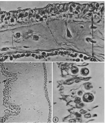
The zooxanthellae in Zoanthus sociatus are located exclusively in the gastro- dermal cell layer (Figs 4-6). This is similar to the situation found in corals, alcyona- rians and sea anemones (see DRool, 1963, McLAuGHLIN & ZAHL 1966, Ti~ENCH 1971a), but is in contrast to that found in some other zoanthids, e.g. ProtopaIythoa grandis, PaIythoa caribbea, Palythoa sp. and Isaurus sp. which have zooxanthellae in the epidermis and in gastrodermal cells lining the lacunae of the mesoglea as well (TI~ENCH
I969, 1971, HER~Er, TS 1970).
In Z. sociatus the highest concentration of algae is found in the tentacles and the oral disc. However, zooxanthellae can also be found in the cells of the gastroderm of the column, and in the "hypertrophied" gastroderm cells of the mesenteries proximal to the mesenteric filaments, where "degenerate-appearing" (pycnotic) zooxanthellae
are especially abundant (Fig. 19).
The ultrastructure of the phycobionts
N u t r i t i o n a l potential in Zoanthus sociatus 183
Fig. 4: Light micrograph of the oral disc of Zoanthus sociatus. The epithelium is ciliated and electron microscopy shows the presence of extensive microvillation, epi = epidermis; m =
mesoglea; g = gastroderm; zx = zooxanthella, (660:1)
Fig. 5- Light micrograph of the body wall of Zoanthus sociatus, c = cuticle; between the cuticle and the very thick mesoglea is a very thin epidermis, ge = gastroenteron, boardered
by the gastroderm. (130:1)
Fig. 6: Light micrograph of the gastroderm celis of Zoanthus sociatus containing zooxanthellae. m ~ mesoglea; ge = gastroenteron. (1500:1)
(Fig. 8). The algae show typical gymnodinioid morphology (TAYLOR 1968, 196%, KEVlN et al. 1969) and, based on morphology alone, appears to be Gymnodinium
184 R . K . TR~Ncl-t
Fig. 7: Electron micrograph of Gymnodinium microadriaticum (?) in a gastrodermal celI in the tentacle of Zoanthus sociatus. N = nucleus; cr = polytene chromosomes; m = mitochondrion;
cp = &loroplast; s = starch; ac . . . . accumulation body". (12 250:1)
Fig. 8: Electron micrograph of a dividing zooxanthella in a gastroderm cell of the orat disc of Zoanthus soc~atus. (6700:1)
Fig. 9: The single-stalked pyrenoid of a zooxanthella in Zoanthus sociatus, py = pyrenoid; cp = chloroplast; s = star& cap. Magnification (15 500:1)
Nutritional potential in Zoanthus sociatus 185
Fig. 10: The periplast membrane system (pm) composed of five unit membranes, the chloroplast envelope (ce) composed of three unit membranes, and chloroplast thylakoids (th) in groups
of three. (33 500:1)
Fig. 11: The nucleus of a zooxanthella showing the polytene chromosomes. OsO4 fixed. (27500:1)
Fig. 12: The chloroplast (cp) and nucleus (N) of a zooxanthella. The nuclear envelope (ne) is composed of two unit membranes. Th = thylakoids. (28 000:1)
Fig. 13: The "accumulation body" of a zooxanthella in Zoanthus sociatus. Note the abundance of membranes and "crystalline-appearing" particles. (20 650:1
186 R . K . TRENCH
chromosomes) (Fig. 11), a characteristic feature of dinoflagellate nuclei (DODGE 1966, 1971). A prominent granu!ar nucleolus has also been observed. The pyrenoid (Fig 9) is vase shaped and its stroma is continuous with that of the chloroplast to which it is attached, most oflcen at a single point, but frequently with double attach, merits (see TAYLOR 1968). No pyrenoid lamellae are present. In addition to the starch cap asso- ciated with the pyrenoid, several free starch grains are distributed through the cyto- plasm (Fig. 7). Each alga is bounded by a system of 5 or 6 unit membranes, the peri- plast membrane system (Fig. 10), which can more readily be discerned during division (see TAYI~OR 1968). The major inclusion is the "accumulation" body (Fig. 13) which appears to be much more complex than has heretofore been recognized, containing an abundance of smooth endoplasmic reticulum and other unrecognizable structures. The precise functions of this organelle remain obscure, although accumulation of waste (TAYLOR 1969c) and storage of material for subsequent reassimilation (ScH~ITTER
1971) have been proposed.
Transfer of photosynthetic products from algae to hosts in vivo
When specimens of Zoanthus sociatus were incubated in H14CO3 in the light and analysed for incorporation of 1~C by tissue autoradiography, it readily became apparent that incorporation occurred in two distinct zones, (a) in the gastrodermis where the zooxanthellae are abundant and (b) on the surface of the cuticle (Fig. 14).
Radioactivity associated with the zooxanthellae was high even afLer very brief "pulse" exposures of the animals to H~CO~ in the light (see Fig. 18). Control incu- bations conducted in the dark, or in the light in the presence of DCMU, showed that non-photosynthetic fixation of 14C by zooxanthellae was negligible (Fig. 17).
During "chase" experiments, the radioactivity was found to diffuse to different regions of the poIyps, including those regions towards the base of the coelenteron, where very few zooxanthellae are found, and within 48 hours could be found asso- ciated with the cuticle, particularly that portion of the cuticle juxtaposed to the epi- dermis. This process is very rapid, and does not have any correlation with breakdown, degeneration or digestion of zooxanthellae. In the hypertrophied regions of the mesenteries, no algae incorporated ~4C during the "pulse" labelling period (Fig. 19, see also p. 202).
N u t r i t i o n a l potential in Zoanthus sociatus 187
Fig. 14: Light micrograph of an autoradiograph of Zoanthus sociatus atter incubation in sea water containing NaHi4CO~ in the light for 45 minutes. Note the presence of intense labelling over the gastroderm of the tentacles and oral disc as well as over the surface of the cuticle.
Magnification (3.3 :t)
Fig. 15: Light micrograph of an autoradiograph of the cuticle of Zoanthus sociatus showing intense labelling after 60 minutes incubation in NaHlaCO3 in the light, cu = cuticle.
Magnification (4660:1)
Fig, 16: Electron micrograph of the cuticle of Zoanthus showing the bacteria associated with it. These organisms are partly responsible for the intense Iabelling associated with the cuticle
188 R . K . TRENCH
Fig. 17: Light micrograph of an autoradiograph of Zoanthus sociatus incubated in the light in the presence of NaHI"*CO.% and DCMU (5 X 10-SM). Note the absence of silver grains
over zooxanthellae. Tissue stained with toluidine blue through the emulsion. (960:1) Fig. 18: Light micrograph of an autoradiograph of Zoanthus sociatus incubated in NaH~4COa
in the light for 4 hrs. Tissue unstained. (960:1)
Fig. 19: Light micrograph of an autoradlograph of mesentery of Zoanthus sociatus incu- bated in NaH14CO8 in the light for 4 hrs. Tissue stained through the emulsion with toluidine blue. Note the absence of silver grains over the tissue and over the pycnotic zooxanthellae
Nutritional potential in Zoanthus sociatus 189
Since the results obtained through the use of the autoradiographic technique were only qualitative, a study was conducted in order to quantify the level of translocation between the zooxanthellae and the animal hosts. The technique used was essentially that described by TRENCH (1971a). The epizoic photosynthetic bacteria and blue-green algae on the cuticle of Z, sociatus initially caused very erratic results. The reason was that it was not possible to separate the bacteria and blue-green algae from the host
i •
5O
i
-___ 40 • •
0 30
• o
l:n
E •
ml6.~ 20 o o
E
u 10 o
i i i ! I i ~ I V I ii I J J
2 4 6 8 I0
Time(hrs)
Fig. 20: Photosynthetic fixation and translocation of 14C from zooxanthellae to animal tissues
in Zoanthus sociatus. • O, 14C in algae; O O, 14C in animal tissues; I ' V, 1~C
in whole DCMU-treated controls
tissue during centrifugation, and observation of the algal pellet showed varying amounts of contamination with bacterial aggregates and filaments of blue-green algae. It was obvious therefore that epizoic photosynthetic 14C was contributing to both "zooxanthellar" and "host" fractions in a non-reproducible manner. I therefore decided to first treat the animals with an antiboitic mixture*. Autoradiographic ana- lysis showed that after six hours of pre-treatment the antibiotics did not eliminate the bacteria nor the blue-green algae, but did eliminate their photosynthesis without affecting that of the endozoic zooxanthellae. Antiboitic treated specimens were there- fore used in subsequent experiments.
Antibiotic-treated animals were incubated in filtered sea water containing NaHI4CO~ for up to 10 h. ControI specimens were incubated in the presence of
190 R . K . TRENCH
DCMU. Sample specimens were withdrawn at hourly intervals and homogenized in a Virtis blender. The algae and animal tissues were separated by sucrose density gra- dient centrifugation (see FRANKER 1971). The supernatant was regarded as animal cell debris. The radioactivity in the algae and in the animal tissues was measured by liquid scintillation spectrometry. Protein was determined by the method of LowRY et al. (1951).
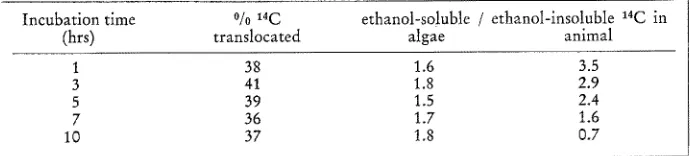
Figure 20 shows that the rate of net '4C fixation by the experimental organisms was essentially constant throughout the experiment. DCMU-treated organisms incor- porated very low levels of i4C. The proportion of photosynthetically fixed 14C trans- located to the animals under these conditions was 3 6 - 4 1 % .
The distribution of 14C in hot 80 % ethanol soluble and insoluble fractions in algae and in animal tissues (see Table 1), shows that in the algae, the ratio of soluble to insoluble 14C remained relatively constant, while in the animal tissues the ratio
Table 1
Assimilation of photosynthetic 14C from zooxanthellae by Zoanthus sociatus
Incubation time °/o ~aC
(hrs) translocated
ethanol-soluble / ethanol-insoluble 14C in
algae animal
1 38 1.6 3.5
3 41 1.8 2.9
5 39 1.5 2.4
7 36 1.7 1.6
I0 37 t.8 0.7
decreased. This decrease is a result of the increase in the insoluble 14C in the animal cells. No estimate of the loss of 14CO2 through animal respiration was made. The major insoluble 14C in the animal appears to be protein or muco-protein (see MUSCATINE & CERNICHIARI 1969, TRENCH 1971a). These data demonstrate that photosynthetic i4C from the algae is incorporated by the animal hosts (see also y o n HOLT & YON HOLT 1968a).
Release of photosynthetic products by zooxanthellae in vitro
When incubated in vitro in the presence of an homogenate of animal tissues, zooxanthellae from Z. sociatus fixed 14C photosynthetically at a constant rate. These algae also released organic i4C to the incubation media at a constant rate, representing 45-48 °/0 of the total 14C fixed. These results are similar to those obtained in previous studies on zooxanthellae from other coelenterates (see MUSCATINE 1967, MtrSCATINE & CERNICHIARI 1969, MtrSCATINE et al. 1972, TRENCH 1971b, c).
N u t r i t i o n a l potential in Zoanthus sociatus 191
~ n ~ e
Fig. 21: Autoradiograph of a two-dimensional radiodaromatogram of the soluble 14C com- pounds released to the incubation medium by zooxanthellae form Zoanthus sociatus. P.W. =
phenol-water; B.P.A.W. = butanol-propionic acid-water
Fig. 22: Autoradiograph of a two-dimensional radiochromatograph of ethanol soluble intra-
14
192 R . K . TRENCH
and TRENCH (1971b, c), but are different from those of VON HOLT & VON HOLT (196810).
Analysis of the ethanol soluble photosynthetic products synthesized by the algal symbionts (Fig. 22) demonstrates that several soluble compounds are synthesized but not released. Comparison of Figures 2I and 22 shows that the appearance of photosynthetic products in the incubation media was riot as a result of indiscriminate release brought about through cell lysis, but was more likely a selective process. The assumption is being made, based on similar previous studies, that the zooxanthettae release the same or similar photosynthetic products in vivo and in vitro, during short term experiments (see TRENCH 1971@
T h e h e t e r o t r o p h i c c o m p o n e n t The feeding behaviour of Zoanthus sociatus
The feeding behaviour of Z. sociatus has been a paradox for several years. In early experiments, freshly collected specimens of Z. sociatus showed no response to live brine shrimp or homogenates or particles of other invertebrates (.see GOREAU et al. 1971). How-ever, in a series of experiments conducted in 1971 at Discovery Bay, what
Table 2
Feeding response of Zoanthus sociatus to homogenates and particulate matter derived from various marine organisms 1
Origin of homogenate Percent Z. sociatus Percent Z. sociatus No. of and/or suspension giving a positive giving a negative experiments
response ~ response 8 Fresh Lobster muscle ~
(particulate) 0 100 7
Fresh crushed crab muscle
(Pachygrapus sp.) 0 100 13
Fresh clam (Donax)
(particulate) 0 100 7
Arternia nauplii (live) 0 100 15
Artemia nauplii (fresh
homogenate) 0 100 20
17 Sea urchin eggs (Echinometra)
(fresh homogenate) 99.5 0.5
I 10 specimens of Zoanthus were used in each experiment.
2 A positive response is defined as the sum of all feeding activities (REIMER 1971b) cul- minating in the uptake of particles into the cells of the digestive epithelium.
A negative response is defined as the sum of all activities resulting in the removal of food particles from the region of the oral disc, and never resulting in uptake of material into the coetenteron.
N u t r i t i o n a l p o t e n t i a l in Zoanthus sociatus 193
®
®
®
Fig. 23: Longitudinal section of a mesentery of Zoanthus sociatus fixed 20 minutes aPcer the animal had fed on a mixture of sea urchin eggs and india ink. Arrows indicate india ink
particles in the cells of the "digestive-excretory" zone of the mes entery. (660:1) Fig. 24. Higher magnification micrograph of tissue in Figure 23 showing india ink particles within the ceils of the mesentery. Arrows indicate two su& aggregates. A few zooxanthellae
can be seen in this tissue (zx). (16 700:1)
Fig, 25: LongitudinaI section of a mesentery of Zoanthus sociatus fixed 6 hours a~er having been fed with a mixture of sea urchin eggs and india ink. Arrows indicate india ink particles.
(800:1)
194 R . K . TRENCH
appeared to be a discriminatory feeding behaviour was observed (see Table 2), in that the specimens of Zoanthus in question rejected homogenates and particulate matter from a variety of sources, but accepted homogenates of Echinometra eggs with a high degree of reproducibility. The significant point was that the eggs of Echinometra could be followed into the cells of the "digestive-excretory" lobes of the mesenteries (see Figs 23-26), which strongly suggested phagocytosis and intracellular digestion.
The inital response of Z. sociatus to homogenates of Echinornetra eggs was eleva- tion of the tentacular margins of the oral disc and the collection of the particles into mucus-bound aggregates. Ciliary activity of the oral disc initially began to move the particles towards the edge, but very abruptly reversed direction and simultaneously the mouth opened and the mucus-laden particles then moved towards the mouth and into the actinopharynx.
In instances of rejection, the tentacular margins were depressed to form a convex oral disc, and particles were moved to the edge and allowed to fall off.
Since the data in Table 2 suggested some form of taste discrimination to be operating, a series of experiments were designed in which homogenates of an acceptable food and those of a non-acceptable food were offered separately and simultaneously. The results (Table 3) show that Z. sociatus accepted homogenates of Echinornetra eggs at all times and rejected the homogenates of Arternia nauplii. When the Echinometra
homogenates were offered first, the normal response was elicited and the mucus laden particles began moving towards the mouth and into the actinopharynx. At this point an aliquot of Artemia homogenate was applied to the oral disc and this too began moving towards the mouth. However, as soon as the Arternia particles made contact with the mouth, a violent contraction, reversal of the direction of ciliary beat and regurgitation even of the Echinometra homogenate was observed.
On the basis of these results, it was concluded that Z. sociatus could feed on exogenously supplied materials, and that the organism may have a weli-developed taste discriminatory mechanism. It was nontheless difficult to explain this apparent
Table 3
Response of Zoanthus sociatus 1 to homogenates of sea urchin eggs and of Arternia nauplii offered simultaneously and separately
Percent Z. sociatus Percent Z. sociatus No. of
Experiment giving positive giving negative experiments
response response
Mixture of Artemia and sea
urchin egg homogenate 0 100 5
2 Homogenate of sea urchin
eggs first applied ... 100 0 7
followed by aliquot of
Artemia homogenate 0 100
1 5 specimens used in each experiment.
N u t r i t i o n a l potential in Zoantbus sociatus 195 preference for sea urchin eggs, since it was not possible to conceive how sea urchin eggs could be i m p o r t a n t as a source of nutrition under natural conditions on the reef.
On returning tO Discovery Bay in 1972, these experiments were repeated, and this time, in addition to the other homogenates tested, live and freshly homogenized z o o p l a n k t o n taken at dusk on the shallow reef, were offered to specimens of Zoanthus t h a t were previously collected and held in running sea water aquaria for four days. I t was not surprising to find that the animals continued to show no response to any live p r e y offered, but it was surprising to observe them ingest homogenates of zoo- p l a n k t o n ( p r e d o m i n a n t l y crustaceas, but also containing small worms and a v a r i e t y of invertebrate larvae), of Artemia nauplii as well as Echinometra eggs. Since the Arternia eggs used in these later experiments were the same batch as those used in the previous experiments, it is difficult to explain these contradicting observations. N o n - theless, these d a t a do show t h a t these coelenterates possess a co-ordinated feeding response which appears to be more closely allied to muco-ciliary feeding as in small polyped, short tentacled corals (YoNcE 1930a), than to the tentacle-feeding coelen- terates, e.g. Palythoa sp. which catch live p r e y (Artemia nauplii) with nematocysts and subsequently transfer the p r e y to the mouth (GoREAtr & TRENCH 1963 unpublished, see also GOREAU et al. 1971 & REIMER 1971).
Since it is now well established that some amino acids a n d / o r the tripeptide glutathione (reduced) elicits the feeding response in several coelenterates (see L~NHOFF 1968, 1971, LINDST~DT 1971, RmM~R 1970, 1971) a study was conducted to determine the response of Z. sociatus to G S H and a few selected amino acids. Since REI~ER (1971a, b) had a l r e a d y studied the behaviour of Z. pacificus, attempts were made to determine to w h a t extent Z. sociatus and Z. pacificus were similar in their feeding behaviour.
The results in Table 4 show that filter p a p e r imbibed with G S H at concentrations of 10-4M to 10-~M elicited a positive response which eventually resulted in the swal-
Table 4
Response of Zoanthus sociatus to filter paper imbibed with GSH, glycine or proline at the concentrations indicated
Chemicals and No. of polyps Percentage
concentrations tested positive Type of response response
GSH 10 --~ 15 20 mouth opened only
10 -a 15 60 mouth opened only
I0 -4 20 83 mouth opened-swallowed paper
I0 -s 25 99 mouth opened-swallowed paper
Gtycine 10 -2 10 15 slight mouth opening
10 -3 I0 0 - -
t0 -4 10 0 - -
10 -5 10 0 - -
Proline 10 -1 15 23 mouth opened-tentacles
10 -~ I0 13 closed over paper
10 -~ 10 0 - -
10 -4 10 0 - -
196 R . K . TRENCFI
lowing of the paper. Untreated filter paper was always rejected. When the animals were placed in sea water containing GSH at 10-SM and a suspension of carmine in sea water pipetted on to the oral disc, the carmine was always taken into the mouth. In sea water alone, the carmine was rejected.
Filter paper experiments with glycine (10 -2 - 10-SM) and proline (10 -1 - 10-SM) gave similar results. Both substances caused mouth opening, and in some instances the paper was closed over by the oral disc and tentacles. The filter paper was however never taken into the mouth. When animals were placed in sea water containing glycine or proline separately, each at 10-eM, the polyps responded by opening the mouth, but when carmine was pipetted on to the oral disc it was always rejected. Animals never responded positively, to alanine, aspartic acid or glutamic acid at any concentrations tested,
The morphology and histochemistry of the mesenteries
Figure 19 shows the general morphology of the mesentery. The mesenteries are supported by a "midrib" of mesoglea. Distally, a zone of secretory cells is located, and these comprize the mesenteric filament. Proximally are found the gastroderm cells which show active phagocytosis. These gastroderm cells appear hypertrophied, and form the zone referred to as the "digestive-excretory" zone of the mesenteries (see YONGE t931).
Electron microscopy conducted on individually dissected mesenteries demon- strated that this tissue is well differentiated. The filaments are composed of several flagellated cells, the flagella being associated with flagellar pits (see Fig. 27) similar to those observed in corals by GOReAU & PHILPOTT (1956), VANDEltmmEN (1972) and GOREAU, N. I. & TRENCH (unpublished). Prominent are also large flagellar root fibers (see Figs 28 and 29). Two types of granular secretory cells are present (Fig. 28). In one form, the granules are about half the diameter of those of the other cells. These smaller granules are orthochromatic when stained with toluidine blue; the larger gran- ules stain green. There are also mucus cells (Fig. 27), and some of these show strong y-metachromasia with toluidine blue while others are orthochromatic. In Weigert's Trichrome some granules stain orange, others blue or green.
The filaments also contain nematocysts (Fig. 29), which appear to be functional and completely differentiated holotrichous isorhizas. These nematocysts were however only very infrequently encountered.
The cells of the "digestive-excretory" zone are microvillated and flagellated (see Figs 30-32), and are highly vacuolar. Similar flagellar pits and flagellar root fibers are found in this tissue.
N u t r i t i o n a l p o t e n t i a l in Zoanthus sociatus 197
Fig. 27: Electron micrograph of a portion of a mesenteric filament of Zoanthus sociatus
showing cellular differention. Mucus glands (rag) and "zymogen" granules can be seen in different cells. Flagellar pits (fp) are very common. Note the tight junction between cells.
198 R . K . TRENCH
Fig. 28: Electron micrograph of a portion of the mesenteric filament of Zoanthus sociatus showing the two size categories of the "zymogen" granules within secretory ceils. 5,000:1) Fig. 29' Electron micrograph of a nematocyst (holotrichous isorhiza) in the mesenteric filament of Zoanthus sociatus. Note also the flag.ellar root fibers (frf) which are very common in this
nssue (10,500:1)
Digestion and assimilation of exogenously supplied protein
N u t r i t i o n a l potential in Zoanthus sociatus 199
Fig. 30: The distribution of acid phosphatase in the mesenteries of Zoanthus sociatus. Acid phosphatase associated with animal cell vacuole. (3,350:1)
Fig. 3t: Acid phosphatase associated with the microvilli of the cells of the "digestive-excre- tory" zone of the mesentery of Zoanthus sociatus. (5,200:1)
Fig. 32: Acid phosphatase positive bodies in cells of the "digestive-excretory" zone of the mesentery of Zoanthus sociatus. Note also the presence of the flagellar pit-flagetlar root fiber
200 R . K . TRENCH
stomodeaum. Initial experiments were therefore conducted in which animals were pre- pared for light microscopy at intervals after the completion of food intake ranging from a few minutes to 12 hours.
Examination of the specimens by light microscopy showed that the food bolus was taken into the gastric cavity, and within 30 rain. india ink particles (presumably with adsorped organic matter) could be found in the "digestive-excretory" zone of the mesentery. India ink suspensions alone were always rejected. This zone of cells (see Figs 23-26) continued their phagocytic activity up to 12 hours after feeding, by whi& time most of the aggregates of food particles in the coelenteron had been con- verted to amorphous masses. The gastroderm cells of the column showed no phagocytic activity.
In several instances between 8 and 12 hours after feeding, india ink particles, aggregated in mucus bails, were found in the actinopharynx. These probably represent particles exocytosed by the "digestive-excretory" celIs afl:er removal of organic matter which had been sorbed. This sequence of phagocytosis and expulsion is consistent with that described by YONGE (1931) in corals.
Since the results described above indicated that Z, sociatus was capable of feeding on exogenously supplied materials, experiments were designed to determine the rate of digestion and assimilation of exogenously supplied protein.
Animals were fed with asS-protein, (prepared as described in Materials and Methods) mixed with homogenates of Echinornetra eggs. After given intervals, the animals were assayed for cold TCA-soluble and insoluble asS.
Figure 33 shows that the TCA-insolubte proteins fed to the animals were rendered soluble rapidly. This process very likely represents the hydrolytic conversion of pro- teins to amino acids or peptides, and probably takes place predominantly in the coelenteron as extracellular digestion. The reconversion of soluble to insoluble 35S, representing the assimilation of 35S-amino acids was a m u & more gradual process, and continued over the 48-hr period of the experiment.
These data are interpreted as demonstrating that Z. sociatus is capable of di- gesting and assimilating proteins, and that digestion is a rapid process followed by a slower process of assimilation.
In other experiments, the animals were fed protein labelled with aH-leucine, and at given intervals after feeding they were fixed and subsequently prepared for light microscope autoradiography.
Examination of the animals showed that radioactivity could be detected very rapidly in the "digestive-excretory" zone of the mesenteries where phagocytosis of labelled particles had taken place. However, within 3 hrs after feeding, radioactivity could be detected within the gastroderm cells of the column, and later (8-12 hrs) could be found in other gastroderm cells including those of the tentacle. Radioactivity accumulated in the "digestive-excretory" zone of the mesenteries throughout the experiment.
Nutritional potential in Zoanthus sociatus 201
appearance of label in the gastroderm cells of the column indicates that although these cells do not appear to phagocytize particles, they may take up small molecular weight substances from the coelenteron. Therefore, a portion of digestion in Zoanthus sociatus
must be extracellular. In fact, when the results of the two experiments are taken to-
3 0 -
20
0
!
10
/
!
0
• 1° • o
o o]
O
i t
I I I i , i ~ , ~ ii '
i !
1 2 3 4 5 6 7 B 9 10 24 48
Time(hrs) after feeding
Fig. 33: The transformation of insoluble 35S-protein fed to Zoanthus sociatus. • = % soluble ~S protein in the food offered to the animals. Open and closed circles represent respectively
data from two separate experiments
gether, it appears that both extracellular and intracellular digestion occurs. It also seems reasonable to conclude that the products of extracellular digestion are trans- ported throughout the organism probably under the influence of the large ciliary tracts of the mesenteries.
Uptake and incorporation of ~H-Ieucine and SH-glucose
When specimens of Z. sociatus were incubated in sea water containing aH-Ieucine or SH-glucose and subsequently analysed for uptake and incorporation of label, it was found that with both substrates, uptake was demonstrable, and that the principal site of uptake appeared to be the epithelium of the oral disc, which electron microscopy
showed to be composed of highly microvillated cells.
202 R . K . TRENCH
the oral disc. It was surprising to find however, that shortly aiter the start of the "chase'period, radioactivity was found associated with gastrodermal cells deep within the coelenteron. This observation was only made in the case of animals incubated in leucine, where, by the end of the "chase" period, the radioactivity was distributed essentially throughout the polyp. In several instances, the specimens used were found to be in a reproductive state, mostly producing sperm, but in two instances also producing eggs. In either case, the sites of egg or sperm production on the mesenteries
accumulated 3H-leucine. This was not observed with ~H-glucose.
The apparently rapid transport of 3H-leucine through the polyp is probably not a diffusion process. The most logical explanation is that during the "pulse" period some stimulant in the sea water caused the organisms to open their mouths, and the coelenteric currents would tend to transport ambient sea water into the coelenteric cavity from which the amino acid was absorbed randomly by the gastrodermal cells. This is analogous to the gastroderm cells absorbing the products of extracellutar digestion (p. 200).
Although these studies are not quantitative, they do demonstrate that Z. sociatus,
like a variety of other marine invertebrates (particularly reefdwelling invertrebrates) possess the necessary morphological and physiological adaptations to effect utilization of nutrient resources in the form of dissolved organic matter.
The ultimate fate of zooxanthetlae in Zoanthus sociatus
The observation that the "digestive-excretory" zone of the mesenteries contained large numbers of "pycnotic" zooxanthellae suggested that a careful analysis of the mor- phological and functional aspects of these algae should be undertaken. Therefore the ultrastructure and capacity for 14C fixation by "pycnotic" zooxanthellae was determined.
Electron microscope observations on the "pycnotic" algae found in the cells of the mesenteries showed a wide range of morphological integrity, from completely "normal" appearing algae to totally disorganized cells (see Figs 34-37). Several attempts were made to demonstrate acid phosphatase activity associated with these degenerating algae using different substrates (e.g. /5-glycerophosphate and p-nitro- phenylphosphate). In no instance could a phosphatase-specific reaction be detected in or closely associated with the "pycnotic" algae (cf. FAN~O~NER 1971).
N u t r i t i o n a l p o t e n t i a l in Zoanthus sociatus 203
Fig. 34: Possible sequence of events in the degradative process in zooxanthelIae in the "dige- stive-excretory" zone ceils of the mesenteries of Zoanthus sociatus. Arrows indicate intact perialgal membrane system. The development of vacuoles within the alga. At this stage the chloroplasts are still intact, but the mitochondrion has begun to be degraded, ac = "accu-
mulation body"; py = pyrenoid; cp = chloroplast. (6,000:1)
Fig. 35: The enlargement of vacuoles within the algal cytoplasm and the development of "myelin" figures within the vacuoles of the mesenteries of Zoanthus sociatus. Note that the
chloroplast still appears intact. (6,330:1)
Fig. 36: Onset of chloroplast degradation. Note the disarray of the chloroplast thylakoids and the chloroplast envelope membranes. The mitochondrion is now also disorganized, but the
perialgal membrane system is still intact. (10,000:1)
204 R . K . T R E N C H
Fig. 38: Electron micrograph of a portion of a zooxanthella in the early stages aiter the onset of degradation showing some newly formed vacuoles, a myelin figure and crystalline bodies
frequently found within vacuoles. (30,000:1)
to approximately half the original (Fig. 37), and at this final stage, the alga contains a conglomerate of membranes derived from cell organelles, and the "accumulation body" (see also TAYLor. 1969c).
Nutritional potential in Zoanthus sociatus 205
lc
o
-tJ Oa .,O
"5 d Z
• , ! I
5 10
Time(days)
Fig. 39: The appearance of 14C-labelled zooxanthellae in the "digestive-excretory" zone of the mesenteries of Z o a n t h u s s o c i a t u s atier a "pulse" incubation of 4 hours
expulsio
Ur)assir
n
f rag ~n'e
I,
"PYcIgq
Zooxar
expulsion of
mUCus
bolus
z g ~ ~ e p i c l e r m i
s
~
,-.gaStr, ocler m
With Zoo~.lthellae
(~ z~
/ exocyto~i
~ _.
~mer) feror)
' " q e / / a e• ...:
C~ic/e~
" mesogle ~
. ,
, ~]...~(
°xanfhetfa e
Fig. 40" Diagramatic representation of the pathway of zooxanthellae during the onset of senescence and degradation. (1) exocytosis of zooxanthellae by the gastroderm cells of column and tentacles. (2) phagocytosis of zooxanthellae by the cells of the "digestive-excretory" zone ~f the mesentery. (3) degradation of zooxanthellae within the mesentery cells, exocytosis of
206 R . K . TRENCH
sea water for up to 15 days. At daily intervals, specimens were fixed and processed for analysis by light microscopic autoradiography. ~l'~e number of zooxanthellae con- taining radioactivity within the cells of the "'digestive-excretory" zone of the raes- enteries were counted.
The results (Fig. 39) show that the number of zooxanthetlae in the mesenteries containing radioactivity is initially low and increases to a maximum about 10-12 days aRer the initial pulse of 14C. In the early stages, all the radioactivity in the mesenteries was associated specifically with the zooxanthellae. However, progressively more activity became associated with the animal cells.
Since initially almost no algae in the mesenteries fixed '4C, it was concluded that the algae which were finally found containing labeI were derived from other regions of the polyp where the i4C fixation originally occurred. A reconstruction of the pos-
sible pathway of zooxanthellae is depicted in Figure 40 (see also HAI)DEN 1968).
DISCUSSION
Nutritional potential in Zoantbus sociatus 207
the symbiotic unit can therefore potentially exploit more resources, thus occupying a polytrophic status within the ecosystem.
In the case of Z. sociatus, several factors have played important roles in fostering the concept that the nutrition of this organism was different from that of corals. It was difficult to demonstrate a feeding response in Zoanthus, and the morphological attributes of a typical carnivorous coelenterate, i.e. the presence of nematocysts and differentiated mesenteric filaments with digestive function, were thought to be either absent or poorly developed. The analogy was therefore made that Zoanthus, like
Xenia, was principally dependent on the photosynthetic products of its zooxanthellae as a source of nutrition. However, it now appears that Zoanthus in fact has the behavioural and physiological capabilities to utilize exogenous substances just as well as some corals do, and like these corals, still utilizes the photosynthetic contribution made by their zooxanthellae.
T h e a u t o t r o p h i c m o d e o f n u t r i t i o n
The zooxanthellae in the cells of Zoanthus sociatus function in a similar manner to chloroplasts in a plant cell, in ~hat they fix carbon photosynthetically, and release a large proportion of this reduced carbon to the animal tissue. The proportion of photosynthetic carbon released was estimated as 36-48 0/0. This value is within the range of that reported by VON HOLT & YON HOLT (1968a), who calculated that this level of production could be of great significance to the animal hosts. It is very clear that the animals utilize the carbon contributions of the algae.
From in vitro studies of the release of products by the algae from Zoantbus, it is clear that these products in no way differ from those reported for zooxanthellae from other hosts (see MUSCATINE 1967, MUSCATINE & CERNICmAI~I 1969, TRENCH 1971b, c), except for the release of leucine. On this basis then, it must be concluded that the numerous released substances found by YON HOLT & YON HOLT (1968b) are probably artefacts of over-extended incubations or cell lysis during incubation.
i08 R . K .
TRENCH
For example, it has been observed
(TRENCH,
unpublished), that in Palythoa sp. expul- sion of zooxanthellae occurs when the animals are experimentally placed under osmotic stress (see also GOI~EAU 1964). The algae are rapidly expelled in mucus strands via the mouth. Examination of these extruded algae by electron microscopy, and ex- posure to H14CO~, showed that they retained both morphological and biochemical integrity. By contrast, the brown mucus masses intermittently produced by Zoanthusunder normal conditions contained pycnotic zooxanthellae similar in appearance to that shown in Figure 37. Such defunct algae were never observed in the gastroderm cells of the column or the tentacles. Again, in the marine hydroid Myrionema amboinense, pycnotic algae occur in the gastrodermal cells, but these are always distally located, while the intact zooxanthelIae are proximally displaced (TRENCH un- published). Cook & MUSCATINE (personal communication) have found a similar situa- tion in Hydra viridis (see also PARDY & MVSCXTINE 1973). Apparently senescence and death of the zooxanthellae occur in morphologically distinct regions in Zoanthus,
while in the hydroids, the compartmentalization of functional from non-functional algae occurs within the same cell.
FANKBONNER (197t) interpreted results of electron microscopic histochemistry as demonstrating intracellular digestion of zooxanthellae by blood amoebocytes in Tri- dacna. The presence of acid phosphatase near pycnotic zooxantheltae was regarded as evidence in support of digestion. In this study, no enzyme specific reaction was detected in association with the pycnotic algae in the cells of the mesenteries. Nonethe- less, the algae did appear to undergo degradation, this process apparently being initiated from within. The breakdown products of autolysis, excluding the "accumula- tion body" and the numerous membrane systems of the algae, may become assimilated by the animals.
It is interesting that although zooxanthellae are rejected by Zoanthus when offered orally, when within the coelenteron, they are phagocitized by the cells of the mesenteries. HADDEN (1968) injected l~C-labelled zooxanthellae into the coelenteron of Z. sociatus and later, by autoradiography, detected these labelled algae in the cells comprising the "digestive-excretory" zone of the mesenteries. Similarly, the "pulse- chase" experiments reported here show clearly that zooxanthellae which initially fix carbon in one region of the polyp may be translocated to the mesenteries where they undergo autolysis. This interpretation is, from the point of view of mechanism, different from that proposed by YONG~ (t968), who suggested that the algae in corals were transported from cell to cell in the gastroderm and finally to the mesenteries by wandering amoebocytes. Such amoebocytes have not been observed, and their pro- gression through the tissues could only be effected if the tissue were syncytial, which electron microscopy has clearly demonstrated it is not.
Nutritional potential in Zoanthus sociatus 209
T h e h e t e r o t r o p h i c m o d e o f n u t r i t i o n
tt is quite apparent that Zoanthus sociatus possesses the necessary morpho- logical behavioural and biochemical mechanisms to utilize exogenous food re- sources in the form of protein. The feeding behaviour is, as in several other coelen- terates, based on chemical activation by the reduced form of the tripeptide glutathione (see LENHO.FF 1968, REIM~R 1971a, b, c, d, LINDSrrEDT 1971). Unlike
Z. pacificus, however, Z. sociatus showed a feeding response with glycine (REIM~R 1971a) as well as proline (cf. LEHMAN ~x~ PO~TER 1973, FULTON 1963, MARISCAL ~x~ LENHOFF 1968), but in both instances, although mouth opening was elicited, particles placed on the oral disc were removed by the ciliary currents. Neither aspartic, glutamic acid nor alanine elicited a positive feeding response. It is therefore possible to conclude that Z. sociatus, like so many other coelenterates tested, does possess the sensory mechanisms which are important in the capture of prey (see LENHOFF 1968a). However, the mechanism for food collection used by Zoanthus is more similar to that used by small polyped-short tentacle bearing corals such as some species of Fungia, Herpetolitha, Polyphyllia, Coeloseris and Pachyseris (YoNoE 1973), which involves ciliary reversal mechansims. It is assumed that the latter organisms feed on detrital material of animal origin which falls out of suspension. The extent to which this might apply to Z. sociatus is open to speculation. The following observations bear on this topic. During exhaustive observations on the reef crest where Z. soc&tus occurs as extensive carpets (see GOREArJ 1959), and elsewhere on the reef, Z. sociatus were never observed feeding. On the reef crest the water mass is constantly in motion, and such turbulence reduces the possibility that particles would settle out of suspension. In reef lagoons where the sediment may be almost at the level of the oral discs of the polyps, it might be possible that sediment borne detritus could come into contact with the animals' feeding apparatus. However, to test the possibility that Zoanthus might utilize detritus, zooplankters were repeatedly extracted in sea water, and the residue offered to the polyps. Such preparations were always rejected. Again, it could be argued that the animals fed at night. Numerous observations at night, using under- water lights, showed that the zooplankters which concentrated in the light beam often came into contact with the tentacles of Zoanthus, but were never captured. This is consistent with laboratory experiments in which Z. sociatus showed no response to the presence of live Artemia nauplii nor live zooplankton. It is therefore not possible to state how these organisms acquire animal protein in nature.
210 R . K . TRENCH
very likely produced by the "zymogen" containing cells of the mesenteric filaments (Fig. 27). Phagocytosis and intracellular digestion is the exclusive function of the cells of the mesenteries proximal to the filament, the same cells wherein the pycnotic zoo- xanthellae occur. This situation is therefore identical to that reported in the Sclerac- tinia and Actiniaria (YoNoE 1968, NICOL 1959). It is then no wonder that many investigators interpreted the presence of these pycnotic algae in the cells of the mesenteries as indicating digestion by the animals (see BOSCHMA 1926, HADDZN 1968). The other food resource that Z. sociatus may exploit is dissolved organic matter. Since the original work of P/0TT~r< (1909), several investigators have studied the uptake and utilization of dissolved organic matter by marine invertebrates (see STrVHrNS 1967, 1968). SCHLIC~tTrR (1973) demonstrated the uptake of dissolved amino acids in the anthozoan Anemonia sulcata. GOREAU et al. (1971), FANK~ONN~I~ (1971) and JOHANNES (1967) have proposed that such organic resources are of great importance to reef-dwelling invertebrates.
Morphologically, Z. sociatus possesses the necessary adaptation for the uptake of dissolved organic matter, i.e. a microvillated epithelium on the oral disc. This structure is usually associated with transport systems (e.g. see RAMSEY 1971, TI~rNCH & GOODAY 1973, TRENCH et al. 1974, GOREAU et al. 1973). From autoradiographic analysis the site of uptake of dissolved glucose and leucine appears to be the oral disc. The efficiency of uptake might well be high, since the animals were able to remove these metabolites from very low concentrations indeed (10 - 6 - 10-TM). TAYLo~. (1974) found half saturation constants of 7.7-8.2 #M for the uptake of glucose, glycerol and alanine by the anemone Aiptasia pallida. Although similar parameters have not been measured for Z. sociatus, the prediction is that these organisms would probably do as well.
In summary, Z. sociatus possesses all the mechanisms described in other reef- dwelling coelenterates with endosymbiotic zooxanthellae for the utilization of primary photosynthate, animal protein and dissolved organic matter. Although this may assist in a clearer understanding of the place of Z. sociatus within the trophic structure ot the reef ecosystem, the nutritional limits that define the niche occupied by this species still remain unclear.
T h e s i g n i f i c a n c e o f t h e p o l y t r o p h i c h a b i t i n c o r a l r e e f e c o s y s t e m s
Reef-dwelling coelenterates and other invertebrates with photosynthetic endo- symbionts, e.g. Tridacna or Tridachia, by virtue of their symbioses possess an auto- trophic mode of nutrition. Nonetheless these organisms retain their "normal" abilities to derive nutrition hete¢otrophically. It has not been possible to determine quantita- tively which nutritional mode contributes most to the total requirements of the animals concerned. What is apparent is that these different potentials tend to act in a comple- mentary and synergistic manner.