Open Access
Methodology
Development of automated brightfield double
In Situ
hybridization
(BDISH) application for
HER2 g
ene and chromosome 17
centromere (CEN 17) for breast carcinomas and an assay
performance comparison to manual dual color
HER2
fluorescence
In Situ
hybridization (FISH)
Hiroaki Nitta*
1, Beatrice Hauss-Wegrzyniak
2, Megan Lehrkamp
2,
Adrian E Murillo
2, Fabien Gaire
2, Michael Farrell
3, Eric Walk
1,
Frederique Penault-Llorca
4, Masafumi Kurosumi
5, Manfred Dietel
6,
Lin Wang
7,8, Margaret Loftus
7,8, James Pettay
7,8, Raymond R Tubbs
7,8and
Thomas M Grogan
1,9Address: 1Office of Medical Affairs, Ventana Medical Systems, Inc, Tucson, AZ, USA, 2Advanced Staining, Ventana Medical Systems, Inc, Tucson,
AZ, USA, 3Discovery, Ventana Medical Systems, Inc, Tucson, AZ, USA, 4Département de Pathologie, Centre Jean Perrin, Clermont-Ferrand Cédex,
France, 5Pathology and Laboratory Medicine Institute, Saitama Cancer Center, Saitama, Japan, 6Institute of Pathology, Charité-University Medicine
Berlin, Berlin, Germany, 7Pathology and Laboratory Medicine Institute, Cleveland Clinic Foundation, Cleveland, OH, USA, 8The Cleveland Clinic
Lerner College of Medicine, Cleveland, OH, USA and 9Department of Pathology, College of Medicine, the University of Arizona, Tucson, AZ, USA
Email: Hiroaki Nitta* - [email protected]; Beatrice Hauss-Wegrzyniak - [email protected]; Megan Lehrkamp - [email protected]; Adrian E Murillo - [email protected];
Fabien Gaire - [email protected]; Michael Farrell - [email protected]; Eric Walk - [email protected]; Frederique Penault-Llorca - [email protected]; Masafumi Kurosumi - [email protected];
Manfred Dietel - [email protected]; Lin Wang - [email protected]; Margaret Loftus - [email protected]; James Pettay - [email protected]; Raymond R Tubbs - [email protected]; Thomas M Grogan - [email protected]
* Corresponding author
Abstract
Background: Human epidermal growth factor receptor 2 (HER2) fluorescence in situ hybridization (FISH) is a quantitative assay for selecting breast cancer patients for trastuzumab therapy. However, current HER2 FISH procedures are labor intensive, manual methods that require skilled technologists and specialized fluorescence microscopy. Furthermore, FISH slides cannot be archived for long term storage and review. Our objective was to develop an automated brightfield double in situ hybridization (BDISH) application for HER2 gene and chromosome 17 centromere (CEN 17) and test the assay performance with dual color HER2 FISH evaluated breast carcinomas.
Methods: The BDISH assay was developed with the nick translated dinitrophenyl (DNP)-labeled HER2 DNA probe and DNP-labeled CEN 17 oligoprobe on the Ventana BenchMark® XT slide
processing system. Detection of HER2 and CEN 17 signals was accomplished with the silver acetate, hydroquinone, and H2O2 reaction with horseradish peroxidase (HRP) and the fast red and
naphthol phosphate reaction with alkaline phosphatise (AP), respectively. The BDISH specificity Published: 22 October 2008
Diagnostic Pathology 2008, 3:41 doi:10.1186/1746-1596-3-41
Received: 29 September 2008 Accepted: 22 October 2008
This article is available from: http://www.diagnosticpathology.org/content/3/1/41
© 2008 Nitta et al; licensee BioMed Central Ltd.
was optimized with formalin-fixed, paraffin-embedded xenograft tumors, MCF7 (non-amplified HER2 gene) and BT-474 (amplified HER2 gene). Then, the BDISH performance was evaluated with 94 routinely processed breast cancer tissues. Interpretation of HER2 and CEN 17 BDISH slides was conducted by 4 observers using a conventional brightfield microscope without oil immersion objectives.
Results: Sequential hybridization and signal detection for HER2 and CEN 17 ISH demonstrated both DNA targets in the same cells. HER2 signals were visualized as discrete black metallic silver dots while CEN 17 signals were detected as slightly larger red dots. Our study demonstrated a high consensus concordance between HER2 FISH and BDISH results of clinical breast carcinoma cases based on the historical scoring method (98.9%, Simple Kappa = 0.9736, 95% CI = 0.9222 – 1.0000) and the ASCO/CAP scoring method with the FISH equivocal cases (95.7%, Simple Kappa = 0.8993%, 95% CI = 0.8068 – 0.9919) and without the FISH equivocal cases (100%, Simple Kappa = 1.0000%, 95% CI = 1.0000 – 1.0000).
Conclusion: Automated BDISH applications for HER2 and CEN 17 targets were successfully developed and it might be able to replace manual two-color HER2 FISH methods. The application also has the potential to be used for other gene targets. The use of BDISH technology allows the simultaneous analyses of two DNA targets within the context of tissue morphological observation.
Background
The human epidermal growth factor receptor 2 (HER2) oncogene, located on the long arm of chromosome 17 (17q12-q21), is over-expressed or amplified in approxi-mately 20% of breast carcinoma cases [1,2]. HER2 status in breast cancer is used as a prognostic factor, a predictive factor, and a therapy selection factor [3] for the human-ized monoclonal antibody trastuzumab (Herceptin®; Genentech), which is an FDA approved drug for use as monotherapy or combined chemotherapy for treatment of breast cancer patients with amplified HER2 status. Tras-tuzumab adjuvant treatment for early HER2 positive breast cancer is effective for improving patient survival and cost-effectiveness analyses of such treatment have shown acceptable ratios [4-7]. However, there is a nega-tive aspect to trastuzumab therapy, namely cardiac toxic-ity [3], which is possibly due to myocardial HER2 gene over-expression associated with anthracycline treatment [8] and substantial cost.
Quantitative HER2 fluorescence in situ hybridization (FISH) analyses for detecting HER2 gene amplification and semi-quantitative HER2 immunohistochemistry (IHC) analyses for detecting over-expressed HER2 protein are performed to determine the HER2 status of breast can-cer patients. The optimal scoring method for determina-tion of HER2 gene status is the use of chromosome 17 centromere (CEN 17) enumeration for calculating the
HER2/CEN 17 ratio [9]. One study showed that chromo-some 17 polysomy (13%) and chromochromo-some 17 mono-somy (2%) were confirmed among 147 breast cancer cases with 2+ and 3+ HER2 IHC scores [10]. Also, chro-mosome 17 polysomy is a key prognosis indicator for breast cancer patients. Patients with chromosome 17
polysomy and no HER2 gene amplification have better prognosis compared to patients with HER2 gene amplifi-cation [11]. Dual color FISH for HER2 and CEN 17 targets is recommended especially for borderline IHC cases [12]. However, there are additional drawbacks to conducting
HER2 FISH assays beyond the requirement for a special-ized fluorescence microscope and the difficulty of preserv-ing FISH signal durpreserv-ing a long term storage. For example,
HER2 FISH testing has exhibited a higher assay failure rate in the hands of some investigators when compared to HER2 IHC testing (5% vs. 0.08%), the FISH assay proce-dure time is longer than the IHC assay (36 hours vs. 4 hours), and the FISH interpretation time is longer than IHC interpretation time (7 minutes vs. 45 seconds) [1]. Another disadvantage of the FISH assay is the difficulty of correlating cytomorphological aspects of the tissue sam-ple with the gene status [13]. Furthermore, tissue slides for the dual color FISH test are still processed manually in most laboratories, which practice can introduce human errors during the lengthy assay. In fact, FISH assays may not always be performed accurately [14]. An international
HER2 proficiency testing study showed that there was 20% (4 out of 20 samples) discordance with HER2 FISH testing among 5 experienced laboratories [15]. On the other hand, some reports using proficiency testing surveys conducted by the College of American Pathologists have demonstrated a much higher concordance for FISH [16].
slides can be interpreted using an ordinary brightfield microscope without oil-immersion lenses and can pro-vide visible tissue morphology for correlation with the
HER2 gene signal. However, with the current CISH method, the assessment of the HER2/CEN 17 ratio is
con-ducted by enumerating HER2 and CEN 17 separately
using two different tissue sections.
Other brightfield microscopy in situ hybridization (ISH) methods use autometallography and enzyme metallogra-phy: 1) Nanogold® with gold enhancement in situ hybrid-ization (GOLDFISH) [23,24] and 2) enzyme metallography or silver in situ hybridization (SISH) [25-27]. The GOLDFISH procedure utilizes the tyramide sig-nal amplification principle and produces large clusters for amplified HER2 gene signal. On the other hand, the SISH method produces discrete metallic silver black signals. Horseradish peroxidase (HRP) of the detection system reacts with silver acetate, hydroquinone, and H2O2 and deposits metallic silver particles at the reaction site. The reaction product can be seen as discrete black dots under a brightfield microscope. Advantages of SISH include the high sensitivity for detection of single gene copies, the high resolution for quantifying DNA targets, and the high contrast with tissue counterstaining for visual separation of the signal and tissue morphology [27].
Recently, an automated HER2 SISH assay was evaluated for assessing the inter-observer interpretative reproduci-bility of the HER2 gene status of 99 clinical cases when compared against the reference standard FISH results [28]. Overall concordance between dual color HER2 FISH and single color HER2 SISH was 96.0% (kappa = 0.754, 95% CI = 0.518–0.993) and the discrepancies were mainly observed among tumors with the heterogeneity of tumor cell populations [28]. Advantages of the SISH assay com-pared to the CISH assay include that the SISH assay pro-duces signal clusters and separately visualized discrete black dots that are easier to count in the majority of clini-cal samples. With SISH, the endogenous gene copies present in non-neoplastic stromal cells are also routinely and reproducibly visualized. However, like the CISH assay, the detection of CEN 17 signal cannot be performed on the same tissue section. Thus, it would be ideal to vis-ualize both HER2 and CEN 17 on the same tissue section like two-color FISH assays. Dual ISH staining for HER2
gene and CEN 17 would be beneficial for analyzing chro-mosome 17 aneusomy and for delineation of cases dis-playing genotypic intratumoral heterogeneity.
One prerequisite for testing HER2 status reproducibly is the use of automation for conducting the test in the same manner among different laboratories located in different parts of the world. Thus, as a step toward standardizing
HER2 testing, our objective was to develop an automated
brightfield double in situ hybridization (BDISH) assay for simultaneous detection of HER2 and CEN 17 DNA targets on formalin-fixed, paraffin-embedded breast cancer tissue samples. Using this method, HER2 status testing can be conducted in a simplified manner for more accurately identifying the patients who are eligible for trastuzumab therapy and potentially leading to the improvement of breast cancer patient care in the future.
Methods
Tissue samplesMCF7 and BT-474 xenograft tumors were utilized for opti-mizing the BDISH assay. MCF7 is a breast adenocarci-noma cell line with non-amplified HER2 status and BT-474 is a breast ductal carcinoma with amplified HER2 sta-tus (50–60 copies of HER2) and chromosome 17 polys-omy [29]. Paraffin sections (4 μm) containing tissue cores of formalin-fixed, paraffin-embedded MCF7 and BT-474 xenograft tumors were placed onto Superfrost® Plus glass slides (Erie Scientific Company, Portsmouth, New Hamp-shire).
Ninety-four (94) breast cancer cases were used from the Cleveland Clinic Foundation and the Cleveland Clinic Lerner College of Medicine, Cleveland, OH, USA under IRB approved protocol. Tissue samples were routinely processed for paraffin-embedding after fixing with an alcoholic formalin fixative. All breast cancer cases had been previously tested for HER2 status by FISH using the PathVysion® HER-2 DNA Probe Kit (Abbott Molecular, Des Plaines, Illinois) at the Cleveland Clinic Foundation. However, it should be noted that non-consecutive tissue sections were used for FISH and BDISH analyses.
Brightfield in situ hybridization
Sequential ISH procedures for HER2 and CEN 17 signal detection were conducted for a complete BDISH assay (Figure 1). Reaction Buffer was used for the washing steps during immunological detection. Liquid Coverslip™ (LCS, a hydrophobic reagent, Ventana) was used for controlling liquid evaporation throughout the assay. For HER2 gene detection, the INFORM® HER2 DNA Probe (Ventana), a dinitrophenyl (DNP)-labeled, nick-translated repeat deleted DNA probe was applied to the glass slide for co-denaturing the probe and target at 95°C. Then, the hybridization step was conducted at 52°C for 2 hours. After 3 stringency wash steps were performed at 72°C with 2× SCC (Ventana), tissue sections were incubated with monoclonal rabbit anti-DNP antibody (Ventana) for
20 minutes and then with HRP-conjugated anti-rabbit antibody for 16 minutes at 37°C. The metallic silver deposit for HER2 ISH signal was developed using silver acetate, hydroquinone, and H2O2 reaction in the presence of HRP using the ultraView™ SISH Detection Kit (Ven-tana). For CEN 17 detection, the INFORM Chromosome 17 Probe (Ventana), a DNP-labeled oligoprobe, was applied to the tissue sections, denatured at 95°C and hybridized at 44°C for 2 hours. Then, after 3 stringency wash steps at 59°C with 2× SSC, tissues were incubated with rabbit monoclonal anti-DNP antibody for 20 min-utes and then with an alkaline phosphatase (AP)-conju-gated anti-rabbit antibody for 12 minutes at 37°C. Finally, the signal for CEN 17 was visualized with a fast
Brightfield double in situ hybridization (BDISH) signal detection scheme with a sequential in situ hybridization method Figure 1
Brightfield double in situ hybridization (BDISH) signal detection scheme with a sequential in situ hybridization method. HER2 gene signal was detected with a DNP-labeled nick translated DNA probe hybridization followed by silver signal detection system (silver acetate, hydroquinone, and H2O2 reaction). Then, chromosome 17 centromere (CEN 17) signal was
red and naphthol phosphate reaction using ultraView Red ISH Detection Kit. Diaminobenzidine (DAB) chromogen and H2O2 substrate reagents from the ultraView Universal DAB Detection Kit (Ventana), 5-bromo-4 chloro-3-indolyl phosphate (BCIP) substrate and nitro blue tetra-zolium (NBT) oxidant reagents from the ISH iVIEW™ Blue Detection Kit (Ventana), and a ready-to-use tetramethyl benzidine (TMB) solution (Fitzgerald Industries Interna-tional, Concord, Massachusetts) were also evaluated for CEN 17 signal detection of the BDISH application. Because both HER2 and CEN 17 probes were labeled with the same DNP hapten, CEN 17 signal detection was com-pleted without DNP-labeled CEN 17 probe after HER2
signal detection to ensure that the anti-DNP antibody of CEN 17 detection didn't recognize the DNP hapten of
HER2 probe.
Single or double stained tissue sections for HER2 and/or CEN 17 targets were counterstained with Hematoxylin II (Ventana) for 4 or 8 minutes and Bluing Reagent (Ven-tana) for 4 minutes. Counterstained slides were first rinsed with distilled water containing DAWN® (Proctor & Gamble Company, Cincinnati, Ohio) for removing LCS from slides and then rinsed with distilled water until soap was removed completely from the slide. Slides were blot-ted very gently with paper towels and completely dried at 45°C or 65°C in the oven for at least 15 minutes. One drop of Cytoseal™ 60 (Richard-Allen Scientific) was applied onto a dried slide and a glass coverslip was care-fully placed onto the slide. Excess mounting media was removed from the slides by gently pressing the slides against paper towels. Different coverslipping methods were also evaluated for preserving the fast red staining during the assay development. BDISH results were observed with a Nikon ECLIPSE 90i microscope (Nikon Instruments Inc., Melville, New York) equipped with Nikon digital camera DXM1200F (Nikon) without oil immersion objective lenses, up to 60×. However, for pres-entation purposes, mainly comparing to FISH images taken with a 100× objective lens, brightfield photographs contained in this report were obtained using a 100× oil immersion objective lens.
FISH
PathVysion HER-2 DNA Probe Kit was used for the FISH test for HER2 and CEN 17 targets of xenograft tumor con-trols as previously described [30]. Photographs of FISH images were taken with Zeiss Axioplan 2 microscope (Carl Zeiss MicroImaging, Inc., Thornwood, New York) with Metasystems JAIM4+ CCD1 Charge Coupling Imaging Camera (MetaSystems Group Inc., Watertown, Massachu-setts) at 100× using an oil-immersion lens.
BDISH performance test
Performance of the BDISH assay was compared to FISH results as the reference standard using the historical crite-ria for HER2 amplification with the PathVysion assay (Negative: HER2/CEN 17 < 2.0 and Positive: HER2/CEN
17 ≥ 2.0) and using the ASCO/CAP guideline criteria
(Negative: HER2/CEN 17 > 1.8, Equivocal: 1.8 ≤ HER2/ CEN 17 ≤ 2.2, and Positive: HER2/CEN 17 > 2.2) with or without the equivocal cases. Scoring BDISH slides was conducted by 4 observers (MK, MD, FPL, and RRT), who were experienced with scoring HER2 FISH slides. Scoring occurred at different sites and at different occasions using different microscopes. Each individual observer evaluated the set of slides at their own pace and judgement. No scores were provided by the observers when the staining quality was deemed not adequate. There was no commu-nication among observers regarding their scoring experi-ence of the BDISH slides. Concordance data of FISH scores vs. consensus BDISH scores among 4 observers and FISH scores vs. individual BDISH scores by 4 observers were determined using SAS 9.1 (SAS Institute Inc, Cary, North Carolina) in calculating frequency tables and Kappa statistics. The consensus among observers was defined as the agreement of three or more observers on a given observation. Scoring of BDISH assays was also ana-lyzed for the sensitivity and specificity against FISH scores with the historical scoring method and the ASCO/CAP scoring method without the equivocal cases. Discordant cases were investigated by a non-observer (HN) for possi-ble causes using BDISH slides.
Results
BDISH assay optimization
Images of HER2 single ISH, CEN 17 single ISH, and HER2
and CEN 17 BDISH results with formalin-fixed, paraffin-embedded xenograft tumor sections are presented in Fig-ure 2. Single copies of HER2 signal were recognized as black discrete dots in the nuclei with MCF7 xenograft tumor (Figure 2A) while amplified HER2 gene signals were visualized as either an increased number of HER2
signals, clusters of black dots with BT-474 tumor, and/or both (Figure 2B). Single CEN 17 copies were observed as red dots that were slightly larger than the black dots for
HER2 genes in the nuclei with MCF7 tumor (Figure 2C) and BT-474 tumor (Figure 2D). After single staining for
HER2 gene or CEN 17 was optimized, the BDISH applica-tion with sequential detecapplica-tion for HER2 targets followed by CEN 17 targets was tested on xenograft tumors. Single copies of HER2 genes and CEN 17 were stained in the nuclei of MCF7 tumor cells (Figure 2E) and amplified
HER2 genes and single copies of CEN 17 were visualized in the nuclei of BT-474 tumor cells (Figure 2F). Because of the size difference and color contrast of black dots for
co-Brightfield in situ hybridization and dual color fluorescence in situ hybridization (FISH) for HER2 and CEN 17 Figure 2
localized in the nuclei of MCF7 tumor cells (arrowheads, Figure 2E). When CEN 17 probe was omitted from the complete BDISH assay, there was no fast red staining on xenograft tumor sections (data not shown). Thus, the anti-DNP antibody used for CEN 17 signal detection (the second ISH detection) didn't recognize the DNP-hapten of HER2 probe signal (the first ISH detection) even though the same hapten was used for the sequential hybridization method. For image comparison of BDISH and FISH for HER2 gene and CEN 17, FISH images with MCF7 tumor and BT-474 tumor are presented in Figure 2G and Figure 2H, respectively. HER2 genes are seen as red-orange dots and CEN 17 targets are seen as green dots.
One successful way to preserve the fast red staining was the use of a toluene-based Cytoseal 60 mounting medium placed onto completely dried tissue sections prior to cov-erslipping with glass coverslips. We also confirmed that the red signal was successfully preserved with the Tissue-Tek® film coverslipper method (Sakura Finetek Japan, Tokyo, Japan) after air-drying slides (data not shown). The most common method of coverslipping tissue sec-tions stained with fast red is the use of an aqueous mount-ing medium. However, this method did not produce crisp fast red staining for quantitative analyses of BDISH signals (data not shown). For the second color for the BDISH application, DAB, BCIP/NBT, and TMB detection systems that produce brown, blue, and green to blue final product, respectively, were evaluated. However, they did not pro-vide sufficient contrast against the HER2 ISH black signal (data not shown).
BDISH assay performance
After optimizing the BDISH assay for HER2 gene and CEN 17 with formalin-fixed, paraffin-embedded xenograft tumor sections, we applied the assay to 94 breast carci-noma cases and scoring BDISH slides was conducted by 4 observers (MK, MD, FPL, and RRT). The consensus among observers was defined as the agreement of three or more observers on a given observation. With the historical scor-ing method (Negative: HER2/CEN 17 < 2 and Positive:
HER2/CEN 17 ≥ 2.0) (Table 1), the consensus
concord-ance rate was 98.9% (Simple Kappa = 0.9736, 95% CI = 0.9222 – 1.0000), the sensitivity was 96.3%, and the spe-cificity was 100%. Individual concordance ranges were between 97.8% (Simple Kappa = 0.9466, 95% CI = 0.8736 – 1.0000) and 100% (Simple Kappa = 01.0000, 95% CI = 1.0000 – 1.0000). With the ASCO/CAP scoring method (Negative: HER2/CEN 17 > 1.8, Equivocal: 1.8 ≤ HER2/CEN 17 ≤ 2.2, and Positive: HER2/CEN 17 > 2.2) (Table 2), the consensus concordance rate was 95.7% (Simple Kappa = 0.8993%, 95% CI = 0.8068 – 0.9919). Individual concordance ranges were between 92.5% (Simple Kappa = 0.8275, 95% CI = 0.7102 – 0.9448) and 95.7% (Simple Kappa = 0.9069, 95% CI = 0.8206 –
0.9933). With the ASCO/CAP scoring method without the FISH equivocal cases (Table 3), the consensus con-cordance rate was 100% (Simple Kappa = 1.0000%, 95% CI = 1.0000 – 1.0000). The sensitivity was 100% and the specificity was also 100%. Individual concordance ranges were between 97.7% (Simple Kappa = 0.9442, 95% CI = 0.8678 – 1.0000) and 100% (Simple Kappa = 1.0000, 95% CI = 1.0000 – 1.0000).
Representative images of BDISH staining on clinical sam-ples are presented in Figure 3. Cancer cells were easily identified based on the tissue morphology and assess-ments of HER2/CEN 17 ratios could be readily conducted. Non-amplified HER2 gene cases showed 0–4 copies of
HER2 genes and 0–4 copies of CEN 17 depending on cell cycle stage and how each cell was cut within a tissue sec-tion (Figure 3A), while amplified HER2 gene cases showed multiple copies or clusters of HER2 genes and a few copies of CEN 17 (Figure 3B). Besides non-amplified and amplified HER2 cases, cases with a single copy of
HER2 gene due to centromere 17 monosomy or a monoallelic deletion of HER2 gene (Figure 3C) and mul-tiple copies of CEN 17 due to chromosome 17 polysomy (Figure 3D) were also observed.
Table 1: Performance of brightfield double in situ hybridization (BDISH) with clinical samples based on the historic scoring method
FISH Total
Positive Negative
BDISH Positive 26 0 26
Negative 1 66 67
Total 27 66 93
Frequency missing = 1 Sensitivity 96.3% Specificity 100% Concordance 98.9% Kappa 0.9736
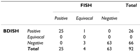
Table 2: Performance of brightfield double in situ hybridization (BDISH) with clinical samples based on the ASCO/CAP method with FISH equivocal cases
FISH Total
Positive Equivocal Negative
BDISH Positive 25 1 0 26
Equivocal 0 0 0 0
Negative 0 3 63 66
Total 25 4 63 92
All discordant cases, that we defined it even by one observer disagreement between the BDISH and FISH scores, were re-examined for possible causes of conflicting results and there were 9 discordant cases. All nine (9) dis-cordant cases of the BDISH slides presented at least some degree of the genotypic heterogeneity of tumor cell popu-lations. In general, there were two types of the tumor cell heterogeneity with HER2 gene status within the same tis-sue section: 1) variegated different genotype tumor cell populations in the same area of tissue section (Figure 4A) and 2) segregated tumor populations in different areas of tissue section (Figures 4B&C). Breast cancers with obvious tumor cell heterogeneity are shown as examples in Figure 4. However, the subtle genotype heterogeneity of tumor cell populations is often seen among the equivocal cases, and it also can be seen in Figures 3D &4B which show less obvious variegated tumor cell heterogeneity. Three (3) of 9 discordant cases demonstrated the segregated tumor cell heterogeneity while the other 6 cases showed various degrees of the variegated tumor cell heterogeneity.
Discussion
Accurate HER2 status testing is important for identifying breast cancer patients who may benefit from receiving trastuzumab therapy. Currently, in the United States, HER2 IHC methods are most commonly used for primary screening for HER2 status, and borderline cases are sub-jected to dual FISH for HER2 and CEN 17 to determine the HER2/CEN 17 ratio. Because the discordance rate between local and central/reference HER2 status testing with IHC and FISH is significantly high [14,31-33], the standardization of diagnosing breast cancer cases is recog-nized as a very important task for improving personalized cancer patient care [3,34]. The American Society of Clini-cal Oncology and the College of American Pathologists has published a guideline recommendation for testing
HER2 status in breast cancer [3] and the Canadian National Consensus has updated the Canadian HER2/neu
testing guideline [35]. Two potential solutions for improving the standardization of HER2 status testing
include: 1) automating the entire process for slide staining [36] and slide reading [36-39] and 2) consolidating the
HER2 testing process within experienced laboratories and pathologists that perform large numbers of HER2 tests [15].
One way to improve the accuracy of HER2 status testing is to automate the assay procedure for HER2 IHC and HER2
FISH assays so that human errors can be diminished. HER2 IHC assays can be performed using an automated slide staining system, but HER2 FISH assays remain tech-nically challenging and time consuming manual molecu-lar diagnostic assays in most laboratories. An evaluator of FISH slides must have access to specialized fluorescence microscopy in a dark room. Because of unstable FISH staining characteristics, the signals of FISH slides can be bleached easily, even while reviewing and enumerating signals. Furthermore, digital images of the FISH slide need to be captured with a sensitive camera system for each patient case for the HER2 gene status record. Therefore, it is desirable to automate a tissue-based HER2 gene status test that can be observed with a regular brightfield micro-scope and that produces stained slides that can be archived.
While the concept of multi-color brightfield ISH applica-tions was published in 1990's [40,41], it was a recent achievement to visualize HER2 and CEN 17 targets within the same nuclei of tissue sections with a manual dual brightfield ISH application [42]. This dual ISH applica-tion utilized TMB chromogen for HER2 gene staining. However, based on published images [28,42], TMB stain-ing does not provide discrete signals when compared to the SISH application. The advantages of the BDISH appli-cation for HER2 gene and CEN 17 presented in the current study are: 1) the automation of the ISH application; 2) the visualization of both HER2 gene and CEN 17 targets in the nuclei of the same cell; 3) the generation of discrete
HER2 gene signals; 4) the ability to reproducibly detect endogenous HER2 and CEN 17 signals in the stromal tis-sues and lymphocytes as a reliable internal assay control; 5) the ability to visualise signal with brightfield micros-copy with non-oil immersion lenses; and 6) the capability to permanently archive the slides.
HER2 and CEN 17 probes are co-hybridized for dual color HER2 FISH. However, for the BDISH assay, because the stringency conditions for the nick-translated HER2 probe and the CEN 17 oligoprobe were different, it was neces-sary to conduct sequential ISH staining steps for HER2
gene and CEN 17 targets. For CEN 17 ISH, we have opti-mized a new detection system with an alkaline phos-phatase-conjugated antibody and fast red chromogen and naphthol phosphate substrate reaction. The fast red-based detection was selected to obtain a good contrast of CEN
Table 3: Performance of brightfield double in situ hybridization (BDISH) with clinical samples based on the ASCO/CAP method without 4 FISH equivocal cases
FISH Total
Positive Negative
BDISH Positive 25 0 25
Negative 0 63 63
Total 25 63 88
17 ISH signal against the discrete black dots of HER2 SISH signal. DAB, BCIP/NBT, and TMB detection systems did not provide sufficient contrast against HER2 ISH black sig-nal (data not shown). Because fast red precipitate is solu-ble in organic solvents, in general, aqueous mounting medium is used for coverslipping. However, the standard coverslipping method with aqueous mounting medium on wet tissue sections did not produce tissue sections with high resolution and therefore detailed tissue structure could not be observed (data not shown). A successful method to preserve fast red staining for CEN 17 and high resolution tissue morphology was, after completely dry the slides, to apply a toluene-based tissue mounting medium (Cytoseal 60) for coverslipping with cover glass
or to use a film coverslipper (Tissue-Tek® film coverslip-per). Incomplete drying resulted in faint red background staining particularly around the fast red precipitate sites with this method. Interestingly, the use of aqueous mounting medium onto the dried tissue slides produced yellowish background staining on tissue sections and this method did not produce satisfactory results (data not shown).
The specificity of single ISH for HER2 gene or CEN 17 and BDISH for both targets was evaluated with xenograft tumors. HER2 and CEN 17 copy numbers have been doc-umented previously using the FISH assay [29]. MCF-7 cells are characterized as non-amplified HER2 and
chro-Brightfield double in situ hybridization (BDISH) for HER2 and chromosome 17 centromere (CEN 17) on formalin-fixed, paraf-fin-embedded clinical breast cancer cases
Figure 3
mosome 17 polysomy (3 copies of chromosome 17 per nucleus) while one of chromosome 17 with HER2 dele-tion (2 HER2 copies per nucleus) [29]. BT-474 cell line presents HER2 amplification with 50–60 copies of HER2
genes and 4–6 copies of CEN 17 per nucleus [29]. Ampli-fied HER2 genes are located not only on chromosome 17, but also are translocated on other chromosomes [29].
HER2 and CEN 17 copy numbers produced with the sin-gle target ISH and BDISH methods matched with previ-ously reported results. As both probes are labeled with the same DNP hapten, our first concern was to determine if detecting specific signal for each probe was feasible. We confirmed that the fast red chromogen detection reagents did not produce red signal when the fast red ISH was per-formed without the CEN 17 probe after detection of HER2
by SISH (data not shown). Thus, the SISH detection and the fast red detection can be combined to perform a sequential double ISH assay with 2 probes labeled with the same hapten. Because the sequential BDISH applica-tion uses 2 specific stringency condiapplica-tions based on the length and sequences of 2 probes, it is not necessary to design 2 probes that require the same stringency for co-hybridization, like double color FISH assays.
Concordance rates between a set of gold standard dual color HER2 FISH scores and HER2 and CEN 17 BDISH scores by 4 observers were calculated for assessing the per-formance of the BDISH assay. There were 9 discordant cases (9.6% of the total cases) based on BDISH score dis-agreement with FISH scores, even by one observer. We have found that the number of equivocal cases influences the concordance rate with the ASCO/CAP scoring method. There were 4 equivocal cases based on FISH scores and all cases showed the BDISH score disagreement by at least 2 observers. A similar observation was reported with an international HER2 testing proficiency study [15]. In their study, the discordant cases (20%) were caused by the specimen having FISH HER2/CEN 17 ratios between 1.7 and 2.3 that are close to the 'equivocal' defined by ASCO/CAP HER2 scoring method (1.8 – 2.2). They also stated "equivocal cases are difficult to interpret, even highly experienced and validated laboratories" [15]. In one study, when the FISH assay was used as the primary test for HER2 status assessment of breast carcinoma cases, heterogeneity of HER2 gene status was observed in 40 of 742 cases (5%) [43]. It has been speculated that genomic and phenotypic heterogeneity of tumor cells is the main reason for the inconsistency of HER2 testing results [44].
The heterogeneity of breast tumor cell populations Figure 4
With current study, all of our discordant cases (9/9 or 100%) displayed the tumor cell population heterogene-ity: three samples showed significant segregated tumor cell population heterogeneity (Figures 4B&C) and other cases showed subtle heterogeneity of tumor cell popula-tions that are seen among equivocal cases (4/4 or 100%). Nonetheless, since consecutive tissue sections were not used for the FISH and BDISH analyses, one can speculate that the tissue sections for the FISH and BDISH tissue sec-tions contained tumor cell populasec-tions with different
HER2 status. Further clinical evaluations of HER2 and CEN 17 BDISH application with patient treatment out-come data are required for more accurate HER2 status assessment of breast cancer patients to be obtained.
Conclusion
We have successfully developed an automated BDISH application for HER2 gene and CEN 17 targets in forma-lin-fixed, paraffin-embedded tissue sections that is highly concordant to the FISH and is reproducibly interpreted among observers. Assessment of HER2 gene status can be conducted without the use of a specialized fluorescence microscope and the time required for completing HER2
gene status assessment can be shortened significantly. Fur-thermore, this application has the potential to be used for other gene targets, any combination of a gene and its chro-mosome centromere, and tissue section-based gene assessment tests including gene translocation studies. The use of BDISH technology allows the simultaneous analy-ses of two DNA targets within the context of tissue mor-phology observation.
Competing interests
HN, BHW, ML, AEM, FG, MF, EW, TM are employed by Ventana Medical Systems, Inc. RRT and MD received grant support and honorarium for speaking from Ventana Med-ical Systems, Inc.
Authors' contributions
LW, ML, JP, and RRT were responsible for identifying and prequalification of the clinical cases used in this study. HN and TMG were responsible for the BDISH assay devel-opment and feasibility studies, staining the clinical sam-ples, and preparing the manuscript draft and image data. BHW and ML were responsible for the final assay develop-ment. AEM conducted all statistical analyses for the per-formance of BDISH assay. FG was the study coordinator and contributed intellectual content of the study. MF designed the probes. EW, MK, MD, RRT, and TMG cri-tiqued the assay performance with their molecular histol-ogy expertise during the assay development. FPL, MK, MD, and RRT were the observers for scoring the clinical samples. All authors contributed intellectual inputs to the study. All authors read and approved the final manuscript.
Acknowledgements
We would like to thank Vu Nguyen for instrument maintenance and Drs. Raymond B. Nagle and Guadalupe Manriquez for slide reviewing during the assay development.
References
1. Yaziji H, Goldstein LC, Barry TS, Werling R, Hwang H, Ellis GK, Gralow JR, Livingston RB, Gown AM: HER-2 testing in breast can-cer using parallel tissue-based methods. JAMA 2004, 291:1972-1977.
2. Owens MA, Horten BC, Da Silva MM: HER2 amplification ratios by fluorescence in situ hybridization and correlation with immunohistochemistry in a cohort of 6556 breast cancer tis-sues. Clin Breast Cancer 2004, 5:63-69.
3. Wolff AC, Hammond ME, Schwartz JN, Hagerty KL, Allred DC, Cote RJ, Dowsett M, Fitzgibbons PL, Hanna WM, Langer A, McShane LM, Paik S, Pegram MD, Perez EA, Press MF, Rhodes A, Sturgeon C, Taube SE, Tubbs R, Vance GH, Vijver M van de, Wheeler TM, Hayes DF: American society of clinical oncology/college of American pathologists guideline recommendations for human epider-mal growth factor receptor 2 testing in breast cancer. J Clin Oncol 2007, 25:118-145.
4. Garrison LP Jr, Lubeck D, Lalla D, Paton V, Dueck A, Perez EA: Cost-effectiveness analysis of trastuzumab in the adjuvant setting for treatment of HER2-postive breast cancer. Cancer 2007, 110:489-498.
5. Norum J, Olsen JA, Wist EA, Lønning PE: Trastuzumab in adju-vant breast cancer therapy. A model based cost-effec-tivemess analysis. Acta Oncol 2007, 46:153-164.
6. Fagnani F, Colin X, Arveux P, Coudert B, Misset JL: Cost/effective-ness analysis of adjuvant therapy with trastuzumab in patients with HER2 positive early breast cancer. Bull Cancer
2007, 94:711-720.
7. Millar JA, Millward MJ: Cost effectiveness of trastuzumab in the early breast cancer: a lifetime model. Pharmocoeconomics 2007, 25:429-442.
8. de Korte MA, de Vries EG, Lub-de Hooge MN, Jager PL, Gietema JA, Graaf WT van der, Sluiter WJ, van Veldhuisen DJ, Suter TM, Sleijfer DT, Perik PJ: 111Indium-trastuzumab visualizes myocardial human epidermal growth factor receptor 2 expression shortly after anthracycline treatment but not during heart failure: a clue to uncover the mechanisms of trastuzumab-related cardiotoxicity. Eur J Cancer 2007, 43:2046-2051. 9. Dal Lago L, Durbecq V, Desmedt C, Salgado R, Verjat T, Lespagnard
L, Ma Y, Veys I, Di Leo A, Sotiriou C, Piccart M, Larsimont D: Cor-rection for chromosome-17 is critical for the determination of true Her-2/neu gene amplification status in breast cancer. Mol Cancer Ther 2006, 5:2572-2579.
10. Salido M, Tusquets I, Corominas JM, Suarez M, Espinet B, Corzo C, Bellet M, Fabregat X, Serrano S, Solé F: Polysomy of chromosome 17 in breast cancer tumors showing an overexpression of ERBB2: a study of 175 cases using fluorescence in situ hybrid-ization and immunohistochemistry. Breast Cancer Res 2005, 7:R267-R273.
11. Torrisi R, Rotmensz N, Bagnardi V, Viale G, Curto BD, Dell'orto P, Veronesi P, Luini A, D'Alessandro C, Cardillo A, Goldhirsch A, Colle-oni M: HER2 status in early breast cancer: relevance of cell staining patterns, gene amplification and polysomy 17. Eur J Cancer 2007, 43:2339-2344.
12. Cayre A, Mishellany F, Lagarde N, Penault-Llorca F: Comparison of different commercial kits for HER2 testing in breast cancer: looking for the accurate cut-off for amplification. Breast Can-cer Res 2007 in press.
13. Lambros MB, Simpson PT, Jones C, Natrajan R, Westbury C, Steele D, Savage K, Mackay A, Schmitt FC, Ashworth A, Reis-Filho JS: Unlocking pathology archives for molecular genetic studies: a realizable method to generate probes for chromogenic and fluorescent in situ hybridization. Lab Invest 2006, 86:398-408.
Publish with BioMed Central and every scientist can read your work free of charge
"BioMed Central will be the most significant development for disseminating the results of biomedical researc h in our lifetime."
Sir Paul Nurse, Cancer Research UK
Your research papers will be:
available free of charge to the entire biomedical community
peer reviewed and published immediately upon acceptance
cited in PubMed and archived on PubMed Central
yours — you keep the copyright
Submit your manuscript here:
http://www.biomedcentral.com/info/publishing_adv.asp
BioMedcentral 15. Dowsett M, Hanna WM, Kockx M, Penault-Llorca F, Rüschoff J,
Gut-jahr T, Habben K, Vijver MJ van de: Standardization of HER2 test-ing: results of an international proficiency-testing ring study. Mod Pathol 2007, 20:584-591.
16. Persons DL, Tubbs RR, Cooley LD, Dewald GW, Dowling PK, Du E, Mascarello JT, Rao KW, Wilson KS, Wolff DJ, Habegger-Vance G: HER-2 fluorescence in situ hybridization: results from the survey program of the College of American Pathologists. Arch Pathol Lab Med 2006, 130:325-331.
17. Arnould L, Denoux Y, MacGrogan G, Penault-Llorca F, Fiche M, Treil-leux I, Mathieu MC, Vincent-Salomon A, Vilain MO, Couturier J: Agreement between chromogenic in situ hybridization (CISH) and FISH in the determination of HER2 status in breast cancer. Br J Cancer 2003, 88:1587-1591.
18. Zhao J, Wu R, Au A, Marquez A, Yu Y, Shi Z: Determination of HER2 gene amplification by chromogenic in situ hybridiza-tion (CISH) in archival breast carcinoma. Mod Pathol 2002, 15:657-665.
19. Peiró G, Mayr D, Hillemanns P, Löhrs U, Diebold J: Analysis of HER-2/neu amplification in endometrial carcinoma by chro-mogenic in situ hybridization. Correlation with fluorescence in situ hybridization, HER-2/neu, p53 and Ki-67 protein expression, and outcome. Mod Pathol 2004, 17:277-287. 20. Park K, Kim J, Lim S, Han S, Lee JY: Comparing fluorescence in
situ hybridization and chromogenic in situ hybridization methods to determine the HER2/neu status in primary breast carcinoma using tissue microarray. Mod Pathol 2003, 16:937-943.
21. Hanna WM, Kwok K: Chromogenic in-situ hybridization: a via-ble alternative to fluorescence in-situ hybridization in the HER2 testing algorithm. Mod Pathol 2006, 19:481-487. 22. Todorović-Raković N, Jovanović D, Nesković-Konstantinović Z,
Nikolić-Vukosavljević D: Prognostic value of HER2 gene ampli-fication detected by chromogenic in situ hybridization (CISH) in metastatic breast cancer. Exp Mol Pathol 2007, 82:262-268.
23. Tubbs R, Pettay J, Skacel M, Powell R, Stoler M, Roche P, Hainfeld J: Gold facilitated in situ hybridization (GOLDFISH): A bright field autometallographic alternative to fluorescence in situ hybridization for detection of HER-2/neu gene amplification. Amer J Pathol 2002, 160:1589-1595.
24. Tubbs R, Skacel M, Pettay J, Powell R, Myles J, Hicks D, Sreenan J, Roche P, Stoler MH, Hainfeld J: Interobserver interpretative reproducibility of GOLDFISH, a first generation gold-facili-tated autometallographic bright field in situ hybridization assay for HER-2/neu amplification in invasive mammary car-cinoma. Am J Surg Pathol 2002, 26:908-913.
25. Tubbs R, Pettay J, Hicks D, Skacel M, Powell R, Grogan T, Hainfeld J: Novel bright field molecular morphology methods for detec-tion of HER2 gene amplificadetec-tion. J Mol Histol 2004, 35:589-594. 26. Downs-Kelly E, Pettay J, Hicks D, Skacel M, Yoder B, Rybicki L, Myles J, Sreenan J, Roche P, Powell R, Hainfeld J, Grogan T, Tubbs R: Ana-lytical validation and interobserver reproducibility of Enz-Met GenePro: a second-generation bright-field metallography assay for concomitant detection of HER2 gene status and protein expression in invasive carcinoma of the breast. Am J Surg Pathol 2005, 29:1505-1511.
27. Powell RD, Pettay JD, Powell WC, Roche PC, Grogan TM, Hainfeld JF, Tubbs RR: Metallographic in situ hybridization. Hum Pathol
2007, 38:1145-1159.
28. Dietel M, Ellis IO, Höfler H, Kreipe H, Moch H, Dankof A, Kölble K, Kristiansen G: Comparison of automated silver enhanced in situ hybridization (SISH) and fluorescence ISH (FISH) for the validation of HER2 gene status in breast carcinoma accord-ing to the guidelines of the American Society of Clinical Oncology and the College of American Pathologists. Virchows Arch 2007, 451:19-25.
29. Grushko TA, Blackwood MA, Schumm PL, Hagos FG, Adeyanju MO, Feldman MD, Sanders MO, Weber BL, Olopade OI: Molecular-cytogenetic analysis of HER-2/neu gene in BRCA1-associated breast cancers. Cancer Res 2002, 62:1481-1488.
30. Hicks DG, Tubbs RR: Assessment of the HER2 Status in Breast Cancer by Fluorescence In-Situ Hybridization (FISH): a Technical Review with Interpretative Guidelines. Hum Pathol
2005, 36:250-261.
31. Paik S, Bryant J, Tan-Chiu E, Romond E, Hiller W, Park K, Brown A, Yothers G, Anderson S, Smith R, Wickerham DL, Wolmark N: Real-world performance of HER2 testing-National Surgical Adju-vant Breast and Bowel Project experience. J Natl Cancer Inst
2002, 94:852-854.
32. Roche PC, Suman VJ, Jenkins RB, Davidson NE, Martino S, Kaufman PA, Addo FK, Murphy B, Ingle JN, Perez EA: Concordance between local and central laboratory HER2 testing in the breast intergroup trial N9831. J Natl Cancer Inst 2002, 94(11):855-857.
33. Dowsett M, Hanby AM, Laing R, Walker R, National HER2 Consulta-tion Steering Group : HER2 testing in the UK: consensus from a national consultation. J Clin Pathol 2007, 60:685-689.
34. Ross JS, Symmans WF, Pusztai L, Hortobagyi GN: Standardizing slide-based assays in breast cancer: hormone receptors, HER2, and sentinel lymph nodes. Clin Cancer Res 2007, 13:2831-2835.
35. Hanna W, O'malley FP, Barnes P, Berendt R, Gaboury L, Magliocco A, Pettigrew N, Robertson S, Sengupta S, Têtu B, Thomson T: Undated recommendation from the Canadian National Consensus Meeting on HER2/neu testing in breast cancer. Curr Oncol
2007, 14:149-153.
36. Tubbs RR, Pettay JD, Swain E, Roche PC, Powell W, Hicks DG, Gro-gan T: Automation of manual components and image quanti-fication of direct dual label fluorescence in situ hybridization (FISH) for HER2 gene amplification: a feasibility study. Appl Immunohistochem Mol Morphol 2006, 14:436-440.
37. Ciampa A, Xu B, Ayata G, Baiyee D, Wallace J, Wertheimer M, Edmis-ton K, Khan A: HER-2 status in breast cancer: correlation of gene amplification by FISH with immunohistochemistry expression using advanced cellular imaging system. Appl Immunohistochem Mol Morphol 2006, 14:132-137.
38. Theodosiou Z, Kasampalidis IN, Karayannopoulou G, Kostopoulos I, Bobos M, Bevilacqua G, Aretini P, Starita A, Lyroudia K, Pitas I: Eval-uation of FISH image analysis system on assessing HER2 amplification in breast carcinoma cases. Breast 2007, 17:80-84. 39. Stevens R, Almanaseer I, Gonzalez M, Caglar D, Knudson RA, Ketter-ling RP, Schrock DS, Seemayer TA, Bridge JA: Analysis of HER2 gene amplification using an automated fluorescence in situ hybridization signal enumeration system. J Mol Diagn 2007, 9:144-150.
40. Speel EJ, Jansen MP, Ramaekers FC, Hopman AH: A novel triple-color detection procedure for brightfield microscopy, com-bining in situ hybridization with immunocytochemistry. J His-tochem CyHis-tochem 1994, 42:1299-1307.
41. Hopman AHN, Claessen S, Speel EJM: Multi-colour brightfield in situ hybridization on tissue sections. Histochem Cell Biol 1997, 108:291-298.
42. Laakso M, Tanner M, Isola J: Dual-colour chromogenic in situ hybridization for testing HER-2 oncogene amplification in archival breast tumors. J Pathol 2006, 210:3-9.
43. Tubbs RR, Hicks DG, Cook J, Downs-Kelly E, Pettay J, Hartke MB, Hood L, Neelon R, Myles J, Budd GT, Moore HC, Andresen S, Crowe JP: Fluorescence in situ hybridization (FISH) as primary methodology for the assessment of HER2 status in adeno-carcinoma of the breast. Diagn Mol Pathol 2007, 16:207-210. 44. Tuma RS: Inconsistency of HER2 test raises questions. J Natl