C A S E R E P O R T
Open Access
In-depth investigation of the molecular
pathogenesis of bladder cancer in a unique
26-year old patient with extensive multifocal
disease: a case report
Tahlita CM Zuiverloon
1,2, Cheno S Abas
1, Kirstin A van der Keur
1, Marcel Vermeij
1, Stephen S Tjin
1,
Angela G van Tilborg
1, Martijn Busstra
2, Ellen C Zwarthoff
1*Abstract
Background:The molecular characteristics and the clinical disease course of bladder cancer (BC) in young patients remain largely unresolved. All patients are monitored according to an intensive surveillance protocol and we aim to gain more insight into the molecular pathways of bladder tumors in young patients that could ultimately contribute to patient stratification, improve patient quality of life and reduce associated costs. We also determined whether a biomarker-based surveillance could be feasible.
Case Presentation:We report a unique case of a 26-year-old Caucasian male with recurrent non-muscle invasive bladder tumors occurring at a high frequency and analyzed multiple tumors (maximal pTaG2) and urine samples of this patient. Analysis includedFGFR3mutation detection, FGFR3 and TP53 immunohistochemistry, mircosatellite analysis of markers on chromosomes 8, 9, 10, 11 and 17 and a genome wide single nucleotide polymorphism-array (SNP). All analyzed tumors contained a mutation inFGFR3and were associated with FGFR3 overexpression. None of the tumors showed overexpression of TP53. We found a deletion on chromosome 9 in the primary tumor and this was confirmed by the SNP-array that showed regions of loss on chromosome 9. Detection of all recurrences was possible by urinaryFGFR3mutation analysis.
Conclusions:Our findings would suggest that the BC disease course is determined by not only a patient’s age, but also by the molecular characteristics of a tumor. This young patient contained typical genetic changes found in tumors of older patients and implies a clinical disease course comparable to older patients. We demonstrate thatFGFR3mutation analysis on voided urine is a simple non-invasive method and could serve as a feasible follow-up approach for this young patient presenting with anFGFR3mutant tumor.
Background
Bladder cancer (BC) is a disease of the elderly with a peak incidence in the sixth decade of life. Tumors are sporadically found under the age of 40 (1-4%) and most young patients present with tumors of low stage and grade [1-4]. Conflicting results have been found con-cerning the natural history and prognosis of bladder tumors in young patients. The small number of cases and the definition of “young” with age ranging from 5-45 years may be responsible for this variation [2,3,5,6].
Evidence is accumulating that there is a difference in the natural history of patients under the age of 20 and patients between 30-50 years of age. Patients <20 years mostly have tumors with a low recurrence rate, a favor-able clinical outcome and few genetic alterations, while patients between 30-50 years have a disease course com-parable to older patients [7].
Almost 80% of the BC patients will present with non-muscle invasive disease (NMI-BC). Treatment is by trans-urethral resection of the tumor, but almost 70% of the patients will have at least one recurrence within five years and 10-20% will progress to muscle-invasive dis-ease. After the first tumor resection all age groups of
* Correspondence: [email protected] 1
Department of Pathology, Erasmus MC, Rotterdam, the Netherlands
patients are monitored according to an intensive surveil-lance protocol that includes 3-monthly cystoscopies the first two years, followed by less frequent observations if a patient stays recurrence free. The main disadvantages of the current protocol are life-long invasive and costly cystoscopic monitoring of patients causing physical dis-comfort and sexual dysfunction [8].
To our knowledge there are only few studies that investigated the molecular changes in bladder tumors of young patients. Identifying the molecular pathways of these tumors could define a subset of patients and redir-ect patient management towards a new patient friendly and individualized follow-up protocol. One of the most promising markers associated with NMI bladder tumors is the mutation status of the Fibroblast Growth Factor Receptor-3 (FGFR3). Mutations in FGFR3 have been associated with BC tumors of low stage and grade and patients having a favorable prognosis [9]. We have recently shown thatFGFR3 mutation analysis on voided urine of NMI-BC patients with a mutation inFGFR3is a non-invasive inexpensive tool for patient surveillance (Zuiverloonet al. submitted). Additionally, multiple stu-dies report on the use of microsatellite analysis (MA) for detection of loss-of-heterozygosity (LOH) as a diag-nostic marker. LOH detected by MA is mainly located on chromosomes 8, 9, 10, 11, 13 and 17 and these losses have been associated with stage, grade, invasive growth, recurrent disease and progression. In the present study we analyzed multiple tumor and urine samples of a unique young patient forFGFR3mutation status, LOH, FGFR3 and TP53 expression and performed a genome wide single nucleotide polymorphism-array (SNP). Since this patient presented with multiple recurrences within a short time-span we determined retrospectively whether FGFR3mutation detection could be a feasible follow-up approach.
Case presentation
We report a unique case of a 26-year-old Caucasian male with recurrent non-muscle invasive bladder tumors occurring at a high frequency. The patient presented at first in March 2007 with macroscopic hematuria for a few weeks. He received a total of 5 trans-urethral resec-tions of the multifocal bladder tumors within 2 years, highest stage and grade being TaG2. The first three resections included an average of 15 papillary tumors and the last two resections included 3 tumors. The patient received two direct post-operative intravesical instillations of epirubicin. Additionally, our patient initi-ally received adjuvant intravesical immunotherapy with bacillus calmette-guérin (BCG) in 2008, but switched to mitomycin-C (MMC) in 2009 for maintenance due to complications. A CT-scan of the pelvis and abdomen demonstrated no evidence of upper urinary tract lesions,
no signs of urolithiasis, nodal or distant metastases. There was no family history of bladder cancer and intoxications included a smoking status of 5.5 pack-years (one pack-year = one pack of cigarettes a day for one year ~20 cigarettes a day for one year). There was no indication of any contact with aromatic amines. The patient worked as a soldier in Bosnia in 2001 and 2003 where he was part of a recovery team driving a diesel armed-truck. Tasks of the recovery team included clean-ing of remainclean-ing ammunition enforced with depleted uranium (DU) and military equipment wreckage.
Methods Tissue samples
Tumor tissue was obtained from formalin-fixed, paraf-fin-embedded samples. Tumor sections were selected by pathological examination to contain a minimum amount of 80% tumor cells and sections were manually dissected from 4μ slides. Samples were first deparaffinized and DNA was extracted using the Qiagen Dneasy blood and tissue kit (Qiagen, GmbH, Hilden, Germany) according to the manufacturer’s protocol.
Urine samples
Freshly voided urine (10-100 ml) was collected prior to a cystoscopy or trans-urethral resection of the tumor and stored at 4°C until transportation to the department of Pathology at Erasmus MC, Rotterdam. Urine was spun down for 10’at 3000 rpm (1500 × g). Cell pellets were washed twice with 10 ml of Phosphate-buffered saline (PBS) and spun down for 10’at 3000 rpm. Pellets were resuspended in 1 ml of PBS, transferred to an eppendorf vial and spun down for 5’at 6000 rpm (3000 × g). Supernatant was discarded and the cell pellet was stored at -20°C until DNA isolation. DNA was extracted using the QiAamp DNA mini-kit (Qiagen GmbH, Hilden, Germany) according to the manufacturer’s protocol.
Microsatellite analysis
Microsatellite analysis was performed as described by van der Aa et al. [11]. Markers used for detection of LOH were: D8S1130, D81125, D8S1107, D8S1109, D8S1145, D9S1118, D9S252, D9S304, D9S299, D9S752, D9S930, G10693, D10S1225, D11S1999, D11S1981 and D17S969.
FGFR3 and TP53 immunohistochemistry
Bladder tumors were fixed in 10% buffered formaldehyde, embedded in paraffin, 4 micrometer sections were mounted on a coated glass slide (Starfrost Knittel-Glaeser D38114 Braunschweig Germany). Haematoxylin-Eosin was used as a general stain and immunohistochemistry was performed with rabbit polyclonal antibody anti-human FGFR3 (Santa Cruz Biotechnology INC) and monoclonal mouse anti-human P53 protein clone D-07 (Dakocytomation, Denmark A/S). Antigen retrieval by microwave heating in TRIS-EDTA buffer pH 9.0 was used and endogenous peroxidase was removed by 0.30% H2O2in PBS/TWEEN. Pretreatment with blocking buffer
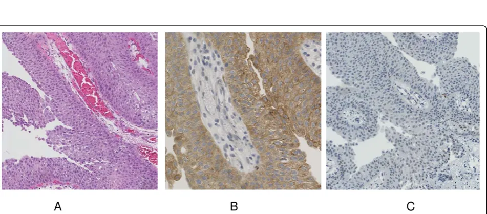
PROTIFAR 0.5% in PBS/TWEEN was used prior to the overnight incubation at 4°C with the primary antibody. Visualization was done by Dako Real Envision Detection system peroxidase/DAB+ (Dakocytomation, Denmark A/ S), containing anti-mouse and anti-rabbit antibodies, according to the manufacturer’s protocol. Counterstain-ing was done with haematoxylin (Klinipath 6921 GX, Duiven, The Netherlands). Expression of FGFR3 was scored in a semi-quantitaive scoring system: 0 = all tumor cells negative, 1 = faint positivity of in some or all cells, 2 = weak but extensive positivity and 3 = strong positivity/overexpression (regardless of extent). TP53 overexression was scored if >10% of the cells stained positive. As a reference normal urothelium was analyzed for FGFR3 and TP53 protein expression. Reference sec-tions of known high and low expressions levels of FGFR3 and TP53 were included in the staining runs.
Infinium HumanHap370CNV Genome wide SNP array For Infinium HumanHap370CNV Genotyping BeadChip SNP array analysis we used 750 ng of patient DNA and followed the protocol as described by the manufacturer (Illumina Inc., San Diego, CA, USA). We used Illumina BeadStudio software to extract data. The Nexus CGH Plug-in for CNV Analysis from BioDiscovery (BioDis-covery Inc., El Segundo, CA, USA) was used to export Illumina CNV data to Nexus Copy Number version 4.0. Arrays were processed using the built-in Rank Segmen-tation algorithm.
Results and discussion
Young patients rarely present with BC and there are multiple studies that indicate a good clinical disease
course where patients present with solitary tumors and a low recurrence and progression rate. Since BC patients are monitored cystoscopically according to an intensive surveillance protocol, gaining more insight into the molecular pathways of BC tumors in young patients could define a subset of patients that can be monitored less frequently, hereby improving patient quality of life and reducing associated costs.
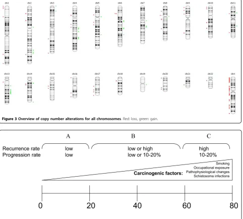
We presented a unique case of a 26-year-old male with multiple multifocal NMI bladder tumors recurring at a high frequency. After starting intravesical mainte-nance therapy with MMC the recurrence rate decreased and up to date the patient stayed recurrence free. Mole-cular analysis of the primary tumor revealed an S249C mutation in FGFR3 (Figure 1B) and overexpression of FGFR3 (Figure 2B). LOH on chromosome 9 was detected by MA and confirmed by the genome wide SNP array analysis. We also found other regions of loss and gain that are considered minor when compared to tumors of the same stage and grade (Figure 3). There was no increased expression of TP53 (Figure 2C). Hence, this young patient appears to have the typical genetic changes found in older patients with NMI-BC. This implies that the patient could have a disease course comparable to older patients and warrants regu-lar controls due to the risk of additional recurrences or progression. These findings combined with previous studies suggest that not only a patient’s age, but also the molecular characteristics of the tumor determine the clinical disease course. Since it takes time to accu-mulate typical genetic changes involved in BC - e.g.
mutations in FGFR3and TP53 and LOH on
Figure 1FGFR3mutation detection on urinary derived DNA.FGFR3mutation analysis of anFGFR3tumor without a mutation (A), mutation S249C on tumor DNA (B) and urinary derived DNA (C) from the same patient used in panel B.
Exogenous risk factors that could have contributed to BC in our patient are smoking, exposure to diesel exhaust and depleted uranium (DU). First our patients smoking status is 5.5 pack-years, which is known to be associated with an increased risk of BC. Secondly, a meta-analysis of BC and diesel exhaust exposure demonstrated a relative risk of 1.44 for occupations exposed to high diesel fume levels [14]. Our patient worked as a driver of a diesel armed truck and was exposed to diesel exhaust fumes, working in a valley were the fumes were retained in a cloud of exploded
ammunition. Lastly, although some believe that there is a link between exposure to DU and cancer develop-ment no hard evidence has been found to support this hypothesis. While evidence from Hiroshima data shows a latency period of 10-15 years to develop can-cer this concan-cerns an acute high-dose exposure and other studies were not able to demonstrate this link in Balkan veterans [15,16]. On the other hand two studies by Miller et al. demonstrated in vitro tumorigenic transformation of osteoblasts when exposed to DU [17,18].
Figure 3Overview of copy number alterations for all chromosomes. Red: loss, green: gain.
Since Van der Aa et al. demonstrated that specifically young patients perceive a cystoscopic investigation as burdensome this emphasizes the need for patient strati-fication [19]. To determine whether a young BC patient should be monitored according to the standard follow-up protocol or can be monitored less frequently by cystoscopy, we propose to determine the FGFR3 muta-tion status of the primary tumor. One possibility is that the tumor will have few genetic changes and secondly that the tumor will have genetic changes comparable to those found in older patients (Figure 4B, 4C). Mutations inFGFR3 are tumor-specific and are not found in nor-mal tissue, meaning that detection of a mutation in voided urine indicates the presence of tumor cells in the urinary tract. The results of the follow-up in time for our patient are indicated in Additional file 1. We
demonstrate that the S249C mutation in FGFR3
detected in the tumor was also detected in the urine (Figure 1C), indicating that the detected tumor cells were shed by the resected tumor. Our results demon-strate that urine cytology does not detect the tumor in two cases, which is in concordance with previous studies that demonstrate a low sensitivity of urine cytology for the detection of tumors of low stage and grade [20,21]. Although this concerns just one patient, our results imply that patient monitoring byFGFR3 mutation ana-lysis could be a feasible non-invasive method in the fol-low-up of young NMI-BC patients presenting with a mutation in FGFR3and indicate that future research is required to investigate this.
Conclusions
Multiple studies demonstrate the relatively benign dis-ease course of bladder tumors in young patients, but there are some cases with a high recurrence rate and a progression rate comparable to older patients. We pre-sent a young patient having multiple multifocal recur-rent bladder tumors with molecular characteristics found in older patients. It would be of interest to per-form molecular studies in a larger subset of patients to elucidate whether these tumors comprise a biologically distinct group. Since BC tumors rarely present in young patients, a multicenter collaboration would be needed for investigation. This could ultimately lead to stratification of patients that need close monitoring and patients with favorable molecular characteristics of the tumor that can be monitored less frequently, hereby decreasing the number of cystoscopies per-formed in young patients who specifically perceive a cystoscopy as burdensome. For the follow-up of young
patients presenting with an FGFR3 mutant NMI
tumor, FGFR3mutation analysis could be a feasible alternative for recurrence detection.
Consent
Written informed consent was obtained from the patient for publication of this case report and any accompany-ing images. A copy of the written consent is available for review by the Editor-in-Chief of this journal.
Additional file 1: Patient follow-up schedule in time. Patient’s surveillance schedule starting at the primary tumor. Next rows represent tumor histology or urine cytology,FGFR3mutation status and type of intravesical treatment.
Click here for file
[ http://www.biomedcentral.com/content/supplementary/1471-2490-10-5-S1.DOC ]
Acknowledgements
We would like to thank Dr. W. Kirkels for bringing this case to our attention and thank Dr. W. B. J. Nathrath for providing material of the primary tumor. This study was funded by a grant from the Koningin Wilhelmina Cancer foundation (KWF): 2006-3672.
Author details
1Department of Pathology, Erasmus MC, Rotterdam, the Netherlands. 2Department of Urology, Erasmus MC, Rotterdam, the Netherlands.
Authors’contributions
TCMZ and ECZ designed the study. TCMZ performed the literature review and drafted the manuscript. CSA, MB and TCMZ collected the tissue and urine material. CSA, SST and KvdK performedFGFR3mutation analysis and MA analysis. AvT performed the SNP-array and interpreted the results. MV performed immunohistochemical analysis and reviewed histopathological diagnosis. ECZ revised the manuscript for important intellectual content. All authors read and approved the manuscript.
Competing interests
The authors declare that they have no competing interests.
Received: 10 September 2009 Accepted: 26 February 2010 Published: 26 February 2010
References
1. Wan J, Grossman HB:Bladder carcinoma in patients age 40 years or younger.Cancer1989,64(1):178-181.
2. McCarthy JP, Gavrell GJ, LeBlanc GA:Transitional cell carcinoma of bladder in patients under thirty years of age.Urology1979,13(5):487-489. 3. Migaldi M, Rossi G, Maiorana A, Sartori G, Ferrari P, De Gaetani C,
Cittadini A, Trentini GP, Sgambato A:Superficial papillary urothelial carcinomas in young and elderly patients: a comparative study.BJU Int
2004,94(3):311-316.
4. Erozenci A, Ataus S, Pekyalcin A, Kural A, Talat Z, Solok V:Transitional cell carcinoma of the bladder in patients under 40 years of age.Int Urol Nephrol1994,26(2):179-182.
5. Yossepowitch O, Dalbagni G:Transitional cell carcinoma of the bladder in young adults: presentation, natural history and outcome.J Urol2002,
168(1):61-66.
6. McGuire EJ, Weiss RM, Baskin AM:Neoplasms of transitional cell origin in first twenty years of life.Urology1973,1(1):57-59.
7. Wild PJ, Giedl J, Stoehr R, Junker K, Boehm S, van Oers JM, Zwarthoff EC, Blaszyk H, Fine SW, Humphrey PA,et al:Genomic aberrations are rare in urothelial neoplasms of patients 19 years or younger.J Pathol2007,
211(1):18-25.
8. Aa van der MN, Bekker MD, Kwast van der TH, Essink-Bot ML, Steyerberg EW, Zwarthoff EC, Sen FE, Elzevier HW:Sexual function of patients under surveillance for bladder cancer.BJU Int2009,104(1):35-40. 9. Hernandez S, Lopez-Knowles E, Lloreta J, Kogevinas M, Amoros A, Tardon A,
mutations as a prognostic factor in nonmuscle invasive urothelial bladder carcinomas.J Clin Oncol2006,24(22):3664-3671.
10. van Oers JM, Lurkin I, van Exsel AJ, Nijsen Y, van Rhijn BW, Aa van der MN, Zwarthoff EC:A simple and fast method for the simultaneous detection of nine fibroblast growth factor receptor 3 mutations in bladder cancer and voided urine.Clin Cancer Res2005,11(21):7743-7748.
11. Aa van der MN, Zwarthoff EC, Steyerberg EW, Boogaard MW, Nijsen Y, Keur van der KA, van Exsel AJ, Kirkels WJ, Bangma C, Kwast van der TH:
Microsatellite Analysis of Voided-Urine Samples for Surveillence of Low-Grade Non-Muscle-Invasive Urothelial Carcinoma: Feasibility and Clinical Utility in a Prospective Multicenter Study (Cost-Effectiveness of Follow-Up of Urinary Bladder Cancer Trial [CEFUB]).Eur Urol2008,55(3):659-67. 12. Johnson DE, Hillis S:Carcinoma of the bladder in patients less than 40
years old.J Urol1978,120(2):172-173.
13. Ozbey I, Aksoy Y, Bicgi O, Polat O, Okyar G:Transitional cell carcinoma of the bladder in patients under 40 years of age.Int Urol Nephrol1999,
31(5):655-659.
14. Boffetta P, Silverman DT:A meta-analysis of bladder cancer and diesel exhaust exposure.Epidemiology2001,12(1):125-130.
15. Finch SC:Radiation-induced leukemia: lessons from history.Best Pract Res Clin Haematol2007,20(1):109-118.
16. Storm HH, Jorgensen HO, Kejs AM, Engholm G:Depleted uranium and cancer in Danish Balkan veterans deployed 1992-2001.Eur J Cancer2006,
42(14):2355-2358.
17. Miller AC, Bonait-Pellie C, Merlot RF, Michel J, Stewart M, Lison PD:
Leukemic transformation of hematopoietic cells in mice internally exposed to depleted uranium.Mol Cell Biochem2005,279(1-2):97-104. 18. Miller AC, Brooks K, Stewart M, Anderson B, Shi L, McClain D, Page N:
Genomic instability in human osteoblast cells after exposure to depleted uranium: delayed lethality and micronuclei formation.J Environ Radioact2003,64(2-3):247-259.
19. Aa van der MN, Steyerberg EW, Sen EF, Zwarthoff EC, Kirkels WJ, Kwast van der TH, Essink-Bot ML:Patients’perceived burden of cystoscopic and urinary surveillance of bladder cancer: a randomized comparison.BJU Int
2008,101(9):1106-1110.
20. Ramakumar S, Bhuiyan J, Besse JA, Roberts SG, Wollan PC, Blute ML, O’Kane DJ:Comparison of screening methods in the detection of bladder cancer.J Urol1999,161(2):388-394.
21. van Rhijn BW, Poel van der HG, Kwast van der TH:Urine markers for bladder cancer surveillance: a systematic review.Eur Urol2005,
47(6):736-748.
Pre-publication history
The pre-publication history for this paper can be accessed here: http://www.biomedcentral.com/1471-2490/10/5/prepub
doi:10.1186/1471-2490-10-5
Cite this article as:Zuiverloonet al.:In-depth investigation of the molecular pathogenesis of bladder cancer in a unique 26-year old patient with extensive multifocal disease: a case report.BMC Urology
201010:5.
Submit your next manuscript to BioMed Central and take full advantage of:
• Convenient online submission
• Thorough peer review
• No space constraints or color figure charges
• Immediate publication on acceptance
• Inclusion in PubMed, CAS, Scopus and Google Scholar • Research which is freely available for redistribution