R E S E A R C H A R T I C L E
Open Access
Treatment of upper urinary tract stones with
extracorporeal shock wave lithotripsy (ESWL)
Sonolith vision
Kogenta Nakamura
1*, Motoi Tobiume
1, Masahiro Narushima
2, Takahiko Yoshizawa
1, Genya Nishikawa
1,
Yoshiharu Kato
1, Remi Katsuda
1, Kenji Zennami
1, Shigeyuki Aoki
1, Yoshiaki Yamada
1, Nobuaki Honda
1and
Makoto Sumitomo
1Abstract
Background:The aim was to retrospectively assess the results of treatment of upper urinary tract stones with the Sonolith vision manufactured by EDAP, and purchased in 2004.
Methods:The subjects were 226 Japanese patients who underwent extracorporeal shock wave lithotripsy (ESWL) alone as an initial treatment and could be followed up for at least 3 months, selected from 277 candidate patients who underwent this therapy between 2004 and 2006. Treatment effect was evaluated by kidney, ureter, and bladder X-ray or renal ultrasonography at 1 and 3 months after treatment. A stone-free status or status of stone fragmentation to 4 mm or smaller was considered to indicate effective treatment.
Results:At 3 months after treatment, the stone-free rate was 69.4% and the efficacy rate was 77.4% for renal stones, while these rates were 91.5 and 93.3%, respectively for ureteral stones. Assessment of treatment effect classified by the location of stones revealed a stone-free rate of 94.6% and an efficacy rate of 94.6% for lower ureteral stones (4.0 mm or smaller, 1 subject; 4.1-10.0 mm, 31 subjects; 10.1-20.0 mm, 5 subjects: number of treatment sessions, 1 or 2 sessions [mean: 1.03 sessions]). Complications of this therapy included renal subcapsular hematoma and pyelonephritis in 1 case each.
Conclusions:ESWL with the Sonolith vision manufactured by EDAP produced a treatment effect equivalent to those achieved with other models of ESWL equipment. ESWL seems to be an effective first-line treatment also in patients who have lower ureteral stones 10 mm or larger but do not wish to undergo TUL, if measures such as suitable positioning of the patient during treatment are taken.
Background
Extracorporeal shock wave lithotripsy (ESWL) was introduced in clinical practice by Chaussy et al.in the 1980 s [1]. Its usefulness is widely recognized, and it has become the most common treatment for upper urinary tract stones. ESWL has been performed more frequently than TUL for stone treatment in Japan compared with that in Europe and the United States (2008 General Meeting, Seminar of the Japanese Urological Associa-tion), which may be largely attributable to its simplicity
and/or the National Health Insurance System in Japan. Our hospital introduced the Sonolith vision (manufac-tured by EDAP) in March 2004 in place of the old equipment, and has been performing ESWL on patients with upper urinary tract stones. Here we report the favorable results of treatment of lower ureteral stones in cases where lithotripsy was difficult, which were achieved by putting the patient in a suitable position.
Methods
The present study was reviewed and approved by the Ethics Committee of Aichi Medical University School of Medicine (No. 11-015). The subjects were 226 Japanese patients who underwent ESWL alone and could be
* Correspondence: kogenaka@aichi-med-u.ac.jp 1
Department of Urology, Aichi Medical University School of Medicine Nagakute, Aichi 480-1195, Japan
Full list of author information is available at the end of the article
followed up for at least 3 months, selected from 277 candidate patients who underwent this therapy as the initial treatment between March 1, 2004 and December 31, 2006. The subjects included 169 male and 57 female aged from 17 to 86 years (mean: 50.5 years). The stones were on the left side in 128 subjects and on the right side in 98. The locations and sizes of the stones are shown in Table 1.
Treatment was performed during a hospital stay of three days and two nights, and the subjects were only fasted before operation. Preoperative medication included diclofenac suppository, Atarax-P, atropine sul-fate (i.m.), and glycerin enema (60 mL). When subjects complained of severe pain during operation, pentazocine (i.m.) was additionally administered. Renal stones were treated at a maximum energy level of 80%, while uret-eral stones were treated at a level of 100%. Both renal and ureteral stones were treated with up to 3000 shock waves. Treatment effect was evaluated by kidney, ureter, and bladder (KUB) X-ray or intravenous pyelography at 1 and 3 months after operation, and was classified as stone-free status (absence of residual stones), effective (presence of residual stones 4 mm or smaller), inade-quate (status other than the above), or ineffective (no stone fragmentation even after 2 treatment sessions of ESWL).
Treatment was completed if an effective or better response was demonstrated by KUB on the day after the operation. If stone fragmentation was found to be inade-quate, a second ESWL session was performed after about 1 to 4 weeks. If any stone fragmentation was achieved, third and subsequent ESWL sessions were performed. Subjects with an inadequate or no response (ineffective) to ESWL underwent transurethral uretero-lithotripsy (TUL) or percutaneous nephrouretero-lithotripsy (PNL). Subjects with urinary tract infection or with stones 20 mm or larger underwent placement of a dou-ble-J stent. During the procedure, subjects with renal or upper ureteral stones were placed in the ipsilateral supine position, those with middle ureteral stones in the ipsilateral prone position, and those with lower ureteral
stones in the contralateral prone position (Figure 1). Subjects with X-ray-negative stones underwent intrave-nous pyelography (IVP) and ureteral catheter insertion in combination with ESWL.
Results
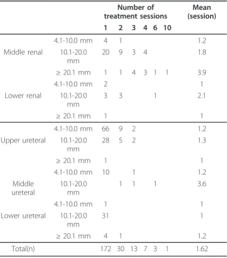
Of the 226 cases, 30 cases underwent placement of a ureteral stent and 14 cases underwent IVP. When the stones were classified by location, middle renal stones were the most frequent (52 cases) among subjects with renal stones, while upper ureteral stones (113 cases) were the most frequent among subjects with ureteral stones. When the stones were classified by size, those 10.1 - 20.0 mm were the most frequent (43 cases) of renal stones, while those 4.1 - 10.0 mm were the most frequent (119 cases) of ureteral stones. One to 10 treat-ment sessions (mean: 1.62 sessions) were performed. Renal stones were treated with a mean of 1.8 sessions, of which middle renal stones of 20.1 mm or larger were treated with the largest number of sessions (mean: 3.9 sessions). Ureteral stones were treated with a mean of 1.44 sessions, of which middle ureteral stones of 10.1 -20.0 mm or larger were treated with the largest number of sessions (mean: 3.6 sessions) (Table 2). One month after operation, the stone-free rate was 48.4% and the efficacy rate was 64.5% for renal stones, while these rates were 70.7 and 80.5%, respectively for ureteral stones. Three months after operation, the stone-free rate was 69.4% and the efficacy rate was 77.4% for renal stones, while these rates were 91.5 and 93.3%, respec-tively for ureteral stones. When the treatment effect was analyzed by the location of stones, a stone-free rate of 94.6% and an efficacy rate of 94.6% were achieved in subjects with lower ureteral stones (≤4.0 mm, 1 subject; 4.1 - 10.0 mm, 31 subjects; 10.1 - 20.0 mm, 5 subjects) with one or two ESWL treatment sessions (mean: 1.03 sessions) at three months after operation (Table 3). All five subjects with stones 10 mm or larger achieved a complete stone-free status.
Fourteen subjects with X-ray-negative stones under-went intravenous pyelography (IVP) and ureteral cathe-ter insertion in combination with ESWL. These patients had a stone-free status with only ESWL therapy.
Of the 25 cases in whom ESWL was ineffective, 8 cases were confirmed to be stone free three or more months after operation, while 7 cases with residual stones were followed up because hydronephrosis improved. Of the subjects with upper ureteral stones in whom ESWL was ineffective, 7 cases underwent TUL and 2 cases underwent PNL. One case with concomitant ureteral stenosis underwent holmium laser incision fol-lowing stone fragmentation with TUL.
The chemical composition of stones could be deter-mined in 128 cases. Calcium oxalate stones were the
Table 1 Location and size of stones
≤4.0 mm
4.1-10.0 mm
10.1-20.0
mm ≥
20.1 mm
Total (n)
Middle renal 5 36 11 52
Lower renal 2 7 1 10
Upper ureteral
77 35 1 113
Middle ureteral
11 3 14
Lower ureteral
1 31 5 37
most frequent (68 cases), followed by mixed calcium oxalate and calcium phosphate stones (49 cases), and stones containing calcium were present in 126 cases.
All subjects experienced postoperative gross hematuria as a complication. One case developed renal subcapsular hematoma which improved with conservative treatment. One case developed fever of 38°C or higher, which resolved with antibiotic therapy. Fifteen subjects required pentazocine for pain during the operative procedure.
Discussion
Since Chaussy et al. [1] reported the application of ESWL with an HM-3 lithotriptor manufactured by Dor-nier Co., Ltd. in clinical practice in the 1980 s, various models of ESWL equipment have been developed and improved. Shock wave generators have been developed, starting with the underwater spark gap type, followed by the electromagnetic conversion type and the piezoelec-tric element type. ESWL is now the first-line treatment for upper urinary tract stones.
Our hospital introduced the Sonolith vision (manufac-tured by EDAP) on March 1, 2004. This model belongs to third generation ESWL equipment. It uses electrical conduction electrodes as the shock wave generator, which generates shock waves in a highly electrical-con-ductive fluid. It provides accurate electrical discharge with high reproducibility, thus producing stable and constant energy. The use of a hydrophone pressure detector allows real-time display of the effective pressure of shock waves on a monitor. The generator is a shallow oval shape, and the output power can be adjusted to 100 different levels. The diameter of the shock-wave head is as large as 22 cm, and the consumptive electrodes can be used in four to five patients. This equipment has a focal depth of 130 mm and a focal size of 3 × 28 mm. The focal point is adjusted using an X-ray C-arm.
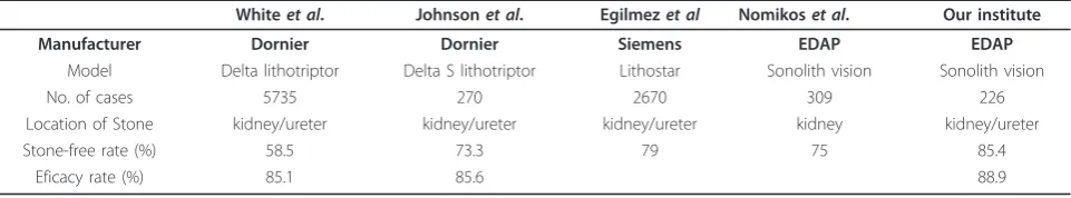
In 226 Japanese cases with renal or ureteral stones who were treated with ESWL in our hospital, the stone-free rate was 85.4% and the efficacy rate was 88.9%, showing similar results to those obtained by other researchers [2-5]. (Table 4).
With regard to lower ureteral stones, Hochreiteret al. [6] treated distal ureteral stones with the HM-3 litho-triptor manufactured by Dornier in 518 patients, and reported a stone-free rate of 97.3% and efficacy rate of 99.4%. Park et al.[7], however, reported that the stone-free rate was 55.6% for lower ureteral stones of 10 mm
Figure 1A) ESWL approaches to middle ureteral stones. (Ipsilateral prone position). B) ESWL approaches to lower ureteral stones. (Contralateral prone position)
Table 2 Location and size stones and number of treatment
Number of treatment sessions
Mean (session) 1 2 3 4 6 10
4.1-10.0 mm 4 1 1.2
Middle renal 10.1-20.0 mm
20 9 3 4 1.8
≥20.1 mm 1 1 4 3 1 1 3.9
4.1-10.0 mm 2 1
Lower renal 10.1-20.0 mm
3 3 1 2.1
≥20.1 mm 1 1
4.1-10.0 mm 66 9 2 1.2
Upper ureteral 10.1-20.0 mm
28 5 2 1.3
≥20.1 mm 1 1
4.1-10.0 mm 10 1 1.2
Middle ureteral
10.1-20.0 mm
1 1 1 3.6
4.1-10.0 mm 1 1
Lower ureteral 10.1-20.0 mm
31 1
≥20.1 mm 4 1 1.2
or larger. Pardalidiset al.[8] also reported that treat-ment was not so effective, with a stone-free rate of 84.6%. Ghalayiniet al. [10] compared laser TUL with ESWL, and reported that laser TUL was significantly more effective than ESWL, with a stone-free rate of 97.5% vs. 81.5% three months after operation. Wu et al. [11] compared holmium yttrium-aluminum-garnet (YAG) laser TUL with ESWL in patients with upper ureteral stones, and reported that there was no signifi-cant difference in the treatment effect on stones 10 mm or smaller, with a stone-free rate of 91.1% for TUL vs. 85.3% for ESWL, while there was a significant difference in the treatment effect on stones 10 mm or larger, with a stone-free rate of 76.8% for TUL vs. 35.2% for ESWL. They stated that laser TUL was superior to ESWL for the treatment of stones 10 mm or larger. In our study, however, all subjects with stones 10 mm or larger achieved a stone-free status. This may be attributable to our new measures: we usually place patients in the con-tralateral prone position to potentiate the treatment effect on lower ureteral stones, in accordance with the report by Köse et al. [12], and before ultrasonography we also apply jelly in a thin layer to the skin surface and to the area on the treatment table around the spot in contact with the skin to avoid exposure to air, thereby reducing minute air bubbles generated from the skin surface and minimizing attenuation of shock waves. ESWL seems to be an effective first-line treatment also in subjects who have lower ureteral stones 10 mm or larger but do not wish to undergo TUL, but it is some-times difficult to treat stones greater than 10 mm by
ESWL monotherapy. So, it is necessary to perform ESWL and TUL combination therapy. In contrast, these subjects with X-ray-negative stones underwent intrave-nous pyelography (IVP) and ureteral catheter insertion in combination with ESWL. These patients of ours had a stone-free status with only ESWL therapy.
Madboulyet al.[13] recently reported that the treat-ment effect of ESWL with the Lithostar Multiline per-formed under general anesthesia was significantly greater when shock waves were delivered at a slow rate (60/min) than at a rapid rate (120/min), with an efficacy rate of 98.7% vs. 90.0%. Paceet al.[14] and Petersonet
al.[15] also reported similar results. Thus, ESWL treat-ment should be performed at a slow rate in the future.
Complications of ESWL include renal subcapsular hematoma, the incidence of which has been reported to be between 0.078% and 0.6% [1,16], and has also been reported to increase up to 32% as a result of perfor-mance of postoperative routine CT and MRI [17]. Risk factors for renal subcapsular hematoma are reported to be 1) hypertension, 2) coagulopathy, and 3) previous ESWL therapy [18]. In our study, renal subcapsular hematoma occurred in one subject. This subject had a history of hypertension, which may have caused this complication.
Conclusions
ESWL with the Sonolith vision manufactured by EDAP produced a treatment effect equivalent to those achieved with other models of ESWL equipment. ESWL seems to be an effective first-line treatment also in patients who
Table 3 Treatment results by location
1 month 3 months
Site No of pts Stone-free rate Efficacy rate Stone-free rate Efficacy rate
Middle renal 52 53.80% 69.20% 71.20% 78.80%
Lower renal 10 20.00% 40.00% 60.00% 70.00%
Total (renal) 62 48.40% 64.50% 69.40% 77.40%
Upper ureteral 113 73.50% 83.20% 89.40% 92.00%
Middle ureteral 14 64.30% 71.40% 100% 100%
Lower ureteral 37 64.90% 75.70% 94.60% 94.60%
Total (ureteral) 164 70.70% 80.50% 91.50% 93.30%
Table 4 Treatment results by model
Whiteet al. Johnsonet al. Egilmezet al Nomikoset al. Our institute
Manufacturer Dornier Dornier Siemens EDAP EDAP
Model Delta lithotriptor Delta S lithotriptor Lithostar Sonolith vision Sonolith vision
No. of cases 5735 270 2670 309 226
Location of Stone kidney/ureter kidney/ureter kidney/ureter kidney kidney/ureter
Stone-free rate (%) 58.5 73.3 79 75 85.4
have lower ureteral stones 10 mm or larger but do not wish to undergo TUL, if measures such as suitable posi-tioning of patients during treatment are taken.
Acknowledgements
Written consent was obtained from the patient and his family prior to publication of this manuscript.
Author details
1Department of Urology, Aichi Medical University School of Medicine
Nagakute, Aichi 480-1195, Japan.2Department of Urology, Meitetsu Hospital Nagoya, Aichi 451-8511, Japan.
Authors’contributions
KN and MT drafted the report, cared for the patient and approved the final version of the manuscript. MN, TY, GN, YK, RK, KZ, SA, YY and NH cared for the patient and approved the final version of the manuscript. MS approved the final version of the manuscript.
Competing interests
The authors declare that they have no competing interests.
Received: 5 April 2011 Accepted: 12 December 2011 Published: 12 December 2011
References
1. Chaussy C, Schuller J, Schmiedt E, Brandl H, Jocham D, Liedl B:
Extracorporeal shock-wave lithotripsy (ESWL) for treatment of urolithiasis.Urology1984,23:59-66.
2. White W, Klen F:Five-year clinical experience with the Dornier Delta lithotripter.Urology2006,68:28-32.
3. Johnson DB, Lowry PS, Schluckebier JA, Kryger JV, Nakada SY:University of Wisconsin experience using the Doli S lithotripter.Urology2003,
62:410-415.
4. Egilmez T, Tekin MI, Gonen M, Kilinc F, Goren R, Ozkardes H:Efficacy and safety of a new-generation shockwave lithotripsy machine in the treatment of single renal or ureteral stones: Experience with 2670 patients.J Endourol2007,21:23-27.
5. Nomikos MS, Sowter SJ, Tolley DA:Outcomes using a fourth-generation lithotripter: a new benchmark for comparison.BJU Int2007,
100:1356-1360.
6. Hochreiter WW, Danuser H, Perrig M, Studer UE:Extracorporeal shock wave lithotripsy for distal ureteral calculi: What a powerful machine can achieve.J Urol2007,169:878-880.
7. Park H, Park M, Park T:Two-year experience with ureteral stones: Extracorporeal shockwave lithotripsy v ureteroscopic manipulation.J Endourol1998,12:501-504.
8. Pardalidis NP, Kosmaoglou EV, Kapotis CG:Endoscopy vs. extracorporeal shockwave lithotripsy in the treatment of distal ureteral stone: ten years’ experience.J Endourol1999,13:161-164.
9. Pearle MS, Nadler R, Bercowsky E, Chen C, Dunn M, Figenshau RS, Hoenig DM, McDougall EM, Mutz J, Nakada SY, Shalhav AL, Sundaram C, Wolf JS Jr, Clayman RV:Prospective randomized trial comparing shock wave lithotripsy and ureterosopy for management of distal ureteral calculi.J Urol2001,166:1255-1260.
10. Ghalayini IF, AI-Ghazo MA, khader YS:Extracorporeal shockwave lithotripsy versus ureteroscopy for distal ureteric calculi: Efficacy and patient satisfaction.Int Braz J Urol2006,32:656-664.
11. Wu CF, Chen CS, Lin WY, Shee JJ, Chen Y, Huang WS:Therapeutic options for proximal ureter stone: Extracorporeal shock wave lithotripsy versus semirigid ureterorenoscope with Holmium: Yttrium-aluminum-garnet laser lithotripsy.Urology2005,65:1075-1079.
12. Köse AC, Demirbas M:The modified prone position: a new approach for treating prevesical stones with extracorporeal shock wave lithotripsy.
BJU Int2004,93:369-373.
13. Madbouly K, El-Tiraifi AM, Seida M, El-Faqih SR, Atassi R, Talic RF:Slow versus fast shock wave lithotripsy rate for urolithiasis: A prospective randomized study.J Urol2005,173:127-130.
14. Pace KT, Ghiculete D, Harju M:Shock wave lithotripsy at 60 or 120 shocks per minute: A randomized, double-blind trial.J Urol2005,174:595-599. 15. Paterson RF, Lifshitz DA, Lingeman JE, Evan AP, Connors BA, Fineberg NS,
Williams JC Jr, McAteer JA:Stones fragmentation during shock wave lithotripsy is improved by slowing the shock wave rate: Study with a new animal model.J Urol2002,168:2211-2215.
16. Newman LH, Saltzman B:Identifying risk factors in development of clinically significant post-shock-wave lithotripsy subcapsular hematomas.
Urology1991,38:35-38.
17. Knapp PM, Kulb TB, Lingeman JE, Newman DM, Mertz JH, Mosbaugh PG, Steele RE:Extra corporeal shock wave lithotripsy-induced perirenal hematomas.J Urol1998,139:700-703.
18. Collado Serra A, Huguet Pérez J, Monreal García de Vicuńa F, Rousaud Barón A, Izquierdo de la Torre F, Vicente Rodríquez J:Renal hematoma as a complication of extracorporeal shock wave lithotripsy.Scand J Urol Nephrol1999,33:171-175.
Pre-publication history
The pre-publication history for this paper can be accessed here: http://www.biomedcentral.com/1471-2490/11/26/prepub
doi:10.1186/1471-2490-11-26
Cite this article as:Nakamuraet al.:Treatment of upper urinary tract stones with extracorporeal shock wave lithotripsy (ESWL) Sonolith vision.BMC Urology201111:26.
Submit your next manuscript to BioMed Central and take full advantage of:
• Convenient online submission
• Thorough peer review
• No space constraints or color figure charges
• Immediate publication on acceptance
• Inclusion in PubMed, CAS, Scopus and Google Scholar
• Research which is freely available for redistribution